13:30
|
 |
Tissue Characterization: Brain 
Sean Deoni
MRI offers of wealth of information that indirectly
informs ontissue microstructure and organization.
Imaging methods, including qualitative T1, T2 and
proton density weighted imaging provide a foundation for
assessing gross brain morphology and cortical
morphometry. Beyond this, quantitative methods,
including diffusion tensor, magnetisation transfer, and
relaxometry can be used to assess more specific
attributes of tissue microstructure and architecture.
In this presentation, we will briefly overview these
methods, with emphasis on relaxometry analysis to
interrogate brain microstructure
|
13:50
|
|
Tissue Characterisation:
Heart
Reza Nezafat1
1Harvard
|
14:10
|
|
Extra-Hepatic Steatosis: New Opportunities and Challenges in
Quantitative MR
Takeshi Yokoo1
1UT Southwestern Medical Center
Abnormal lipid metabolism is associated with obesity,
resulting in accumulation of fat in non-adipose tissues
– a process called steatosis. Steatosis has long been
known to occur in the liver and skeletal muscle, but
also occurs in other organs including the pancreas,
heart, and the kidneys, with potential significant
pathophysiological implications. In this educational
session, we will discuss the clinical significance of
extra-hepatic steatosis and the value of quantitative MR
in its noninvasive evaluation, as well as future
research opportunities and technical challenges.
|
14:30
 |
0985.
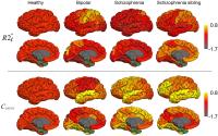 |
Structural and hemodynamical contributions to brain T2*
relaxation in schizophrenia, bipolar disorder and siblings
Jie Wen1, Daniel Mamah2, Jie Luo3,
Xialing Ulrich1, Deanna Barch4,
and Dmitriy Yablonskiy1
1Radiology, Washington University, Saint
Louis, MO, United States, 2Psychiatry,
Washington University, Saint Louis, MO, United States, 3Research
Lab of Electronics, MIT, Cambridge, MA, United States, 4Psychology,
Washington University, Saint Louis, MO, United States
Investigating brain structure and functioning by means
of tissue-specific T2* relaxation properties in vivo can
potentially guide the uncovering of neuropathology in
psychiatric illness. In this abstract, R2* (=1/T2*)
relaxation rate constant was separated into
tissue-specific (R2*t) and hemodynamic BOLD
contributions. 17 control, 17 bipolar disorder, 16
schizophrenia, and 12 unaffected schizophrenia sibling
participants were scanned. A MANOVA of 38 gray matter
regions showed significant group effects for BOLD but
not for R2*t. Our results suggest that
increased baseline activity in certain brain regions is
part of the underlying pathophysiology of specific
psychiatric disorders.
|
14:42
|
0986.
 |
Radial MOLLI sequence for fast, precise and accurate
myocardium T1 mapping
Benjamin Marty1,2, Bertrand Coppa1,2,
and Pierre G Carlier1,2
1NMR laboratory, Institute of Myology, Paris,
France, 2NMR
laboratory, CEA, I2BM, MIRCen, Paris, France
Quantitative cardiac NMR imaging, and more particularly
T1 mapping has become a popular modality to characterize
myocardial tissue. In this work, we developed and
validated a radial variant of the MOLLI acquisition
(raMOLLI) that allows to significantly decrease the
acquisition time down to 5 heart beats, while keeping
high precision on T1 estimation due to a large number of
acquired data-points along the T1 relaxation recovery
curve. Insensitivity of measured T1 values to heart rate
was also demonstrated with this sequence.
|
14:54
|
0987.
 |
Cardiac Magnetic Resonance Reveals Signs of Subclinical
Myocardial Inflammation in Asymptomatic HIV-infected
Patients
Julian Alexander Luetkens1, Jonas Doerner1,
Carolynne Schwarze-Zander2, Jan- Christian
Wasmuth2, Christoph Boesecke2,
Alois M Sprinkart1, Frederic C Schmeel1,
Rami Homsi1, Juergen Gieseke3,
Hans H Schild1, and Claas P Naehle1
1Radiology, University of Bonn, Bonn,
Germany, 2Internal
Medicine I, University of Bonn, Bonn, Germany, 3Philips
Research, Hamburg, Germany
People living with chronic human immunodeficiency virus
(HIV) infection are at an increased risk for
cardiovascular disease. In the present study we
investigated HIV-infected patients, which were
controlled for the disease, using multiparametric
cardiovascular magnetic resonance (CMR). With this CMR
approach we could demonstrate that HIV-infected patients
without cardiac symptoms not only have subtle evidence
of impaired myocardial function, but also elevated
markers of myocardial inflammation and increased
myocardial fibrosis. These findings indicate subclinical
myocardial inflammation in HIV-infected patients despite
effective antiretroviral therapy, and therefore may
contribute to the persistently increased cardiovascular
morbidity and mortality observed in these patients.
|
15:06
|
0988.
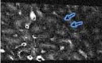 |
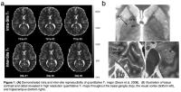
MR Imaging of Liver Microstructure in Hepatic Fibrosis and
Cirrhosis at 11.7 T - Permission Withheld
Mark Valasek1, Qun He2,3, Claude
Sirlin2, Graeme M. Bydder2, and
Nikolaus M. Szeverenyi2
1Pathology, University of California, San
Diego, San Diego, CA, United States, 2Radiology,
University of California, San Diego, San Diego, CA,
United States, 3Ningbo
Jansen NMR Technology Co., Ltd., Cixi, Zhejiang, China,
People's Republic of
We performed MR microscopy at 11.7 T to examine the
tissue structure of normal, fibrotic and cirrhotic liver
samples. Images having 100-1,000 times the spatial
resolution of clinical MR images were obtained in small
tissue samples using an animal imaging system with
appropriately small custom T/R solenoid coils.
Diffusion imaging with three direction of sensitization
revealed sheet like fibrous structures, exhibiting high
signal intensity in regions where the sensitization
direction was orthogonal to a sheet.
|
15:18
|
0989.
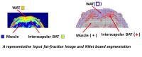 |
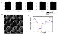
An Automatic Machine learning Approach for multi-parametric
MR based Brown adipose tissue characterization and
Segmentation in mice and rats
Bhanu Prakash KN1, Hussein Srour 1,2,
Sanjay Kumar Verma1, Jadegoud Yaligar1,
Venkatesh Gopalan1, Swee Shean Lee1,
Kai Hsiang Chuang 1,2,
and Sendhil Velan S1,3
1Laboratory of Metabolic Imaging, Singapore
Bioimaging Consortium, Singapore, Singapore, 2Queensland
Brain Institute, Brisbane, Australia, 3MRS
& Metabolic Imaging Group, Singapore Institute for
Clinical Sciences, Singapore, Singapore
We have utilized multiparametric
MR images (fat-fraction (FF), T2 and
T2*) of adipose tissues and evaluated
different segmentation algorithms like multidimensional
thresholding, region growing, clustering, and machine
learning approach for its suitability and efficacy to
separate WAT from BAT depots. A
machine learning algorithm i.e. Neural Network
based segmentation provided increased specificity
compared to other algorithms. This methodology can be
easily extended for multi-parametric human images and
longitudinal studies.
|
15:30
|
|
Adjournment & Meet the
Teachers |
|