16:00
|
|
Approach to Intracranial Hemorrhage. 
Jalal B. Andre1
1Radiology, University of Washington,
Seattle, WA, United States
Intracranial hemorrhage (ICH) has been traditionally
classified as intra- versus extra-axial in location, and
can arise from a variety of etiologies. This talk will
focus on the importance of location, timing, and source
of hemorrhage through a case-based exploration of ICH
and associated complications as they pertain to the
following locations: 1) Intraventricular 2)
Intraparenchymal 3) Subarachnoid 4) Subdural 5) Epidural
|
16:30
|
1064.
 |
Multi-contrast joint intra- and extracranial artery wall
imaging – a feasibility study
Lei Zhang1, Jun Wu2, Lijie Ren3,
Tingting Wang2, Xin Liu1, and Yiu-Cho
Chung1
1Paul C. Lauterbur Center for Biomedical
Imaging, Shenzhen Institutes of Advanced Technology,
Chinese Academic of Sciences, Shenzhen, China, People's
Republic of, 2Neurology,
Peking University Shenzhen Hospital, Shenzhen, China,
People's Republic of, 3Neurology,
Shenzhen Second People’s Hospital, Shenzhen, China,
People's Republic of
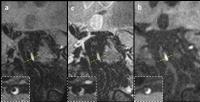
In this study, we evaluated multi-contrast three
dimensional (3D) high resolution black blood vessel wall
imaging technique for joint intra- and extracranial
artery wall imaging in 8 stroke patients. The new
techniques covered both the intra- and extracranial
segments in one scan. T1w, T2w scans were performed.
MPRAGE was included when hemmorrhage was suspected. 8
plaques were identified. Two of them had high signal in
all three sequences, suggestive of intraplaque
hemorrhage. The 3D multi-contrast large coverage black
blood techniques would be a promising tool to the study
on the association between atherosclerotic plaques and
ischemic stroke.
|
16:45
|
1065.
 |
A multiple comparison between 3T intracranial vessel wall
sequences
Arjen Lindenholz1, Anita Harteveld1,
Jeroen Siero1, Jaco Zwanenburg1,
and Jeroen Hendrikse1
1Medical Imaging, UMC Utrecht, Utrecht,
Netherlands
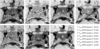
In this study we optimized high resolution magnetic
resonance vessel wall imaging regarding scan duration
signal-to-noise (SNR) and contrast-to-noise (CNR). We
compared the SNRs and CNRs two clinically used
intracranial vessel wall sequences with 5 variants with
various trade-offs between scan time, resolution and
contrast between vessel wall and cerebrospinal fluid
(CSF). Compared to the clinically used sequences, we
developed a sequence which was considerably
faster and had comparable or higher SNRs and CNRs that
resulted in a good visibility of the intracranial vessel
wall.
|
17:00
|
|
Neurovascular Imaging Techniques
Kevin M Johnson1
1Medical Physics, University of Wisconsin -
Madison, WI, United States
This overview talk discusses current imaging techniques
used for the evaluation of patients at risk for or
following hemorrhage. In particular, it provides insight
into the state of imaging techniques used to image
vascular origins and the growing abilities to correlate
vascular structure interactions.
|
17:30
|
1066.
 |
The performances evaluation of 32-channel coil system for
extracranial and intracranial artery wall imaging at 3T
Xiaoqing Hu1, Lei Zhang1, Xiao
Chen1, Xiaoliang Zhang2,3, Xin Liu1,
Hairong Zheng1, Yiu-Cho Chung1,
and Ye Li1
1Paul C. Lauterbur Research Center for
Biomedical Imaging, Shenzhen Institutes of Advanced
Technology, CAS, Shenzhen, China, People's Republic of, 2Department
of Radiology and Biomedical Imaging, University of
California San Francisco, San Francisco, CA, United
States, 3UCSF/UC
Berkeley Joint Graduate Group in Bioengineering, San
Francisco, CA, United States
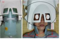
Atherosclerosis is a major cause of ischemic stroke. The
high resolution magnetic resonance imaging (MRI) of
vessel wall can detect nonstenotic atherosclerotic
plaque missed by luminal angiography. To develop a
multi-channel radiofrequency (RF) coil system with high
spatial resolution and large longitudinal coverage for
the intracranial and extracranial arteries vessel wall
imaging in one setting. The high resolution images with
0.6 mm3 are
obtained with the proposed “24+8” channel coil system
from a patient in vivo.
|
17:45
 |
1067.
 |
Vessel wall thickness measurements of the circle of Willis
using 7.0T MRI
Anita A. Harteveld1, Anja G. van der Kolk1,
Nerissa P. Denswil2, Jeroen C.W. Siero1,
Hugo J. Kuijf3, Aryan Vink4, Wim
G.M. Spliet4, Peter R. Luijten1,
Mat J. Daemen2, Jaco J.M. Zwanenburg1,3,
and Jeroen Hendrikse1
1Radiology, University Medical Center
Utrecht, Utrecht, Netherlands, 2Pathology,
Academic Medical Center, Amsterdam, Netherlands, 3Image
Sciences Institute, University Medical Center Utrecht,
Utrecht, Netherlands, 4Pathology,
University Medical Center Utrecht, Utrecht, Netherlands
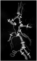
In the last decade, several MRI sequences have been
developed for direct visualization of the intracranial
vessel wall. Although much is known about vessel wall
(intima-media) thickness of extracranial arteries, less
is known about the intracranial arterial vessel wall. In
the current study, vessel wall thickness of major
intracranial arteries was measured in ex
vivo samples
of the circle of Willis, using 7T MRI and histological
validation, to ultimately provide a reference guide for
normal intracranial vessel wall thickness. The results
show that ultrahigh-resolution MRI at 7T enables
accurate measurement of vessel wall thickness in ex
vivoCoW specimens.
|
18:00
|
|
Adjournment & Meet the
Teachers |
|