10:30
|
|
Imaging Membrane & Protein Metabolism 
Kristine Glunde
This presentation will provide an overview of current 1H
and 31P
magnetic resonance spectroscopy (MRS) approaches as well
as chemical exchange saturation transfer (CEST) and
amide proton transfer (APT) techniques that detect
membrane and protein metabolism in cancer, along with a
discussion of the detected molecules in the realm of
cancer diagnosis and treatment monitoring.
|
10:50
|
|
Imaging Neurotransmission - Permission Withheld
In-Young Choi1
1University of Kansas Medical Center
|
11:10
|
|
Imaging Energy Metabolism
Craig R. Malloy1
1University of Texas Southwestern
|
11:30
 |
0890.
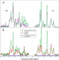 |
Metabolic profiling of in
vivo brain
rodent models by relaxation-enhanced 1H
MRS of the downfield region at 21.1 T
Tangi Roussel1, Jens T. Rosenberg2,
Samuel Colles Grant2,3, and Lucio Frydman1,2
1Chemical Physics, Weizmann Institute of
Science, Rehovot, Israel, 2Center
for Interdisciplinary MR, National High Magnetic Field
Laboratory, Tallahassee, FL, United States, 3Chemical
& Biomedical Engineering, Florida State University,
Tallahassee, FL, United States
This study explores new opportunities that ultra-high
field combined with non-water-suppressed 1H
MRS methodologies make possible regarding the profiling
of signals that resonate downfield from the water peak.
Studies were carried out on rats using a 21.1-T ultra-widebore
system, and focused on quantitatively analyzing the
metabolic concentration changes for ischemic stroke and
glioblastoma tissues. A general decrease in the relative
metabolic concentrations were observed for both
pathologies, certain molecules depart from this trend:
lactate, glutathione (stroke), choline and UDP-Nacetyl
hexosamines (glioma). Potential explanations for these
features and new research avenues opened by these types
of measurements are discussed.
|
11:42
|
0891.
 |
Study of the Mutated Isocitrate Dehydrogenase 1 in Acute
Myeloid Leukemia Using Hyperpolarized [1-13C]a-ketoglutaric
Acid
Eugen Kubala1,2,3, Kim A. Muñoz Álvarez1,
Oliver Dovey4, Steffen J. Glaser2,
Markus Schwaiger1, George S. Vassiliou4,
Roland Rad5,6, Rolf F. Schulte3,
and Marion I. Menzel3
1Department of Nuclear Medicine, Klinikum
Rechts der Isar, Technische Universität München, Munich,
Germany, 2Department
of Chemistry, Technische Universität München, Munich,
Germany, 3General
Electric Global Research, Munich, Germany, 4The
Welcome Trust Sanger Institute, Hinxton/Cambridge,
United Kingdom, 5Department
of Medicine II, Klinikum Rechts der Isar, Technische
Universität München, Munich, Germany, 6Cancer
Consortium (DKTK), German Cancer Research Center (DKFZ),
Munich, Germany
Previous studies suggest that isocitrate
dehydrogenase 1 (IDH1)
mutation plays a significant role in the cancerous
metabolome. Among other alternations, expression of branched
chain amino-acid transaminase 1(BCAT1) is
reduced, causing a decrease of α-ketoglutaric acid (αKG)
to glutamic acid metabolic pathway. More importantly,
the mutated IDH1 catalyzes
a reaction of αKG to the oncometabolite
2-hydroxyglutarate. In this study we proved that these
metabolic changes can be measured using hyperpolarized
[1-13C]α-KG and 13Cmetabolic
magnetic resonance spectroscopy (13CMMRS) in
acute myeloid leukemia cell line in vitro.
|
11:54
|
0892.
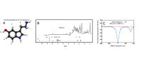 |
CEST Imaging of the Serotonin Pathway
Rafal Janik1, Lynsie A.M. Thomason2,
and Greg J. Stanisz1,2,3
1Medical Biophysics, University of Toronto,
Toronto, ON, Canada, 2Physical
Sciences, Sunnybrook Research Institute, Toronto, ON,
Canada, 3Department
of Nerurosurgery and Pediatric Neurosurgery, Medical
University of Lublin, Lublin, Poland
A novel method for the detection of brain 5-HT,
tryptophan, and 5-HIAA is presented. The method relies
on the chemical exchange of an amide proton which is
shifted outside the normal range for amide protons. This
is demonstrate in-vivo in a rat model of 5-HT increase.
|
12:06
|
0893.
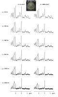 |
Towards “non-invasive histology” of the brain by
diffusion-weighted MR spectroscopy in vivo: comparison
between diffusion-extracted synthetic cells and real
histology in the mouse and primate brain
Marco Palombo1,2, Clémence Ligneul1,2,
Chloé Najac1,2, Juliette Le Douce1,2,
Julien Flament1,2, Carole Escartin1,2,
Philippe Hantraye1,2, Emmanuel Brouillet1,2,
Gilles Bonvento1,2, and Julien Valette1,2
1CEA/DSV/I2BM/MIRCen, Fontenay-aux-Roses,
France, 2CNRS
Université Paris-Saclay UMR 9199, Fontenay-aux-Roses,
France
A new diffusion-weighted MRS paradigm, combining
advanced modeling with metabolites diffusion
measurements at long diffusion times, is applied in the
mouse and macaque brain in vivo. Resulting synthetic
astrocytes and neurons (derived from cell-specific
metabolite diffusion) can be compared with histological
data. Very good agreement between Sholl analysis on real
and synthetic astrocytes validates our approach and
assumptions. We also measure increased size and
complexity of synthetic astrocytes in primate compared
to mouse, while dendritic organization appears better
conserved throughout species. Although still in its
infancy, our strategy opens new perspectives for the
non-invasive evaluation of brain cell morphology.
|
12:18
|
0894.
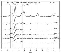 |
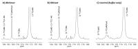
In vivo 1H
and 31P
MR spectroscopy in healthy fibroglandular breast tissue at 7
Tesla. - Permission Withheld
Wybe JM van der Kemp1, Bertine L Stehouwer1,
Vincent O Boer1, Peter R Luijten1,
Dennis WJ Klomp1, and Jannie P Wijnen1
1Radiology, UMC Utrecht, Utrecht, Netherlands
Water and fat suppressed 1H total choline MR
spectroscopy and 31P
MR spectroscopy were performed in healthy fibroglandular
breast tissue of a group of 8 volunteers. 31P T2 values
were determined, and reproducibility of 1H
and 31P
MR spectroscopy were investigated. The 1H
and 31P
data were combined to calculate estimates of absolute
concentrations of PC, GPC and PE.
|
12:30
|
|
Adjournment & Meet the
Teachers |
|