10:00
|
|
Alzheimer's Disease 
Tammie Benzinger1
1Washington University in Saint Louis, School
of Medicine; Mallinckrodt Institute of Radiology
|
10:30
 |
0627.
 |
Neurodegeneration simulation in the connectome: a heuristic
approach to unfold the key white matter pathways in
Alzheimer’s disease
Matteo Mancini1, Marcel A. de Reus2,
Laura Serra3, Marco Bozzali3,
Martijn van den Heuvel2, Mara Cercignani3,4,
and Silvia Conforto1
1Department of Engineering, University of
Rome "Roma Tre", Rome, Italy, 2Department
of Psychiatry, Brain Center Rudolf Magnus, University
Medical Center Utrecht, Utrecht, Netherlands, 3Neuroimaging
Laboratory, IRCSS Santa Lucia Foundation, Rome, Italy, 4Brighton
& Sussex Medical School, Clinical Imaging Sciences
Centre, University of Sussex, Brighton, United Kingdom
In order to identify the white matter impairment that
could lead to Alzheimer’s disease (AD), we combined
computational simulations with a graph theoretical
approach. We reconstructed the structural connectome of
AD patients and healthy controls by means of diffusion
tensor imaging, and characterized the differences
between the two groups using graph theoretical measures.
We then simulated neurodegeneration processes in the
controls using two different heuristic algorithms. We
were able to reproduce the AD disruption pattern in the
controls, and we observed a relevant role of the
connections between hubs and peripheral regions in the
simulated damaging process.
|
10:45
|
0628.
 |
A New Biomarker for Neuroinflammation in Preclinical
Alzheimer’s disease Progression
Yong Wang1,2,3, Qing Wang2,4,
Joshua S Shimony2, Anne M Fagan4,5,
John C Morris5,6, and Tammie L.S. Benzinger2,6,7
1Obstetrics and Gynecology, Washington
University in St. Louis, St. Louis, MO, United States, 2Mallinckrodt
Institute of Radiology, Washington University in St.
Louis, St. Louis, MO, United States, 3Biomedical
Engineering, Washington University in St. Louis, St.
Louis, MO, United States, 4Knight
Alzheimer's Disease Research Center, St. Louis, MO,
United States, 5Neurology,
Washington University in St. Louis, St. Louis, MO,
United States, 6Knight
Alzheimer’s Disease Research Center, St. Louis, MO,
United States, 7Neurosurgery,
Washington University in St. Louis, St. Louis, MO,
United States
The preclinical pathophysiology of Alzheimer’s disease
(AD) is not limited to the neuronal compartments.
Neuroinflammation characterized by activation of
microglia and astrocytes may contribute as much to AD
disease pathogenesis as do amyloid plaques and
neurofibrillary tangles. We demonstrated that a novel
magnetic resonance imaging technique, diffusion basis
spectrum imaging (DBSI), can accurately image
neuroinflammation changes that occur in preclinical AD
patients. DBSI neuroinflammation biomarker can be used
to identify asymptomatic subjects at highest risk of
developing dementia, and lead to effective new AD
disease-modifying therapies targeting neuroinflammation.
|
11:00
|
|
Network-sensitive structural and functional MR imaging
methods
Juan Zhou1
1Center for Cognitive Neuroscience,
Neuroscience and Behavioral Disorders Program,
Duke-National University of Singapore Medical School,
Singapore
Each neurodegenerative disease is defined by selectively
vulnerable neurons, regions, networks, and functions, as
well as genetic risk factors. In the past decade, new
network-sensitive neuroimaging methods have made it
possible to test the notion of network-based
degeneration in living humans. In this talk, the basic
theory/preprocessing/data analyses of these methods
including structural covariance networks (MRI),
functional connectivity (fMRI-BOLD) and structural
connectivity (Diffusion MRI) will be introduced. We will
focus on applications of these network-sensitive methods
on two common causes of dementia, Alzheimer's disease
(AD) and frontotemporal dementia. Lastly, important
frontiers in the field of network-based
neurodegeneration will be reviewed.
|
11:30
|
0629.
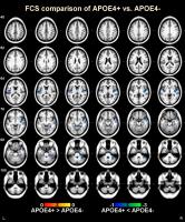 |
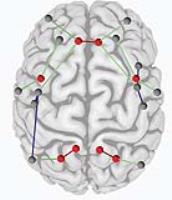
Changed Brain Connectivity in Elderly APOE e4 Carriers: a
Whole-brain Voxel-wise Functional Connectivity Strength
Analysis
Kai Liu1, Teng Zhang1, Yanjia Deng1,
Lin Shi2,3, Defeng Wang4,5, and
ADNI Alzheimer’s Disease Neuroimaging Initiative 6
1Department of Imaging and Interventional
Radiology, The Chinese University of Hong Kong, Hong
Kong, Hong Kong, 2Department
of Medicine and Therapeutics, The Chinese University of
Hong Kong, Hong Kong, Hong Kong, 3Chow
Yuk Ho Technology Centre for Innovative Medicine, The
Chinese University of Hong Kong, Hong Kong, Hong Kong, 4Research
Center for Medical Image Computing, Department of
Imaging and Interventional Radiology, The Chinese
University of Hong Kong, Hong Kong, Hong Kong, 5Shenzhen
Research Institute, The Chinese University of Hong Kong,
Shenzhen, China, People's Republic of, 6Los
Angeles, SC, United States
Apolipoprotein E epsilon 4 allele (APOE-4) is considered
as the strongest genetic risk factor for late-onset
Alzheimer’s disease, and investigation of its
neuropathological effect in normal elderly using
advanced neuroimaging connectivity probes has brought
much research curiosity. In this study, the underlying
abnormal brain connectivity in APOE-4 carriers was
analyzed using the functional connectivity strength
(FCS), which provides a voxel-wise method to explore the
significant connectivity changes at whole-brain level.
The results identified APOE-4 related significant
connectivity decrease in the bilateral insular and left
temporal lobe. We hope these findings could help to shed
light on the APOE-4’s neuropathological mechanism.
|
11:45
|
0630.
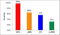 |
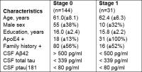
Who will develop Alzheimer’s disease? New insights from
multimodal neuroimaging
Letizia Casiraghi1,2, Fulvia Palesi2,3,
Gloria Castellazzi2,4, Andrea De Rinaldis2,4,
Elena Sinforiani5, Claudia Angela Michela
Gandini Wheeler-Kingshott 2,6,
Egidio D'Angelo1,2, and Carol Di Perri2
1Department of Brain and Behavioral Sciences,
University of Pavia, Pavia, Italy, 2Brain
Connectivity Center, C. Mondino National Neurological
Institute, Pavia, Italy, 3Department
of Physics, University of Pavia, Pavia, Italy, 4Department
of Electrical, Computer and Biomedical Engineering,
University of Pavia, Pavia, Italy, 5Neurology
Unit, C. Mondino National Neurological Institute, Pavia,
Italy, 6NMR
Research Unit, Queen Square MS Centre, Department of
Neuroinflammation, UCL Institute of Neurology,
University College London, London, United Kingdom
Mild cognitive impairment (MCI) is considered a
transitional state between healthy controls (HC) and
Alzheimer’s disease (AD). This study compares the
predictive value of neuropsychological evaluation,
structural magnetic resonance imaging, diffusion tensor
imaging and resting state functional MRI indices able to
identify MCI conversion. AD versus HC and converted MCI
(cMCI) versus non-converted MCI (ncMCI) presented
different features of differentiation. This result
suggests adopting advanced MRI techniques to investigate
early alterations. Due to the clinical heterogeneity of
MCI patients, considering cMCI as AD-like and ncMCI as
HC might be inappropriate when attempting to
distinguishing between cMCI and non-converted MCI.
|
12:00
|
|
Adjournment & Meet the
Teachers |
|