16:00
|
|
Tissue Phase Mapping & more: new Insights into Regional
Cardiac Function 
Bernd Jung1
1University Hospital Bern
The purpose of this talk is the presentation of an
overview of the different MRI approaches to measure
regional cardiac function. Such methods go beyond the
routinely used standard CINE images (providing global
functional parameters such as ventricular volumes) and
include myocardial tagging, DENSE, SENC and Tissue
Phase Mapping. The latter technique measures myocadial
velocities and will be discussed in somewhat more
detail. Some recent studies are presented also including
the determination of strain values from velocity data.
Finally, feature tracking based on SSFP CINE images is
illustrated which can also be used to determine strain
values.
|
16:30
|
0796.
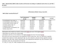 |
Associations between Inflammatory Markers and Global
Systolic Function Measured by MRI: The Multi-Ethnic Study of
Atherosclerosis (MESA)
Amir Ali Rahsepar1, Mohammadali Habibi2,
Cheeling Chan3, Nadine kawel2,
Kiang Liu3, Joao Lima2, and James
Carr1
1Radiology, Northwestern University, Chicago,
IL, United States, 2Cardiology,
Johns Hopkins University, Baltimore, MD, United States, 3Preventive
medicine, Northwestern University, Chicago, IL, United
States
In this cross-sectional study, we investigated the
associations between Inflammatory markers and global
systolic function measured by MRI in The Multi-Ethnic
Study of Atherosclerosis (MESA).
|
16:42
|
0797.
 |
Potential application of tissue phase mapping in early
detection of heart function deficiency in Fabry disease with
cardiac manifestation
Yi-Ting Wu1, Hsu-Hsia Peng2, Meng-Chu
Chang2, Ming-Ting Wu3, and Hsiao-Wen
Chung1
1Graduate Institute of Biomedical Electronics
and Bioinformatics, Taipei, Taiwan, 2Department
of Biomedical Engineering and Environmental Sciences,
National Tsing Hua University, Hsinchu, Taiwan, 3Department
of Radiology, Kaohsiung Veterans General Hospital,
Kaohsiung, Taiwan
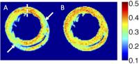
Fabry disease is an X chromosome-linked genetic disease
that can lead to cardiac dysfunction later in life. For
early detection of heart function deficiency, velocity
information in the myocardium obtained in a cardiac
cycle using MR tissue phase mapping (TPM) can
potentially provide a preclinical diagnosis of Fabry
cardiomyopathy. Regional MR TPM analysis was performed
on 7 Fabry disease patients and 22 healthy subjects.
Preliminary results demonstrated significantly delayed
time course as well as decreased velocity amplitudes in
myocardial contractions in the patients. MR TPM may find
useful value in early detection of myocardial defects.
|
16:54
|
|
Innovations in Cardiac Tissue Characterization
Sonia Nielles-Vallespin1,2, Pedro Ferreira2,
Ranil de Silva2, Andrew D Scott2,
Philip Kilner2, Daniel Ennis3,
Eric Aliotta3, Peter Kellman1,
Dimitru Mazilu1, Robert S Balaban1,
Dudley J Pennell2, David N Firmin2,
and Andrew E Arai1
1National Institutes of Health, MD, United
States, 2Imperial
College of London, Royal Brompton Hospital, London,
United Kingdom, 3University
of California, CA, United States
This study shows that helical and laminar
microstructures in the myocardium and their dynamic
reorientations during cardiac contraction can be studied
by in vivo cDTI non-invasively and non- destructively.
Furthermore, it demonstrates in the loaded and beating
heart in vivo that sheetlet reorientation is the
predominant mechanism underlying myocardial LV wall
thickening during systolic contraction. Further study of
the microstructural dynamics of cardiac contraction and
myocardial dysfunction with in vivo cDTI may produce new
diagnostic and prognostic information in human cardiac
disease.
|
17:24
|
0798.
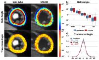 |
Free-breathing Diffusion Tensor Imaging of the In Vivo Human
Heart - Stimulated Echo vs. Spin Echo Acquisition
Constantin von Deuster1,2, Christian T.
Stoeck1,2, Martin Genet2, David
Atkinson3, and Sebastian Kozerke1,2
1Division of Imaging Sciences and Biomedical
Engineering, King's College London, London, United
Kingdom, 2Institute
for Biomedical Engineering, University and ETH Zurich,
Zurich, Switzerland, 3Centre
for Medical Imaging, University College London, London,
United Kingdom
In vivo cardiac Diffusion Tensor Imaging (DTI) using the
Stimulated Echo Acquisition Mode (STEAM) is particularly
challenging during free breathing acquisition. To
address this limitation, spin echo (SE) sequences
employing motion-compensated diffusion gradients may be
used. In this work, scan time, SNR efficiency and
diffusion tensor metrics are compared between the STEAM
method and a second-order motion compensated SE
approach. For SE, SNR and gating efficiency were
increased by 2.65 and 29% relative to STEAM,
respectively. It is concluded that the SE method is an
attractive alternative to STEAM based approaches for in
vivo free-breathing cardiac DTI.
|
17:36
|
0799.
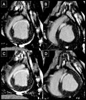 |
Characterization of Myocardial Fiber Orientation to Assess
Therapeutic Exosomes from Cardiosphere-derived Cells (CDCs)
in Myocardial Infarcted Porcine with In Vivo
Diffusion-Tensor CMR on a Clinical Scanner
Christopher Nguyen1, James Dawkins2,
Xiaoming Bi3, Debiao Li1,4, and
Eduardo Marban2
1Biomedical Imaging Research Institute,
Cedars-Sinai Medical Center, Los Angeles, CA, United
States, 2Heart
Institute, Cedars-Sinai Medical Center, Los Angeles, CA,
United States, 3Siemens
Healthcare, Los Angeles, CA, United States, 4Bioengineering,
University of California Los Angeles, Los Angeles, CA,
United States
Diffusion-Tensor cardiovascular magnetic resonance (DT-CMR)
is capable of mapping myocardial fiber orientation. In
myocardial infarction (MI) murine models, DT-CMR can
identify the effects of stem cell therapy on myocardial
fiber orientations. The study illustrated the powerful
potential of DT-CMR in identifying adverse treatment
despite successful delivery of viable stem cells.
However, it remains to be seen if this recent work is
translatable to large animal and clinical studies. In a
MI porcine model, in vivo DT-CMR revealed that
myocardial fiber orientation was preserved with
CDC-derived exosome treatment and adversely changed with
placebo treatment consistent with observed viability and
function changes.
|
17:48
|
0800.
 |
Resolving Microscopic Fractional Anisotropy in the Heart
Irvin Teh1, Henrik Lundell2,
Hannah J Whittington1, Tim Bjørn Dyrby2,
and Jürgen E Schneider1
1Division of Cardiovascular Medicine,
Radcliffe Department of Medicine, University of Oxford,
Oxford, United Kingdom, 2Danish
Research Centre for Magnetic Resonance, Copenhagen
University Hospital Hvidovre, Copenhagen, Denmark
Diffusion tensor imaging (DTI) is widely used for
structural characterization of the heart. However, the
measured fractional anisotropy (FA) is influenced by
diffusion anisotropy as well as orientation dispersion.
In the heart, orientation dispersion is ubiquitous and
stems from the transmural variation in cardiomyocyte
orientation and regions where multiple cell populations
intersect. We propose microscopic FA (µFA) as a more
robust measure of intrinsic diffusion anisotropy that is
insensitive to orientation dispersion, and demonstrate
this with simulations and ex vivo MRI.
|
18:00
|
|
Adjournment & Meet the
Teachers |
|