ISMRM 24th Annual Meeting & Exhibition • 07-13 May 2016 • Singapore |
|
Weekend Educational
Course: An Update on Body MRI Protocols & Applications
Skill Level: Intermediate
Organizers: Ivan Pedrosa, M.D., Lorenzo Mannelli, M.D., Ph.D., Scott B. Reeder, M.D., Ph.D. & Edwin J.R. van Beek, M.D., Ph.D., M.Ed., FRCR
Saturday 07 May 2016 |
Overview
This whole-day course will provide an update of current Body MRI
protocols and clinical applications for evaluation of diseases in the
abdomen and pelvis. Speakers will emphasize on the technical
requirements and the practical aspects in the implementation of
state-of-the-art Body MRI techniques into routine clinical practice. A
comprehensive review of contrast agents for body MRI will be presented.
MRI protocols and indications for assessment of disease in the liver,
bowel, and genitourinary tract will be discussed.Target Audience
This course is aimed at radiologists, imaging scientists and MR
technologists who wish to review the state-of-art MRI protocols and
indications for assessment of disease in the abdomen and pelvis.
Educational Objectives
Upon completion of this course, participants should be able to:
- Convey the equipment and
software requirements to implement a clinical body MRI practice;
- Illustrate an approach to
diagnosing liver lesions;
- Demonstrate the value of MRI
in diagnosis of gastrointestinal disease; and
- Illustrate the use of MRI in the diagnosis of genitourinary
disease.
|
|
PROGRAM |
| |
|
|
Setting Up Your Body MRI Practice |
|
| |
|
|
Moderator:
S. Sendhil Velan |
|
08:00
|
Hardware, Patient Preparation, & Monitoring Considerations
for Body MRI 
Richard Kinh Gian Do1
1Radiology, Memorial Sloan Kettering, New
York, NY, United States
In clinical body MRI, diagnostic radiologists often make
choices in hardware, patient preparation or monitoring
that impact workflow or image quality. In this session,
we will review choices with potential effects in your
day-to-day clinical practice and go through scenarios
centered on body MR protocols.
|
08:30
|
Optimize Your MRI Sequences for Abdomen & Pelvis
Examinations
David J Lomas1
1Radiology, University of Cambridge &
Addenbrooke's Hospital, Cambridge, United Kingdom
The major factors that influence MR sequence
optimisation for abdominal and pelvic exams will be
outlined and discussed. Typical body and pelvic exams
will be used to illustrate the key issues regarding
selection of coils, imaging planes and sequence
parameters.
|
09:00
|
Contrast Agents: Which one Should You Choose?
Ruth P Lim1
1Radiology, Austin Health, Australia
Objectives: 1. To review chemical properties of
commercially available gadolinium based contrast agents
(GBCAs) 2. To review applications of commercially
available GBCAs 3. To review current recommendations
for safe use of GBCAs
|
09:30
|
Break & Meet the Teachers |
|
| |
|
|
Diagnostic Approach to Focal Liver
Lesions |
|
| |
|
|
Moderator: Hebert Alberto Vargas |
|
10:00
|
|
MRI of Lesions in the Non-Cirrhotic Liver
Valerie Vilgrain
|
10:30
|
|
MRI Charaterization of Lesions in the Cirrhotic Liver
Utaroh Motosugi
The most frequent malignant tumor in cirrhotic liver is
hepatocellular carcinoma (HCC). In a typical case, the
imaging-based diagnosis of HCC is simple: hypervascular
in arterial phase and washout in portal-venous/delayed
phase. However, we often encounter atypical cases:
hypovascular HCCs. Gadoxetic acid has advantage in
hepatobiliary phase imaging, which helps distinguish HCC
from pre-malignant lesion. “Hypovascular hypointense
nodule in gadoxetic acid-enhanced MRI” is a new concept
observed in cirrhotic patients, which suggests early HCC
and develop hypervascular (typical) HCC subsequently. In
this lecture, I will cover hypovascular HCCs with a
special emphasis on “hypovascular hypointense nodule”.
|
|
| |
|
|
Gastrointestinal |
|
| |
|
|
Moderators:
Suraj Serai |
|
11:00
|
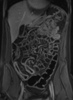 |
MR Enterography - Permission Withheld
Jordi Rimola1
1Radiology, Hospital Clínic Barcelona, Spain
|
11:30
|
|
Rectal CA Staging - Permission Withheld
Bertrand Ang
Highlights · Use of MRI for rectal carcinoma
staging and learn the clinical and therapeutic
implications of rectal carcinoma Target Audience:
Radiologists and MRI technicians.
Objectives: Understand the anatomical and
pathological basis for MRI rectal carcinoma staging and
its impact on therapeutic options.
Methods: Basic anatomy
and pathology of rectal carcinoma will be introduced
followed by case examples.
Results: Participants will be able to understand the
important anatomical and pathological MRI findings in
rectal carcinoma.
|
12:00
|
|
Lunch & Meet the Teachers |
|
| |
|
|
Pelvis MRI |
|
| |
|
|
Moderator: Vikas Gulani |
|
13:30
|
|
Uterus: Benign Disease
Tracy Jaffe
|
14:00
|
|
Uterus: Malignant Disease - Permission Withheld
Yulia Lakhman1
1Radiology, Memorial Sloan Kettering Cancer
Center
This presentation will highlight the value of MRI for
risk-stratification and appropriate treatment selection
in patients with new diagnosis of endometrial and
cervical cancer. It will also illustrate the central
role of MRI prior to fertility sparing treatments in
patients with endometrial and cervical cancer,
respectively. At the end of the presentation, the
attendees will be able to recognize and report
clinically pertinent imaging findings when evaluating
patients with new diagnosis of endometrial or cervical
cancer. This information is important for the
radiologist to serve as an effective consultant to the
referring physician.
|
14:30
|
|
Adnexal Masses
Helen Addley
MR imaging of adnexal masses can optimally characterise
lesions aiding treatment selection. This talk aims to
discuss typical and unusual imaging appearances to guide
the radiologist with discussion of imaging algorithms
and clinical case discussion.
|
15:00
|
|
Break & Meet the Teachers |
|
| |
|
|
Genitourinary MRI |
|
| |
|
|
Moderator: Vikas Gulani |
|
15:30
|
|
Adrenal & Renal MRI
Hersh Chandarana1
1NYU School of Medicine
The role of radiologist and imaging is evolving from
traditional role of identifying renal lesion and
detecting enhancement, to predicting aggressiveness and
biology of the renal tumor as well as providing
operative guidance. MR imaging can play a very important
role not only as a problem solving tool but also as a
‘first-line’ examination for assessment of renal tumors.
Additional information garnered from MRI has a potential
to significantly impact management by guiding
therapeutic decisions.
|
16:00
|
|
MR Urography & Bladder CA Staging
Hebert Alberto Vargas1
1Memorial Sloan Kettering Cancer Center
Urothelial cancer is the most common malignancy of the
urinary tract. Most patients present with hematuria and
undergo initial imaging with CT and/or ultrasound for
the assessment of potential etiologies of this symptom.
A cystoscopy and biopsy are necessary to confirm the
diagnosis of bladder cancer. The potential role of MRI
is to triage patients to different forms of treatment
according to the cancer’s stage.
|
16:30
|
|
Adjourment & Meet the
Teachers |
|
| |
The International Society for Magnetic Resonance in Medicine is accredited by the Accreditation Council for
Continuing Medical Education to provide continuing medical education for physicians. |