08:00
|
|
MR Imaging After Rotator Cuff Repair - Permission Withheld
Young Cheol Yoon1
1Samsung Medical Center
Radiologists should understand and pay great degree of
attention on the technical aspects of the operation,
such as anchor types, suture patterns, suture materials,
and instruments as well as expected and abnormal MR
findings when read the post-operative MRI after rotator
cuff repair surgery.
|
08:30
|
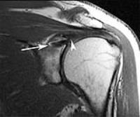 |
Imaging Following Shoulder Instability Surgery 
Laura W. Bancroft, MD1,2
1Diagnostic Radiology, Florida Hospital,
Orlando, FL, United States, 2Diagnostic
Radiology, University of Central Florida College of
Medicine, Orlando, FL, United States
Imaging following shoulder instability surgery depends
on suspected pathology. Direct anatomic repair of labral
tears may be perfomed in conjunction with capsular
shift. There should be no separation of the
labrocapsular complex and glenoid margin with intact
labral repair. Overall accuracy of MR arthrography for
detecting labral tears after prior instability repair is
> 90%. Arthroscopic Bankart repair may be performed in
conjunction with remplissage procedure in patients with
engaging Hill-Sachs lesion. MRI will show reattachment
of posterior structures into the defect, along with
anchor embedded in the trough. Postoperative imaging of
Laterjet procedure must assess incorporation of the bone
block and any recurrent imaging features of instability.
|
09:00
|
|
MRI of the Postoperative Elbow
Hollis Potter, MD1
1Radiology, Hospital for Special Surgery, New
York, NY, United States
|
09:30
|
|
Post Treatment Wrist
Shadpour Demehri1
1Johns Hopkins Medical Institute
|
10:00
|
|
Break & Meet the Teachers |
10:30
|
|
Imaging Following Cartilage Repair
Michael Recht1
1Radiology, New York University School of
Medicine
|
11:00
|
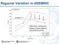 |
Postoperative Hip: FAI & Dysplasia
Young-Jo Kim
dGEMRIC is a biochemical imaging technique that can
assess the charge density of cartilage. Cartilage can
respond to increased mechanical loading by increasing
charge density. In acetabular dysplasia, there is
increased mechanical load due to the shallow acetabulum,
which will normalize after pelvic osteotomy.
Prospective monitoring of the hip cartilage before and
after osteotomy using dGEMRIC demonstrates that
cartilage responds appropriately to alterations in hip
mechanics after osteotomy for dysplastic hips.
|
11:30
|
|
Entrapment Neuropathies of the Pelvis Following Surgery
Gustav Andreisek
Entrapment neuropathies of the pelvis following surgery
are rare but important causes for a negative outcome or
complications after surgery.
|
12:00
|
|
Lunch & Meet the Teachers |
13:30
|
|
Postoperative Ankle
James Linklater
|
14:00
|
|
Postoperative Knee: Menisci
Edwin Oei1
1Radiology & Nuclear Medicine, Erasmus MC
Rotterdam, Rotterdam, Netherlands
In this lecture, the meniscal anatomy and the important
role the meniscus plays in the structure and function of
the knee will reviewed, followed by a discussion of the
three surgical strategies for operative treatment of
meniscal tears (resection, repair, and replacement). MR
protocol choices for postoperative assessment of the
meniscus will be presented as well as normal and
abnormal MR imaging findings in the postoperative
meniscus after each of the different surgical
procedures.
|
14:30
|
|
Postoperative Knee:
Ligaments
James Griffith
|
15:00
|
 |
Postoperative Knee: Total Knee Replacement - Permission Withheld
Florian M Buck1
1University of Zurich, Zurich, Switzerland
The purpose of this presentation is to provide an overview of the
possibilities and restrictions of todays MARS MR imaging
techniques in patients after total knee replacement.
After following this presentation, the learners will
understand the major clinical problems faced by
orthopedists after total knee replacement, how MR
imaging can contribute in these situations and where the
limitations of today’s technical possibilities are in a
clinical setting.
|
15:30
|
|
Adjournment & Meet the
Teachers |