|
Combined Educational & Scientific Session
ORGANIZERS: Harald Kramer, M.D.
Tuesday, 25 April 2017
| Room 315 |
08:15 - 10:15 |
Moderators: Harald Kramer, Sergio Uribe |
Slack Channel: #s_cv
Session Number: CE02
Overview
This session will highlight the rapidly evolving field of volumetric analysis of cardiovascular flow dynamics. It will review technical and clinical implementation of 4D flow imaging techniques and explore possible clinical applications with implications on patient prognosis and/or therapy management. Lectures will also highlight the important aspects of data acquisition and post processing.
Target Audience
This session is targeted to physicians and imaging scientists who are interested in the field of cardiovascular flow dynamics. They will be able to expand their knowledge with respect to new technical approaches and clinical scenarios.
Educational Objectives
As a result of attending this course, participants should be able to:
-Describe general technical principles of 4D Flow MRI;
-Identify potential clinical applications of 4D Flow MRI;
-Recognize the potentials and limitations of image processing of 4D Flow data sets; and
-Predict the possible clinical impact of 4D Flow MRI on patient management.
08:15
|
|
4D Flow MRI: Technical 
tino ebbers
Four-dimensional flow magnetic resonance imaging (4D flow MRI) enables comprehensive access to time-varying and multidirectional blood flow through the cavities of the heart and great vessels. This presentation aims to assist understanding of acquisition and analysis methods of 4D flow MRI with a focus on the heart and greater vessels. Different visualization strategies and computation of derived flow parameters, as wall shear stress, pressure difference, turbulent kinetic energy, and intracardiac flow components, are discussed.
|
08:45
 |
0422.
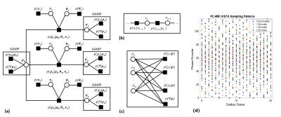 |
A Bayesian Approach to Enable Single Breath-Hold 4D Flow MRI
Adam Rich, Lee Potter, Ning Jin, Yingmin Liu, Orlando Simonetti, Rizwan Ahmad
PC-MRI based 4D flow imaging is a powerful tool to quantify hemodynamics within the heart and the great vessels. We develop a Bayesian technique to greatly accelerate 4D flow image acquisition. Our technique exploits the rich structure in 4D flow images within a joint reconstruction algorithm. We validate the technique using retrospectively accelerated flow phantom data and prospectively accelerated, single breath-hold in vivo data. In the flow phantom, stroke volume differed by ≤9% for R≤28 when compared to fully sampled data. The high acceleration provided by the Bayesian approach could allow for clinical application of single breath-hold 4D flow imaging.
|
09:00
|
0423.
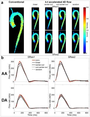 |
k-t accelerated aortic 4D flow MRI in under 2 minutes: feasibility and impact of resolution, k-space sampling patterns, and respiratory navigator gating on hemodynamic measurements
Emilie Bollache, Alex Barker, Jeremy Collins, Pim van Ooij, Rouzbeh Ahmadian, Alex Powell, James Carr, Julia Geiger, Michael Markl
Our objective was to assess the performance of eight highly k-t accelerated non-gated free-breathing aortic 4D flow MRI measurements acquired in under 2 minutes (PEAK GRAPPA R=5; TRes=67.2ms; four ky-kz-space Cartesian fillings: linear, center-out, out-center-out, random; two spatial resolutions=3.5x2.3x2.6mm3, 4.5x2.3x2.6mm3), both in vitro and in 10 healthy volunteers. Despite lower image quality, the significantly shorter k-t accelerated datasets provided aortic hemodynamic indices in agreement with conventional respiratory-gated 4D flow measurements. Differences were non-significant when using linear and out-center-out k-space samplings (absolute differences≤22%). In conclusion, aortic 4D flow MRI in under 2 minutes is feasible with moderate underestimation of flow indices.
|
09:15
|
|
4D Flow: Clinical
Ann Bolger
Four dimensional flow visualization and quantification using phase contrast MRI has expanded the functional assessment of intracardiac and vascular blood flow in health as well as many forms of common disease. The impacts of chamber remodeling, endothelial disruption, material property changes related to infarction, atherosclerosis, or inflammation may all influence site-specific and global flow, and provide clues to their presence and severity. Further, the tolerance for variability in load, heart rate and rhythm may also be predicted by flow-based measures at baseline. Definition of the range of normal flow across the gender and age spectrum is critical to utilizing 4D flow methods in clinical applications.
|
09:45
|
0424.
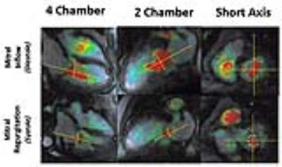 |
Reproducibility and Accuracy of 4D Flow Valve Tracking for Assessment of Mitral Regurgitation
Kanae Mukai, Purvi Parwani, Michael Hope
Quantification of mitral regurgitation (MR) is challenging to accurately perform with echocardiography.1 Recent data suggest that MRI is more accurate than echocardiography, but the conventional approach used requires the combination of data from two different types of MRI sequences.2 This study investigated the direct measurement of mitral regurgitation with 4D Flow using a valve tracking approach. We used both mitral annulus tracking and mitral flow jet tracking, and compared measurements to the conventional MRI approach for calculating mitral regurgitation. Flow jet but not annulus tracking demonstrated good reproducibility of measurement and correlation with the conventional MRI approach.
|
10:00
|
0425.
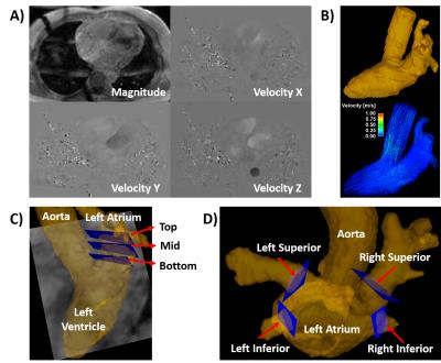 |
Exploration for Novel Markers of Left Atrial Disease and Thrombo-embolic Risk in Patients with Paroxysmal Atrial Fibrillation Using 4D Flow MRI
Julio Garcia, Michael Bristow, Carmen Lydell, Andrew Howarth, Bobby Heydari, Frank Prato, Maria Drangova, Rebecca Thornhill, Pablo Nery, Stephen Wilton, Allan Skanes, James White
This study may be of interest for clinicians and clinical researchers who study or measure left atrial blood flow. This pilot study demonstrates that: 1) 4D flow imaging of LA inflow and vortex formation is clinically feasible, 2) Significant differences in LA flow can be identified in patients with paroxysmal AF versus healthy controls; 3) Asymmetry of pulmonary vein inflow was observed in this population and may be contributory to (or as a result of) alterations in LA vortical flow, and 4) Vortical flow is fractionated in patients with a history of paroxysmal AF. These early observations seed interest for LA 4D flow as a marker of early or established left atrial disease and may provide value for the prediction of thrombo-embolic events.
|
|