|
Combined Educational & Scientific Session
ORGANIZERS: Guanshu Liu, Ph.D., Steven P. Sourbron, Ph.D., & Elena Vinogradov, Ph.D.
Wednesday, 26 April 2017
| Room 310 |
13:45 - 15:45 |
Moderators: Feliks Kogan, Peter van Zijl |
Slack Channel: #s_contrastmechanisms
Session Number: CE07
Overview
This session focuses on the cutting edge aspects of the Chemical Exchange Saturation Transfer (CEST) Imaging. The presentations will cover a range of topics: theoretical background and quantitative interpretation of the results, novel animal studies and translational efforts in humans.
Target Audience
Researchers and clinicians interested in CEST and looking for a background on the novel contrast method and examples of the cutting edge studies.
Educational Objectives
Upon completion of this course, participants should be able to:
-Identify basic physics principles behind the CEST method;
-Recognize basic equations describing CEST and recall quantification metrics employed;
-Describe sub-types of CEST agents;
-Name 2-3 in-vivo applications of CEST; and
-Identify promising areas for clinical translation and recognize potential obstacles.
13:45
|
 |
Introduction to CEST & Quantification 
Phillip Zhe Sun
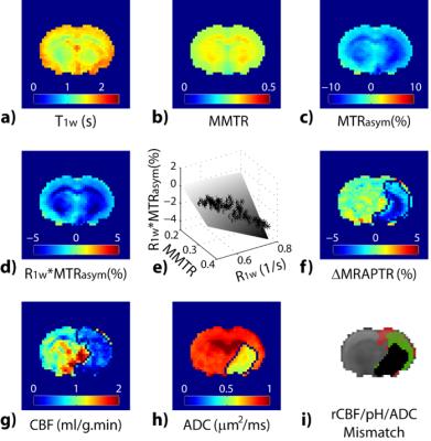
CEST MRI has emerged as a sensitive contrast mechanism for several metabolites such as glucose, glycogen, creatine and glutamate, as well as tissue pH. It has promising applications in a host of disorders including acute stroke, epilepsy and tumor. As we make the transition from CEST-weighted MRI toward quantitative in vivo CEST imaging for improved characterization of the underlying physiology, it is helpful to review persistent progress in the field of CEST imaging from equations, cells, rodents and patients.
|
14:05
|
0856.
 |
Characterization of in vivo Chemical Exchange Parameters Using Chemical Exchange-Sensitive MRI at 9.4 T and 15.2 T
Julius Chung, Wonmin Choi, Tao Jin, Jung Hee Lee, Seong-Gi Kim
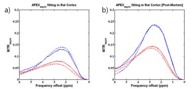
Chemical-exchange sensitive imaging can prove to be complex particularly when imaging intermediate exchanges in-vivo where contrast comes from an amalgamation of different sources. We propose a method that utilizes Z-spectra data from multiple fields to determine apparent exchange parameters that characterize the exchange to which a particular saturation scheme is sensitive. Parameter determination in glutamate phantoms proved commensurate to utilizing an on-resonance dispersion measurement and in the rat brain cortex the kex was measured to be 11,240 s-1 antemortem and 7070 s-1 postmortem. This method is useful for determining exchange parameters relevant to complex systems when signal source is unclear.
|
14:15
 |
0857.
 |
Improving Amide Proton Transfer (APT) MRI Quantification in Acute Human Stroke Patients: Achieving More Pure APT Signals and Higher Detection Sensitivity
Hye-Young Heo, Yi Zhang, Tina Burton, Shanshan Jiang, Peter van Zijl, Richard Leigh, Jinyuan Zhou
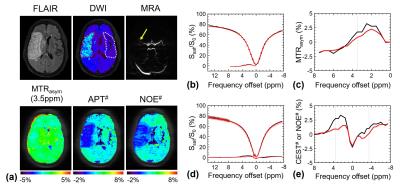
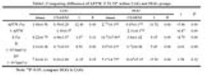
APT-weighted (APTw) imaging based on MTR asymmetry analysis has shown promise for identifying ischemic lesions, but suffers from low accuracy due to small APTw intensity changes, Quantitative APT, nuclear overhauser enhancement (NOE), perfusion and diffusion MRI were performed on acute stroke patients (n=30). The results showed that while APTw MRI for pH analysis based on MTRasym analysis was confounded by upfield NOE effects, NOE-free APT-MRI contrast between normal and ischemic lesions was substantially increased, nearly 3 times larger than that based on MTRasym analysis. Furthermore, noticeable NOE contrast was observed for lesions, explained in terms of a relayed-NOE transfer mechanism.
|
14:25
|
|
DIACEST Exogenous
Assaf Gilad
We will review the principles for designing an optimal bioorganic probes that are based on the specific targets. In addition we will discuss how to design a genetically encoded probe for a specific scientific question.
|
14:45
|
0858.
 |
Spatiotemporal quantification of reporter gene expression in the mouse heart using the Lysine Rich Protein and cardiac chemical exchange saturation transfer
Shelby Meier, Jose Abisambra, J Brandon, Assaf Gilad, Moriel Vandsburger
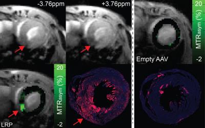
We utilized cardiac chemical exchange saturation transfer MRI and an artificial reporter gene, the Lysine Rich Protein (LRP), to image gene transfer in the mouse heart using 2 routes of viral vector administration.
|
14:55
|
0859.
 |
T1?-weighted Dynamic Glucose Enhanced MRI
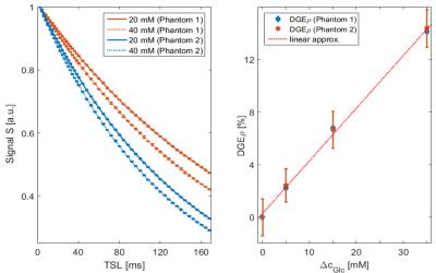
Patrick Schuenke, Daniel Paech, Christina Koehler, Johannes Windschuh, Peter Bachert, Mark Ladd, Heinz-Peter Schlemmer, Alexander Radbruch, Moritz Zaiss
Natural D-glucose can serve as a biodegradable contrast agent for the detection of tumors by means of Chemical Exchange Saturation Transfer (CEST) or Chemical Exchange sensitive Spin-Lock (CESL) Dynamic Glucose Enhanced (DGE) MRI. For application of CESL-based DGE-MRI at a 7T whole-body scanner, we implemented an adiabatic CESL sequence and essentially increased the temporal resolution employing a T1ρ-weighted acquisition scheme. Further, we introduced a simple, robust and quantitative DGE contrast. First application of T1ρ-weighted DGE-MRI in a glioblastoma patient provided a substantial contrast between tumor and healthy brain tissue and showed the dynamic glucose enhancement after a glucose bolus injection.
|
15:05
|
|
Translational CEST
Seth Smith
The goal of this educational presentation is to discuss the important contributions that CEST MRI can have for clinical-translational studies, by highlighting the unique contrasts available and examining the current applications of CEST MRI in the literature. We will further discuss the potential limitations for more clinically viable adoption of CEST and discuss the opportunities to overcome these limitations. We will close by discussing the potential impact of a unique contrast to clinical-translational studies of the human condition.
|
15:25
|
0860.
 |
Differentiating glioma histologic grade preoperatively by two functional modalities: Amide Proton Transfer (APT) and Intravoxel Incoherent Motion (IVIM) MR imaging
Tianyu Zou, Hao Yu, Chunxiu Jiang, Yingjie Mei, Fanheng Huang, Jinyuan Zhou, Zhibo Wen
A correct preoperatively grading of glioma is always the most important issue in clinic. APT and IVIM imaging are designed to assess glioma on the level of cell and molecule. So, in this study we combined these two functional techniques hoping to explore their diagnostic performance in differentiating HGG from LGG.
|
15:35
|
0861.
 |
Clinical Translation of Tumor Acidosis Measurements with AcidoCEST MRI
Kyle Jones, Edward Randtke, Eriko Yoshimaru, Christine Howison, Pavani Chalasani, Robert Klein, Setsuko Chambers, Phillip Kuo, Mark Pagel
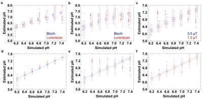
We optimized acidoCEST MRI, a method that measures extracellular pH (pHe), and translated this method for clinical imaging. We fit CEST spectra with the Bloch equations modified to include the direct estimation of pH, and generated parametric maps of tumor pHe in the SKOV3 tumor model, a patient with high grade invasive ductal breast carcinoma, and a patient with metastatic ovarian cancer. AcidoCEST MRI successfully measured a pH 6.58 in a tumor of the patient with metastatic ovarian cancer. The primary breast tumor failed to accumulate sufficient agent to generate pHe measurements.
|
|