ISMRM 25th Annual Meeting & Exhibition • 22-27 April 2017 • Honolulu, HI, USA

Electronic Poster Session: Cardiovascular
3095 -3116 Probing the Myocardial Tissue Composition
3117 -3140 Vascular Imaging: Lumen, Vessel Wall & Function
3141 -3163 Cardiac Function
3212 -3235 Velocity & Flow
3236 -3258 Myocardial Ischemia Imaging
3259 -3282 New Methods
 
Probing the Myocardial Tissue Composition
Electronic Poster
Cardiovascular

 
Monday, 24 April 2017
Exhibition Hall  08:15 - 09:15


    Computer #

 
3095.   
1 Reducing magnetization-transfer-induced T1 estimation error in myocardial T1 mapping for the MOLLI sequence: simulation, phantom and in vivo studies
Jiaxin Shao, Dapeng Liu, Kyung Hyun Sung, Kim-Lien Nguyen, Peng Hu
The modified Look-Locker inversion-recovery (MOLLI) sequence is a widely used myocardial T1 mapping technique for tissue characterization. It is known that the MOLLI sequence underestimates myocardial T1 values (>10%) due to magnetization transfer (MT) effects and short myocardial tissue T2 values. In this work, we demonstrate that in vivo reduction of the inversion factor is predominantly responsible for the MT effect that reduces MOLLI T1 values. Thus, using an alternative T1 estimation algorithm with a measured inversion factor for the MOLLI sequence can reduce the MT effect on T1 estimation error.


3096.   
2 Ultrafast Cardiac Balanced Inversion Recovery with Interleaved Acquisition (UF-CABIRIA) for improved simultaneous T1 and T2 quantification of the heart at 3T.
Francesco Santini, Grzegorz Bauman, Oliver Bieri
CABIRIA is a method for the simultaneous quantification of T1 and T2 values of the myocardium. As other T1-quantification sequences, it relies on a bSSFP readout, and it is thus susceptible to off-resonance effects. In this work, we show an improvement over the CABIRIA method that reduces the TR and therefore increases the robustness towards field inhomogeneities. This method can potentially be extended to other quantification techniques.


3097.   
3 Quantitative Tissue Characterization by Magnetization Transfer-Weighted Cardiac MRI Detects Fibrosis Progression without Gadolinium in Patients with End Stage Renal Disease
Tori Stromp, Rebecca Kidney, Tyler Spear, Kristin Andres, Joshua Kaine, Steve Leung, Moriel Vandsburger
Long term hemodialysis treatment for end stage renal disease (ESRD) is associated with a time dependent increase in cardiac death. The inability to use late gadolinium enhancement (LGE) cardiac MRI (CMR) in patients with renal dysfunction impedes the noninvasive monitoring of fibrosis progression and appropriate treatment selection in the population. We used magnetization transfer (MT) weighted CMR to monitor the progression of ventricular fibrosis over 1 year in patients with ESRD. While no major changes in hypertrophy or contractility were apparent, pervasive increases in myocardial signal was measured by MT-weighted CMR, consistent with reduced MT and progressive fibrosis development.


3098.   
4 Improved Detection of Right Ventricular Myocardial Infarction Using 3D Navigator-Gated Free Breathing Delayed Enhancement Cardiac Magnetic Resonance
Jiwon Kim, Tara Shah, Neil Mehta, Jonathan Weinsaft, Pascal Spincemaille, Yi Wang, Thanh Nguyen
Right ventricular myocardial infarction (RV-MI) is a serious consequence of coronary artery disease that adversely affects outcomes. Conventional CMR employs 2D breath-held imaging (CMRBH) to detect RV-MI, an approach that may sacrifice spatial resolution to enable patient breath-holds, and is thus suboptimal for imaging the RV. This study compared 3D navigator-gated free breathing CMR (CMRNAV) to CMRBH for detection of RV-MI in 75 post-MI patients. Results demonstrated a 2-fold increase in detection rate of RV-MI by CMRNAV, accompanied by higher spatial resolution in 30% less scan time.


3099.   
5 Simultaneous Imaging of Myocardial Fat and Scar using Dark Blood Late Gadolinium Enhancement
Maryam Nezafat, Ahmed Fahmy, Gifty Addae, René Botnar
Dark-Blood Late Gadolinium Enhancement (DB-LGE) sequence has been recently introduced to suppress the blood signal in order to enhance the contrast between blood and scar. However, it is difficult to distinguish scar from fat as both appear bright in DB-LGE images. In this study, we present a DB-LGE sequence that allows quantification of scar and fat volume in the heart in a single imaging sequence. Numerical simulation, phantom and in-vivo scans were performed to evaluate the proposed sequence.


3100.   
6 3D high resolution imaging of human hearts for visualization of the cardiac structure
Julie Magat, Valéry Ozenne, Fanny Vaillant, David Benoist, Marion Constantin, Virginie Dubes, Stephen Gilbert, Mark L Trew, Jérôme Naulin, Louis Labrousse, Mélèze Hocini, Michel Haissaguerre, Olivier Bernus, Bruno Quesson
The motivation of this study is to develop high resolution 3D MR imaging of human hearts to characterize the cardiac structure non-invasively. For this purpose, T1-weighted images and diffusion MRI in 3D from intact and infarcted hearts were acquired and analyzed to compare myocyte and myolaminar orientations in healthy and pathological regions.


3101.   
7 Improving precision of myocardial T1 mapping with 3-parameter fit model using tissue characteristic-based denoising
Jihye Jang, Shiro Nakamori, Reza Nezafat
For the reconstruction of pixel-wise T1 maps, 3-parameter fit model is highly accurate, but is sensitive to noise. Therefore, it is desirable to develop a robust method to reduce sensitivity to noise when 3-parameter fit model is used. In this work, we propose a robust denoising method based on tissue characteristics to improve precision of myocardial T1 mapping. In phantom and in-vivo studies, denoising filtering provided similar T1 measurements with significantly improved precision. This technique will make 3-parameter fit model more favorable by reducing sensitivity to noise and will allow for more accurate and precise myocardial T1 mapping.


3102.   
8 Cine T1 Mapping: Look-Locker inversion recovery for phase resolved T1-Mapping with B1+ Correction At 3T
Sebastian Weingärtner, Chetan Shenoy, Lothar Schad, Mehmet Akçakaya
Conventionally, evaluation of myocardial T1-times is limited to a single snapshot of the cardiac cycle, leaving much of the dependence between functional and tissue characterization unstudied. Here, we propose an ECG-triggered steady-state Look-Locker technique that allows for functional, cardiac phase-resolved native T1-mapping. Integratedly acquired phase-resolved B1+-maps are used for T1-time correction. High accuracy and good consistency of the T1-times across cardiac phases is shown in phantom scans. In-vivo T1-times show slight underestimation and similar precision compared to saturation-recovery T1-mapping. High visual image quality at all cardiac phases is obtained at temporal resolutions up to 40ms in a single breath-hold.


3103.   
9 Active Shape Models Based Motion Correction for Myocardial T1 Mapping
Hossam El-Rewaidy, Shiro Nakamori, Gifty Addae, Warren Manning, Reza Nezafat
A new framework based on Active Shape Models (ASM) is introduced to correct the motion artifacts induced by respiratory and cardiac motion in myocardial T1 mapping. This framework includes three main steps: training ASM model to capture intensity variations of T1 images at different inversion times, segmentation of T1 weighted images using the trained model, and estimating and applying the registration parameters to correct for motion between images. 


3104.   
10 A comparison of STEAM and spin echo diffusion tensor CMR in hypertrophic cardiomyopathy patients
Zohya Khalique, Andrew Scott, Pedro Ferreira, Margarita Gorodezky, Ricardo Wage, Sonia Nielles-Vallespin, David Firmin, Dudley Pennell
Diffusion tensor (DT) cardiovascular magnetic resonance (CMR) can provide myocardial microstructural information using either motion compensated spin echo (SE) or stimulated echo acquisition mode (STEAM) sequences. Here we present the first DT-CMR data from a cohort of hypertrophic cardiomyopathy patients using SE, and compare parameters obtained from SE and STEAM sequences at 3T in two phases of the cardiac cycle. We demonstrate that whilst reliability measures, diffusivity, and anisotropy differ between the sequences, both approaches are able to detect the sheetlet microstructure that is integral to wall thickening during cardiac contraction and shown to be aberrant in this condition.


3105.   
11 Identifying tissue changes of “normal-appearing” myocardium in pediatric myocarditis patients using quantitative T1 and T2 mapping techniques
Haipeng Wang, Bin Zhao, Cuiyan Wang, Tianyi Qian, Guangbin Wang, Jing An, Fei Gao, Andreas Greiser, Junyu Zhao
This study aimed to identify diffuse myocardial tissue changes in pediatric myocarditis patients by applying T1 and T2 mapping techniques. The correlations between T1 and T2 values and cardiac function were assessed. Abnormal post-contrast T1 values and extracellular volume (ECV) of “normal appearing” myocardium in pediatric myocarditis patients were found and were correlated with left ventricular cardiac function and stroke volume (SV) in children with chronic myocarditis. 


3106.   
12 The Effect of Mixing Time on Diffusion Spectrum Imaging of the Ex-vivo Porcine Heart
Claudia Zanella, Christian Stoeck, Constantin von Deuster, Sebastian Kozerke
Higher order diffusion imaging has revealed new insights into myocardial microstructure. At the b-values required for diffusion kurtosis or diffusion spectrum imaging, translation into human in-vivo application requires Stimulated Echo Acquisition Mode imaging (STEAM) at prolonged mixing-times. In this study the effect of mixing time on diffusion parameters is investigated covering the range of current spin-echo and STEAM aproaches. Results show that fractional anisotropy increases and mean diffusivity decreases with mixing-time. Diffusional kurtosis was found to decrease with mixing-time by varying amounts along fiber, sheet and sheet-normal direction which needs to be considered when comparing spin-echo imaging with STEAM acquisitions.


3107.   
13 Whole-heart T1 mapping using a 2D fat image navigator with 100% scan efficiency
Giovanna Nordio, Gastao Cruz, Claudia Prieto, Torben Schneider, Rene M. Botnar, Markus Henningsson
In this study a whole-heart saturation-recovery T1-mapping technique in combination with a fat image navigator (fat-iNAV) is proposed. Myocardial fat is imaged by the fat-iNAV to estimate respiratory motion. Each of the T1-weighted images are subsequently motion corrected prior to reconstruction of the T1 map. Fat-iNAV motion correction let to an improvement in myocardial borders delineation and accuracy of the myocardial T1 values, while there was a general underestimation of myocardial T1 in the non-motion corrected T1 maps (1115.4±110ms vs 998.6±101ms). Further work will investigate non-rigid motion correction and undersampled reconstruction for T1 mapping.


3108.   
14 Accelerating High-Resolution Whole-Heart 3D T2 Mapping
Dan Zhu, Haiyan Ding, Henry Halperin, Daniel Herzka
In this work, we aim to determine an optimal strategy for accelerating high-resolution 3D myocardial T2 mapping. We quantitatively evaluate the performance of diverse methods involving different subsampling patterns and different reconstruction strategies relative to volume-by-volume SENSE reconstruction. Reconstructions which address all volumes as a single reconstruction problem (i.e. joint-sparsity SENSE or model-based SENSE) outperform volume-by-volume approaches, and variable density sampling outperforms equal spacing or CAIPIRIHNA undersampling. The T2 values observed in parametric maps proved to be more sensitive to data corruption than images themselves, limiting the degree of data reduction tolerable. 


3109.   
15 In-vivo comparison of STEAM EPI and STEAM spiral diffusion-weighted sequences
Margarita Gorodezky, Andrew Scott, Pedro Ferreira, Sonia Nielles-Vallespin, Zohya Khalique, Dudley Pennell, David Firmin
A cardiac DTI STEAM spiral sequence with a novel asymmetric zonal excitation and a reduced FOV was implemented and compared to an established STEAM EPI sequence. 10 volunteers were scanned in systole and diastole and standard cDTI parameters were compared for both sequences. The spiral trajectory is both efficient and motion robust and its centre-out nature allows a shorter echo time and, therefore an increase in image SNR over EPI.  The spiral trajectory is also able to take full advantage of the two-dimensional reduced in-plane field-of-view (FOV) made possible by the 3 RF pulses used in the STEAM sequence.


3110.   
16 Biopsy-based calibration of T2* magnetic resonance for estimation of liver iron concentration
Antonella Meloni, Filippo Leto, Daniele De Marchi, Vincenzo Positano, Laura Pistoia, Aurelio Maggio, Alessia Pepe
An excellent linear agreement between R2* values and cardiac iron measured with emission spectroscopy was found. The results further validate the current clinical practice of monitoring cardiac iron in vivo by CMR.


3111.   
17 Myocardial Motion Velocity in Patients with Cirrhotic Cardiomyopathy
Ting-Hsu Chen, Ming-Ting Wu, Mao-Yuan Su, Hsu-Hsia Peng
In this study, we employed MR tissue phase mapping (TPM) to evaluate three-directional left ventricular velocity. The purpose of this study is to establish quantitative indices to comprehend the characteristics of myocardial motion in patients with cirrhotic cardiomyopathy. Compared with normal subjects, patients displayed lower systolic Vz and prolonged systolic TTPz. Lower systolic Vphi was also demonstrated in patient group. In conclusion, to analyze myocardial velocity and TTP can potentially provide helpful information for realizing the myocardial compensatory mechanism in patients with cirrhotic cardiomyopathy.


3112.   
18 Dependence of the Precision of Myocardial Late Gadolinium Enhancement Quantification on Inversion Time Selection Simulated Using Synthetic Inversion Recovery MR Imaging
Akos Varga-Szemes, Rob van der Geest, U. Joseph Schoepf, Carlo De Cecco, Taylor Duguay, Pal Suranyi
Clinical late gadolinium enhancement (LGE) imaging of the myocardium requires inversion time (TI) optimization for adequate image quality. As the optimal TI (TI0) depends on various factors, its influence on the precision of myocardial LGE quantification is of interest. In this study we aimed to prospectively investigate how the precision of LGE quantification varies in the clinically relevant TI range in 53 patients using T1-mapping-based synthetic inversion recovery (IRsynth) approach. We concluded that phase-sensitive IRsynth images provide precise quantification independent of TI, while magnitude IRsynth-based quantification is precise at TI0 or longer TIs, but showing decreased precision below TI0.


3113.   
19 Precision and sensitivity of radial MOLLI sequence for fast myocardial T1 mapping
Benjamin Marty, Bertrand Coppa, Pierre Carlier
Quantitative cardiac NMR imaging, and more particularly T1 mapping has become a most important modality to characterize myocardial tissue. In this work, we evaluated the precision and sensitivity of a fast radial MOLLI sequence by comparison with conventional MOLLI on 14 subjects presenting different heart rates, BMI and cardiac conditions. This sequence demonstrated the same precision on T1 estimates than conventional MOLLI, and was not influenced by heart rate variations. It might represent a good candidate for ultra-fast acquisition of myocardial T1 maps.


3114.   
20 Temporal Diffusion Spectroscopy in the Heart with Oscillating Gradients
Irvin Teh, Jürgen E. Schneider, Hannah J. Whittington, Tim B. Dyrby, Henrik Lundell
Conventional pulsed gradient spin echo, with its longer diffusion time, is poorly sensitive to diffusion at short length scales. Oscillating gradient spin echo enables assessment of diffusion at sub-cellular length scales, providing information about cell size and the intracellular environment. We observed that time dependence of diffusion in the myocardium was more pronounced along the 2nd and 3rd eigenvectors compared to the 1st eigenvector of the diffusion tensor. This is consistent with known anisotropic cardiomyocyte geometry. Furthermore, the measured diffusion at high frequencies still exhibited strong anisotropy that may reflect anisotropy of intracellular organelles such as actin-myosin filaments.


3115.   
21 Cardiac CEST MRI with Dual-Echo Readout for B0 Correction: A Preliminary Reproducibility Study for Assessment of Metabolic Activity in the Heart
Zhengwei Zhou, Xiaoming Bi, Debiao Li
Previous studies have shown that cardiac CEST technique can detect myocardial metabolic abnormalities in chronic myocardial infarction. However, this navigator-gated technique is still sensitive to B0 field variations caused by respiratory motion within the acceptance window. 

In this work, we developed a cardiac CEST dual-echo technique which not only acquires CEST-weighted image, but also enables acquisition of B0 map for each saturation frequency offset. The reproducibility of this technique was also assessed.


3116.   
22 Dynamic Nitroxide-Enhanced MRI Detects Oxidative Stress in the Hearts of Mice Subject to Angiotensin II Infusion
Sophia Cui, Rene Roy, Brent French, Frederick Epstein
Oxidative stress contributes importantly to the pathophysiology of many types of cardiovascular disease. Nitroxides are relatively stable free radicals that have been used as redox-sensitive MRI contrast agents in preclinical studies to assess tumor redox status. We implemented a dynamic nitroxide-enhanced MRI method to test the hypothesis that MRI can detect cardiac oxidative stress in vivo. Imaging was performed in untreated controls and mice infused with angiotensin II for 7 days. The MRI signal decay rate in the heart was significantly higher in the angiotensin II group, indicating that these methods detect cardiac oxidative stress due to angiotensin II infusion.
Vascular Imaging: Lumen, Vessel Wall & Function
Electronic Poster
Cardiovascular

 
Monday, 24 April 2017
Exhibition Hall  08:15 - 09:15


    Computer #

 
3117.   
25 Estimation of motion-corrupted data using parallel imaging for carotid artery vessel wall imaging
Robert Frost, Luca Biasiolli, Linqing Li, Aaron Hess, Peter Jezzard
Carotid artery imaging is hampered by motion artefacts, including those caused by occasional swallowing motion. In this work, we extend previous work on intelligent reacquisition to estimate and replace motion-corrupted data. ‘Bad’ phase-encode lines were synthesized from surrounding ‘good’ lines using parallel imaging techniques. Estimation and replacement of corrupted data reduced background ghosting levels in comparison to the previous reacquisition approach.  A small number of reacquisitions was maintained to ensure good quality data at the centre of k-space because these are essential for image quality and for calibration of the parallel imaging-based estimation.


3118.   
26 Three-dimensional black-blood multi-contrast protocol for carotid imaging using compressed sensing: a repeatability study
Jianmin Yuan, Ammara Usman, Scott Reid, Kevin King, Andrew Patterson, Jonathan Gillard, Martin Graves
Multi-contrast black-blood MRI protocol has demonstrated the ability to assess carotid plaque vulnerability. Current limitation for its wide application is the long scanning time. The purpose of this work is to evaluate the repeatability of a compressed sensing (CS) accelerated multi-contrast protocol at 3T.


3119.   
27 3D black blood T2 mapping of the carotid artery wall with compressed sensing and data-driven parallel imaging
Jianmin Yuan, Ammara Usman, Scott Reid, Kevin King, Andrew Patterson, Jonathan Gillard, Martin Graves
Quantitative MRI has many advantages over traditional contrast weighted methods, which may be more suitable for multi-centre studies across different MRI systems. This study describes the development of a 3D black blood T2 mapping sequence with the combination of compressed sensing and data-driven parallel imaging. Phantom and volunteer experiments were performed to optimise the parameters and patients with atherosclerotic carotid artery disease were scanned using the optimised sequence.


3120.   
28 Whole-brain vessel wall imaging within 5 minutes using compressed sensing accelerated IR-SPACE
Zhaoyang Fan, Xiaoming Bi, Marcel Maya, Qi Yang, Shlee Song, Nestor Gonzalez, Schlick Konrad, Esther Raithel, Christoph Forman, Gerhard Laub, Debiao Li
Inversion-recovery (IR) prepared SPACE was recently proposed as a whole-brain intracranial vessel wall imaging technique. This work aimed to investigate the feasibility of accelerating the scan from 8 min to <5 min using compressed sensing (CS). A prototype CS IR-SPACE sequence was implemented on a 3T system. Wavelet sparse regularization (λ) and iteration (Iter) were optimized for the scenario of sampling 15% of k-space data based on a volunteer study. Image quality was visually comparable between CS IR-SPACE and regular IR-SPACE scans. In addition, CS IR-SPACE and IR-SPACE showed comparable lesion delineation quality in patients despite markedly different scan times. 


3121.   
29 Endothelial permeability in the aortic root of atherosclerotic mice: quantification using 3 dimensional, black blood, self-gated T1 mapping
Alison Pruzan, Arthi Sridhar, Gustav Strijkers, Zahi Fayad, Claudia Calcagno
Atherosclerotic plaques prone to rupture are characterized by endothelial dysfunction and increased endothelial permeability. The aortic root is a vascular territory where permeable atherosclerotic plaques form consistently and reliably.  However, morphological and quantitative parametric imaging of the mouse aortic root is very challenging, due to the small dimensions, rapid blood flow through the valves, and high heart rate. Here we demonstrate feasibility of pre and post-contrast T1 mapping of the mouse aortic root using a 3 dimensional, self-gated fast low angle shot (FLASH) sequence with black blood imaging for improved vessel wall delineation. Future studies will entail further development of this technique for the more accurate quantification of endothelial permeability and fractional blood volume in the mouse aortic root. 


3122.   
30 Quantitative Multi-Contrast Atherosclerosis Characterization (qMATCH): Comprehensive Quantitative Evaluation of Atherosclerosis in a Single-Scan
Yibin Xie, Anthony Christodoulou, Nan Wang, Debiao Li
Although MRI is an attractive imaging modality for the evaluation of carotid atherosclerosis thanks to its versatility and noninvasiveness, its current clinical usage is still limited. Major drawbacks of conventional protocols include long scan time and observer variability due to the qualitative nature of the images. In this work we proposed a fast, 3D, quantitative, multi-contrast MRI technique, qMATCH, for a comprehensive evaluation of carotid atherosclerosis in less than 8 minutes. Preliminary results from phantom and in vivo studies demonstrated excellent image quality and reliable quantification of tissue relaxation times. 


3123.   
31 Aortic Vessel Wall Imaging Using 3D Phase Sensitive Inversion Recovery in Children and Young Adults
Animesh Tandon, Tarique Hussain, Andrew Tran, René Botnar, Gerald Greil, Markus Henningsson
Currently, T2-weighted TSE black blood zoom imaging (T2Z) is used to visualize atherosclerosis, but volumetric, high isotropic resolution datasets would be preferable. We compared aortic luminal sharpness (ALS) of a novel 3D bSSFP phase-sensitive inversion recovery black blood sequence (3DPSIR) to T2Z in 10 children and young adults with familial hypercholesterolemia at 5 aortic locations. 3DPSIR sequence duration was 7.0 minutes compared to 11.4 minutes for T2Z with no significant difference in ALS overall. 3DPSIR high resolution volumetric datasets will enable rapid, detailed 3D vessel wall imaging in children and young adults, allowing improved exploration of vascular health.


3124.   
32 Quantitative T1 and T2* carotid atherosclerotic plaque imaging using 3D multi-echo phase-sensitive inversion recovery sequence: a feasibility study
Yasuhiro Fujiwara, Hirotoshi Maruyama, Kanako Toyomaru, Yuri Nishizaka, Kazuma Yamasaki, Masahiro Fukamatsu
A quantitative evaluation of plaques is required in MR plaque imaging. The purpose of this study was to determine whether the 3D multi-echo Phase-Sensitive Inversion Recovery sequence can improve the T1 intra-plaque hemorrhage (IPH)-to-muscle contrast while simultaneously providing accurate T1 and T2* values for an IPH. This sequence may have potential to improve plaque MR imaging and make it possible to obtain detailed information about the components of carotid atherosclerotic plaques. 


3125.   
33 Quantitative Evaluation of RECS-3D MERGE MR Imaging for Carotid Plaque Assessment
Bo Li, Hao Li, Guofu Huang, Xia Qian, Wei Wang, Li Dong
We sought to quantitatively investigate the performances on blood suppression efficiency, wall-lumen CNR and plaque burden measurements for the RECS-3D MERGE sequence in carotid vessel wall MR imaging.

Twelve patients with carotid artery stenosis were recruited in this study. Lumen SNR and wall-lumen CNR were calculated and plaque burden measurements (i.e., lumen area, wall area, mean wall thickness and maximum wall thickness) were measured.

The RECS-3D MERGE was quantitatively demonstrated to have the ability to provide the comparable blood suppression, black-blood image quality and the highly correlated morphometry with the current plaque imaging protocol. The RECS-3D MERGE could be a promising tool for plaque burden measurement in a short scan time.


3126.   
34 Evaluation of Four Injection Strategies for High Spatial Resolution Gadobenate Dimeglumine-Enhanced MR Angiography
Gregory Wilson, Jeffrey Maki
Four injection profiles were evaluated in 40 subjects (10 each). The profiles were designed to provide a signal intensity plateau for a 20 second contrast-enhanced MR angiography scan, using gadobenate dimeglumine. The injection profiles were: 1) standard, non-diluted, 1.6 mL/sec; 2) diluted, 1.6 mL/sec; 3) diluted, bi-phasic, and 4) diluted, patient-tailored. All subjects received a total dose of 0.1 mmol/kg, divided between a test bolus and a full bolus. The “diluted” protocols included dilution of the total dose to 40 mL with normal saline. Signal intensity profiles were measured and analyzed for width of plateau (FW80M) and peak signal intensity.


3127.   
35 3D segmentation of the Pulmonary Arteries on Magnetic Resonance Angiography
Christopher Johns, Alberto Biancardi, Guilhem Collier, David Capener, Andy Swift, Jim Wild
Contrast enhanced magnetic resonance angiography (MRA) is in common use in the assessment of pulmonary hypertension. We present a novel method for segmentation of the pulmonary arteries allowing rapid assessment of the proximal pulmonary arteries down to the 4th generation. Whilst the strongest correlation with mean pulmonary artery pressure was with the right main pulmonary artery, the torsion of the 4th generation pulmonary arteries was also associated with increasing mean pulmonary artery pressure. We hope quantitative vessel segmentation will improve our understanding of the impact of proximal pulmonary arterial remodelling in pulmonary hypertension. 


3128.   
36 The value of 3T contrast-enhanced whole-heart Coronary MRA in a integrated assessment of Cardiac Magnetic Resonance Protocol for Detection of suspected or known Coronary Artery Disease
lijun zhang , Yi He, Zhan ming Fan, Debiao Li
We try to evaluate the additive diagnostic value of a 3T whole-heart CE-MRCA integration into a CMR-MPI/LGE protocol for the detection of coronary artery disease A total of 38 subjects were examined by CMR (including CMR-MPI, MRCA, and LGE)[j1]  and x-ray invasive coronary angiography (ICA). Diagnostic performances of MRCA, CMR-MPI/LGE, and MRCA+CMR-MPI/LGE integration were determined having XA as standard for coronary artery disease. In per-vessel analysis, integrated protocol (AUC=0.84) performed better than the isolated CMR-MPI/LGE (AUC=0.63). In this suspected or known coronary artery disease population, integration of CE-MRCA significantly improved per-vessel diagnostic accuracy of a comprehensive 3T CMR-MPI/LGE protocol.  


3129.   
37 A novel method for performing percutaneous transluminal renal angioplasty with the guide of non-contrast magnetic resonance angiography overlaid on fluoroscopy images - permission withheld
Yoshiaki Morita, Tetsuya Fukuda, Yoshiaki Watanabe, Tatsuya Nishii, Atsushi Kono, Naoaki Yamada
Herein, we present a new method for the performance of percutaneous transluminal renal angioplasty (PTRA) guided by non-contrast magnetic resonance angiography (NATIVE True FISP) overlaid on intra-procedural fluoroscopy images. This novel overlay system led to sufficient visualization as the overlaid vasculature on the live fluoroscopic image, and enabled the safe completion of PTRA with fewer angiographic procedures, thus requiring lower volumes of iodinated contrast material and shorter fluoroscopic times.


3130.   
38 Velocity-selective unenhanced peripheral MR angiography: Initial clinical testing compared with digital subtraction angiography
Taehoon Shin, Rajiv Menon, Devin Watson, Rajabrata Sarkar, Sanjay Rajagopalan, Robert Crawford
We performed initial clinical testing of velocity-selective magnetization-prepared MR angiography (VS-MRA) in 31 patients with peripheral arterial disease with digital subtraction angiography (DSA) serving as the reference standard. The image quality score of VS-MRA was 2.8±0.6 in the scale of 0-3 (0: non-diagnostic, 3: excellent). The sensitivity and specificity for detecting significant stenosis were 90.1 % and 96.2%, respectively on a per-segment basis (n=286), and were 92.7% and 92.0 %, respectively on a per-region basis (n=105). The Fleiss-Cohen κ value for the agreement between VS-MRA and DSA was 0.91 on a per-segment basis and 0.88 on a per-region basis. 


3131.   
39 Super-Resolution Intracranial Quiescent-Interval Slice-Selective Magnetic Resonance Angiography
Ioannis Koktzoglou, Shivraman Giri, Jianing Pang, Jeremy Collins, Robert Edelman
We evaluated whether super-resolution reconstruction could be used to improve the vascular detail of quiescent-interval slice-selective (QISS) MRA of the intracranial circulation. Results suggest that super-resolution reconstruction can substantially improve intracranial arterial delineation without the signal-to-noise ratio penalty of directly acquiring thin-slice images.


3132.   
40 Simultaneous Bright and Black Blood Whole Heart Phase Sensitive Inversion Recovery (PSIR) for Non Contrast Enhanced Coronary Lumen and Plaque Characterization
Giulia Ginami, Radhouene Neji, Rene Botnar, Claudia Prieto
This study introduces a 3D whole-heart sequence with image-based navigation for non-contrast enhanced coronary lumen and coronary thrombus/haemorrhage characterization. The sequence is based on a phase sensitive inversion recovery (PSIR)-like approach enabling the acquisition of two bright-blood datasets for visualization of the coronary lumen and estimation of motion parameters. Furthermore, a third, fully co-registered dark-blood dataset for thrombus/haemorrhage characterization is obtained from the PSIR reconstruction. With the proposed approach the coronary lumen is visualized with improved SNR/CNR and vessel sharpness when compared to a conventional T2 prepared coronary MRA acquisition. In addition, effective blood signal suppression and feasibility for thrombus visualization is shown with the PSIR reconstructed black-blood dataset.


3133.   
41 Evaluating Circle of Willis using Simultaneous Non-Contrast Angiography and intraPlaque Hemorrhage (SNAP)
Yalun Chen, Ling Tang, Le He, Xihai Zhao, Zhensen Chen, Huijun Chen
The purpose of this study is to evaluate the ability of SNAP to evaluate the CoW by comparing with an established non-contrast MRA technique, time of flight (TOF).We found SNAP have good agreement with TOF and similar intra-reader reproducibility in CoW evaluation. However, the agreement between TOF and SNAP and the reproducibility of each technique is moderate in the anterior communicating artery identification. Thus, SNAP shows great promise for imaging both intracranial artery and vessel wall with a single scan.


3134.   
42 Flow enhanced (FEED) Arterial Spin Labeling Angiography with segmented Zero Echo Time Readout - video not available
Jianxun Qu, Bing Wu, Zhenyu Zhou
ASL based MRA with continuous labeling has the optimal label efficiency. To achieve data acquisition efficiency, a long readout train is frequently used after labeling. Flow void might appear due to the lack of tagged blood entering the imaging volume during readout. A method named hybrid ASL address this problem with additional pulsed inversion. Yet, blood inverted by pulsed ASL still experiences considerable T1 decay during readout. We propose a method termed flow enhanced ASL (FEED). Interleaved labeling modules along with segmented readout was employed. As the results show, FEED could increase the signal level without prolonging scan time.


3135.   
43 Free-breathing non-contrast 3D radial respiratory motion-resolved versus motion-corrected whole-heart coronary MRA at 3T
Jessica Bastiaansen, Lorenzo di Sopra, Jérôme Yerly, Davide Piccini, Ruud van Heeswijk, Matthias Stuber
Coronary MRA performed using respiratory self-navigation techniques that apply retrospective motion-correction suffer from artifacts originating from static anatomical structures surrounding the heart. This becomes even more complicated at 3T due to insufficient fat suppression. Here we implemented free-breathing self-navigated 3D coronary MRA at 3T. Instead of using a motion model, we resolved the motion using 4D k-t sparse SENSE. Resulting images were then quantitatively compared with those reconstructed from the same datasets but using a 1D motion correction. We demonstrate that non-contrast whole-heart coronary MRA can be performed at 3T and that 4D motion-resolved reconstruction effectively minimizes adverse effects of respiratory-motion.


3136.   
44 Dual-Phase Whole-Heart Imaging Using Image Navigation in Congenital Heart Disease
Danielle Moye, Tarique Hussain, Rene Botnar, Animesh Tandon, Gerald Greil, Adrian Dyer, Markus Henningsson
Dual phase whole heart imaging allows accurate depiction of cardiac segmental anatomy for congenital heart disease. We applied image-based navigation (iNAV) to improve its clinical utility. Qualitative and quantitative image quality scoring showed that iNAV gave equivalent image quality compared to the standard respiratory navigator. However, the distal coronary arteries were better visualized and the sequence duration was shorter. Using dual-phase iNAV, complete segmental anatomy was delineated in 27/30 patients (90%). The use of iNAV dual phase 3D SSFP whole heart acquisition allows complete morphological diagnosis for congenital heart disease with high image quality with more clinically acceptable time constraints.


3137.   
45 Improved Golden-Angle Stack-of-Stars Non-Contrast Enhanced 4D Dynamic MR Angiography using Parallel Imaging and Compressed Sensing
Ziwu Zhou, Fei Han, Songlin Yu, Dandan Yu, Stanislas Rapacchi, Danny Wang, Lirong Yan, Peng Hu
In this study, we investigated the feasibility of accelerating a recently proposed arterial spin labeling (ASL) based, time-resolved non-contrast enhanced dynamic MR angiography (NCE-dMRA) technique using parallel imaging and compressed sensing. By taking advantage of the inherent and unique “subtraction sparsity” in ASL type acquisition, we combined a recently developed CS technique, which uses a magnitude subtraction to enhance sparsity in MRA data, with the ESPIRiT method for SENSE type PI reconstruction. Proposed method was compared with the previously used KWIC reconstruction to demonstrate its advantage in reducing temporal blurring caused by view-sharing.  


3138.   
46 Single-Shot Coronary QISS MRA in Children using Radial k-Space Sampling and Compressed Sensing
Daming Shen, Robert Edelman, Joshua Robinson, Hassan Haji-Valizadeh, Shivraman Giri, Ioannis Koktzoglou, Cynthia Rigsby, Daniel Kim
Coronary MRA is complex to set up (e.g., navigator gating to track respiratory motion), typically takes 5-10 min to complete, and often produces inconsistent results. Quiescent-Interval Slice-Selective (QISS) MRA is a promising method for rapid coronary imaging. We sought to develop a single-shot (real-time) coronary QISS MRA using compressed sensing.


3139.   
47 Automated Coronary Artery Ostia Detection in Magnetic Resonance Angiography
Bernhard Stimpel, Christoph Forman, Jens Wetzl, Michaela Schmidt, Andreas Maier, Mathias Unberath
Anomalies of the coronary ostia can have severe consequences. To provide a screening solution, automated ostia detection based on single breath-hold coronary MRA scans is presented. The aorta is segmented in the data sets to serve as an orientation point and vesselness enhancing filters are applied. Searching the aorta surface for high vessel responses by a ray-tracing procedure yields information about the position of the coronary ostia. The proposed approach was successfully validated in 10 volunteers with an average deviation of $$$7.6 \pm 1.0$$$° in angular and $$$1.2 \pm 0.58$$$ mm in superior-inferior direction. 


3140.   
48 Using Non-Contrast-Enhanced Magnetic Resonance Angiography (MRA) in Evaluating Preoperative Facial Artery of Vacularized Submental Lymph Node Flap: Compared to Conventional Contrast-Enhanced MRA
Ming-Chen Wu, Ming-Yi Hsu, Ren-Fu Shie, Yui-Ping Fan, Hsiao-Ching Yu, Chien-Yuan Lin, Sung-Yu Chu
The aim of this study was to determine whether the recently developed non-contrast-enhanced Inhance 3D velocity magnetic resonance angiography (Inhance 3D MRA) technique is capable of evaluating the course of facial artery (branch artery) in preoperative evaluation of vacularized submental lymph node flap. The result shows that Inhance 3D MRA is a promising method for assessing facial artery course without concerning the contrast media (CM) travel time and the venous contamination.
Cardiac Function
Electronic Poster
Cardiovascular

 
Monday, 24 April 2017
Exhibition Hall  08:15 - 09:15


    Computer #

 
3141.   
49 
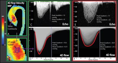
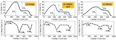
Age-stratified normal left atrial deformation assessed by novel longitudinal strain parameters on a 3T MR scanner
Shuang Leng, Xiaodan Zhao, Angela Su-Mei Koh, Ru San Tan, Liang Zhong
The aim of this study was to examine the age-related normal left atrial (LA) longitudinal deformation by a novel and fast assessable strain parameter with standard cardiac magnetic resonance (CMR) imaging. A total of 60 heathy subjects (30 males, age between 20 and 80 years) were categorized by age into young, middle-aged, and elderly adults. The LA longitudinal strain and strain rate measurements corresponding to reservoir and conduit phases were negatively related to age, while no significant age-dependency was observed for LA contraction strain and strain rate values. These strain parameters can be used to evaluate LA deformation and functionality.


3144.   
52 
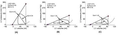
Left ventricular-arterial coupling and mechanical efficiency assessed by pressure-volume loop in pulmonary artery hypertension patients
Xiaodan Zhao, Fei Xu, Xiaoke Shang, Yang Dong, Wen Ruan, Gangcheng Zhang, Ru San Tan, Ju Le Tan, Yucheng Chen, Liang Zhong
Left ventricular end-systolic elastance (Ees), arterial elastance (Ea) and ventricular arterial coupling (VAC) (ratio of Ea/Ees) has been considered “gold” standard to assess ventricular contractility and performance. Ventricular arterial uncoupling due to impaired Ees or augmented Ea impaired ventricular mechanical efficiency (ME) and cardiac output. Ventricular contractility and arterial loading and the degree of their mismatching have yet been studied in pulmonary hypertension (PH). A total of 42 PH subjects who underwent both cardiac magnetic resonance (CMR) and right heart catheterization (RHC) were categorized into three groups – preserved LV ejection fraction (LVEF > 50%) and VAC < 0.8 (group 1); preserved LVEF and VAC > 0.8 (group 2); reduced LVEF (< 50%) (group 3). The results showed that VAC was correlated negatively with ME, which indicated arterial-ventricular uncoupling impaired mechanical efficiency. Importantly, the group 2 with ventricular arterial uncoupling had impaired mechanical efficiency despite its preserved ejection fraction.


3143.   
51 
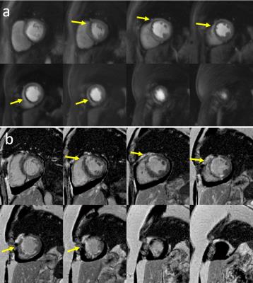
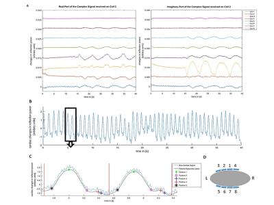
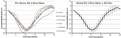
Accelerated Cardiac Cine “Watermark” MRI provides Cardiac Function via Magnitude Cine and 2D Myocardial Strain via Spatially Modulated Phase - permission withheld
Ronald Beyers, Davis Vigneault, Nouha Salibi, David Bluemke, Thomas Denney
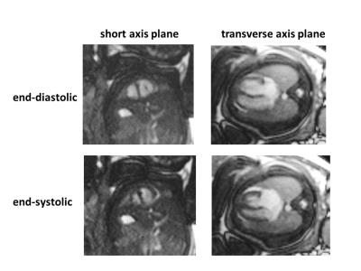
We developed a parallel imaging accelerated Cine Watermark (CWM) sequence to provide normal cine magnitude images plus phase image-only multi-directional spatial encoding for quantitative cine strain -- while requiring no extra operator effort.  Spatial cosine modulation post-processed by complex image sum/differencing produced separate normal magnitude cine and unique phase-only spatial modulation for strain calculation.  In vivo human scans demonstrated good magnitude cine and phase-only quantified displacement. Cardiac strains were tracked by a novel non-linear least squares method combining quadratic b-spline contours, nearest neighbor optimized phase tracking, and physically-motivated regularizers.  Cardiac left ventricular, short-axis, circumferential strain results agreed with previous literature.


3145.   
53 Cardiac function relation to preload variation based on free breathing motion
Teodora Chitiboi, Rebecca Ramb, Eve Piekarski, Li Feng, Anja Hennemuth, Leon Axel
Free-breathing cardiac cine MRI provides novel information on cardiac dynamics. In this work, we investigate the relation between cardiac function and left-ventricular preload variations induced by respiratory motion, in normal subjects and patients. We used the left-ventricular diameter perpendicular to the septum as a measure of preload and the relative change in diameter between end-systole and end-diastole as a measure of functional response. There was a significantly larger change in diameter during the respiratory cycle for normal subjects compared to patients.  We also found a significant correlation between preload and cardiac function for normal subjects, which was reduced for patients.


3146.   
55 
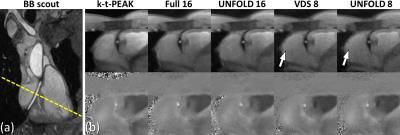
Quantitative Evaluation of Ultra-fast Real Time Imaging Compared to Cine at 1.5 T and Demonstration of Clinical Value in Atrial Fibrillation
Aaron Hess, Betty Raman, Joana Leal, Adam Lewandowski, Jane Francis, Masliza Mahmod, Dirk Voit, Markus Untenberger, Jens Frahm, Stefan Neubauer, Matthew Robson
Ultra-fast real time imaging allows cine imaging of the whole heart without gating or breath-holding in around 1 minute.  Here clinical cardiac metrics are evaluated and compared with those from a standard clinical acquisition in normal volunteers. We find agreement adequate for clinical use.  The real time approach is evaluated in a case of atrial fibrillation and found to provide a more robust evaluation of cardiac parameters in a shorter acquisition time.


3142.   
50 
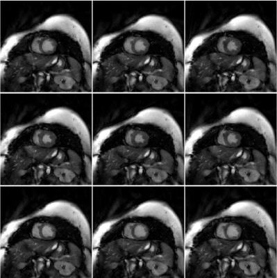
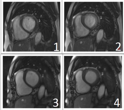
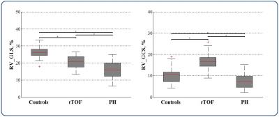
Right ventricular deformation by CMR feature tracking in patients with volume-overload and pressure-overload congenital heart diseases
Shuang Leng, Xiaodan Zhao, Wen Ruan, Ru San Tan, Ju Le Tan, Liang Zhong
Two major patterns of right ventricular (RV) overload are volume overload and pressure overload. This study aimed to investigate the differences of RV myocardial deformation associated with volume and pressure overload congenital heart diseases. Cardiac magnetic resonance feature tracking was applied to RV 4-chamber and short axis view in a subject group comprising 23 patients with repaired tetralogy of Fallot (rTOF), 23 patients with pulmonary hypertension (PH), and 23 age- and gender-matched normal controls. Results indicated that PH patients had most diminished RV longitudinal and circumferential strain. Patients with rTOF had reduced RV longitudinal strain but higher circumferential strain.


3147.   
56 
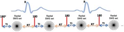
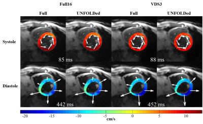
UNFOLDed spiral SPiRIT TPM enables high spatio-temporal resolution analysis of cardiac function - permission withheld
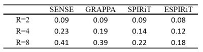
Marius Menza, Moritz Braig, Bend Jung, Daniela Föll, Jürgen Hennig, Axel Krafft
A detailed TPM analysis of cardiac function necessitates high spatio-temporal resolution which leads to prolonged scan durations. These scan times are typically too long for data acquisition within a single breath hold. Respiratory navigator-gating can compensate breathing-related motion, but causes an additional increase in measurement time and images with residual motion artifacts. The aim of this study was to combine UNFOLD with variable density spiral SPiRIT TPM to achieve an additional scan time reduction by a factor of 2 and an effective undersampling factor of 6. UNFOLDed SPiRIT TPM demonstrates good results and enables scan times within very short breath holding.


3148.   
57 
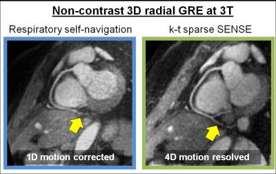
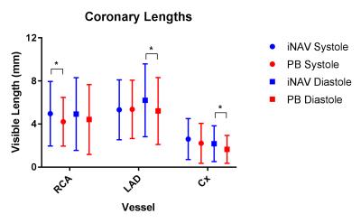
MOTION-RESOLVED 5D IMAGING OF THE HEART: TIME TO GET RID OF THE ECG?
Lorenzo Di Sopra, Davide Piccini, Simone Coppo, Jessica Bastiaansen, Matthias Stuber, Jérôme Yerly
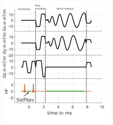
The performance of motion-resolved whole-heart MR imaging strongly depends on the quality of cardiac- and respiratory-gating signals. While navigators or self-navigation can be used to account for respiratory motion, ECG is a mainstay for synchronizing data acquisition with the cardiac cycle. We tested whether physiological motion information, directly extracted from k-space-center, can replace respiratory navigators and ECG signals. The proposed solution was applied in 9 healthy volunteers and results were compared to those obtained with the ECG-signal. Correlation between R-wave time-stamps from the ECG and the cardiac self-gating signal was excellent, while image quality and coronary artery conspicuity remained unchanged.


3149.   
58 
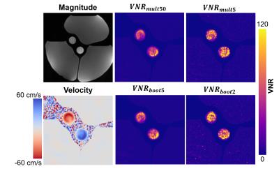
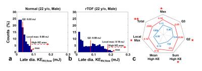
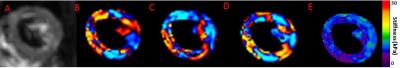
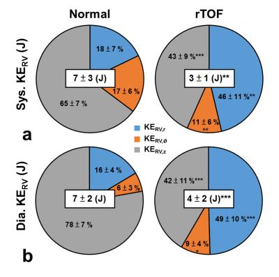
Does Right Ventricular Myocardial Kinetic Energy Correlate With Pressure Overload in Repaired Tetralogy of Fallot Patients?
Meng-Chu Chang, Ming-Ting Wu, Ken-Pen Weng, Mao-Yuan Su, Marius Menza, Hung-Chieh Huang, Hsu-Hsia Peng
The association of right ventricular (RV) myocardium adapted to pressure overload of repaired tetralogy of Fallot (rTOF) patient is still unclear. We evaluated three-directional myocardial kinetic energy (KE) and correlated it with RV-related pressure. The recruited rTOF patients presented decreased peak KERV both in systole and diastole. However, patient group exhibited increased percentage of radial KERV, accompanying with highly positive correlation with RV systolic pressure. In conclusion, the investigation of the correlation between myocardial kinetic energy and RV pressure overload may helpful to comprehend compensatory mechanism and myocardial remodeling in patients with rTOF.


3150.   
59 
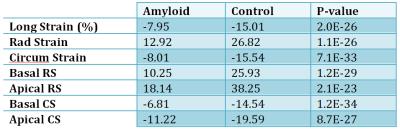
Myocardial Strain Imaging with Feature Tracking MRI in Patients with Cardiac Amyloidosis: Comparison with Normal Control Subjects
James Glockner
Feature tracking (FT) myocardial strain analysis was performed in 87 patients with biopsy proven cardiac amyloidosis and in 64 control subjects using long and short axis ECG-triggered b-SSFP images and commercial software.  Radial, circumferential, and longitudinal peak systolic strain values were all significantly different between amyloid patients and control subjects, suggesting that this technique is feasible in assessing patients with suspected cardiac amyloidosis. 


3151.   
60 
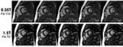
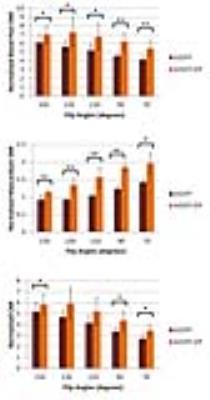
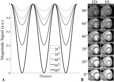
Feature-Tracking Regional Myocardial Strain:  Effects of Tag Strength and Flip Angle
Eric Schrauben, Andreas Greiser, Brett Cowan, Alistair Young
Using feature-tracking combined with variable tag strength and flip angle, myocardial strain is measured and compared against traditional tagging MRI in healthy volunteers . 


3152.   
61 
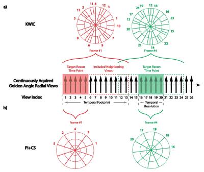
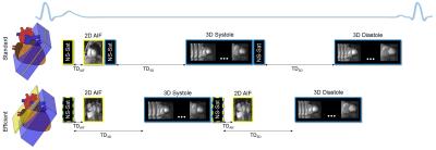
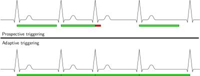
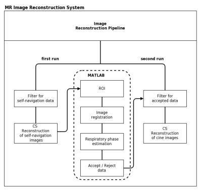
Free-Breathing Self-Navigated Isotropic 3-D CINE Imaging of the Whole Heart using Adaptive Triggering and Retrospective Gating - permission withheld
Jens Wetzl, Felix Lugauer, Randall Kroeker, Michaela Schmidt, Andreas Maier, Christoph Forman
We present a method for free-breathing whole-heart 3-D CINE imaging based on adaptive triggering and retrospective gating and compare it to a previously published method using prospective triggering. We show that our method is simultaneously robust to heart-rate variability during the scan and able to cover the entire cardiac cycle, which is not the case for prospective triggering. A validation in 6 volunteers shows reduced end-diastolic volume bias compared to a gold-standard 2-D reference for our method. Image reconstruction is integrated into the scanner system and takes less than 3 minutes.


3153.   
62 
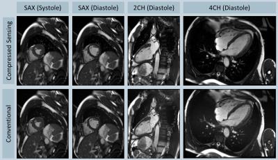
Real-time cardiac cine based on a radial bSSFP sequence and compressed sensing: initial experience on the evaluation of left ventricular function in patients
Xiaoyong Zhang, Zhongzhou Chen, Xiaohai Ma, Lei Zhao, Hui Chen, Caiyun Shi, Shi Su, Xin Liu, Bensheng Qiu, Zhaoyang Fan, Guoxi Xie
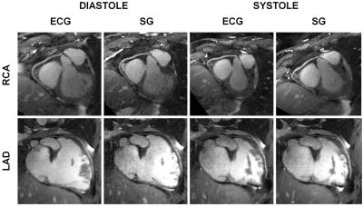
Cardiac cine magnetic resonance imaging is a valuable technique for assessing cardiac function. However, conventional cardiac cine imaging is based on breath-holding and ECG-triggering, which has particularly difficulty to be used for the diagnosis of the patients with arrhythmia. In this work, a novel technique for real-time cardiac cine imaging was developed and used to evaluate the left ventricular (LV) function in patients with arrhythmia. Preliminary experiment results demonstrated that the proposed method can accurately evaluate patient’s LV function without the use of ECG triggering and improve the image quality of the patients with arrhythmia. 


3154.   
63 Global circumferential strain derived from feature tracking imaging can detect early subclinical myocardial disorders in patients with heart failure preserved ejection fraction: Comparison with tagged cine magnetic resonance imaging - permission withheld
Yoshiaki Morita, Naoaki Yamada, Makoto Amaki, Yoshiaki Watanabe, Tatsuya Nishii, Atsushi Kono, Tetsuya Fukuda
The heart failure preserved ejection fraction (HFpEF) accounts for 40–50% of all causes of heart failure. For HFpEF, identification of strain disturbances where systolic function is seemingly preserved is a decisive step toward revealing hidden heart damage. Recently, the novel technique of feature tracking imaging (FTI) was introduced for myocardial strain measurement directly from conventional cine images. We demonstrated that FTI allows for detailed strain assessment with acceptable correlation with the tagging method. In particular, global circumferential strain, which cannot be detected by analysis using cine images, may possibly serve as a sensitive and early marker of cardiac dysfunction.


3156.   
65 Four Chamber Endocardial Surface Reconstruction from Cardiac MRI Data
Xiaoxia Zhang, Steven Lloyd, Himanshu Gupta, James Davies, Nouha Salibi, Louis Dell’Italia, Thomas Denney
Shape analysis of cardiac chambers has important implications for cardiac diseases, most extensively studied in mitral regurgitation (MR). Here we present a novel algorithm for fitting surfaces to the endocardium of all four chambers through the entire cardiac cycle and demonstrate its use in patients with MR. This algorithm, which is based on standard CMR acquisitions, demonstrates improved visualization of the chambers and their function, which can help cardiologists and surgeons in treatment planning in conditions such as mitral regurgitation.


3157.   
66 Regions of spared hypertrophy in pressure overloaded hearts promote severe systolic dysfunction as assessed by comprehensive cardiovascular magnetic resonance
Sebastian Haberkorn, Joachim Schmitt, Christoph Jacoby, Jürgen Schrader, Malte Kelm, Uli Flögel
Regional heterogeneity of contractile function was described in patients with left ventricular (LV) hypertrophy, suggesting a predominant impairment of areas with distinct hypertrophy. Heterogeneity of myocardial hypertrophy therefore may contribute to global and regional abnormalities of LV function. Here, we systematically investigated the spatial and temporal patterning of myocardial hypertrophy in response to experimental pressure overload by cardiovascular MRI. Surprisingly, we identified a specific basolateral LV segment that is frequently spared from the development of myocardial hypertrophy and promotes systolic dysfunction. Moreover, the initial LV geometry and the vascular topology seemed to limit the extent of adaptation to pressure overload.


3158.   
67 Healthy aging of the left ventricle in relationship to cardiovascular risk factors:  The Multi-Ethnic Study of Atherosclerosis (MESA)
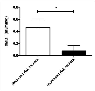
Chia-Ying Liu, Shenghan Lai, Nadine Kawel-Boehm, Harjit Chahal, Bharath Ambale-Venkatesh, Joao Lima, David Bluemke
We used cardiovascular magnetic resonance imaging to measure the LV and aortic structure and function in the Multi-Ethnic Study of Atherosclerosis (MESA). The cohort was divided into groups with or without traditional risk factors. In multivariable analyses adjusting for age, sex and race, individuals with risk factors had significantly larger LV mass index (by 17%) and lower LV contractibility (circumference strain, lower by 14%). LV structure and function are also better preserved in senescent hearts in the absence of traditional cardiovascular risk factors.


3159.   
68 MRI Feature Tracking Strain Provides Incremental Prognostic Information Over Serum Biomarkers in AL Amyloidosis - video not available
Jeffery Illman, James Glockner, Ian Chang, Arvin Arani, Shivaram Arunachalam, Kiaran McGee, Martha Grogan, Angela Dispenzieri, Philip Araoz
The association of MRI feature tracking (FT) strain with all-cause mortality was retrospectively performed on 76 patients with new diagnosis of AL amyloid.  Mean follow-up was 4 years.  MRI FT radial, circumferential, and longitudinal strain were each associated with all-cause mortality in univariate analysis.  In separate multivariate models with serum biomarker stage, radial, circumferential, and longitudinal strain each remained prognostic.  This study shows the incremental prognostic value of MRI FT strain in AL amyloidosis.  


3160.   
69 The impact of heterogeneity on regurgitation classification for pulmonary artery after repaired Tetralogy of Fallot
Pei-Hsin Wu, Hsiao-Wen Chung, Ming-Ting Wu, Cheng-Wen Ko
The degree of pulmonary regurgitation has been a determinant for re-intervention for patients with repaired TOF. However, in practice, the accurate assessment of regurgitation may be interfered by the simultaneous existence of forward flow and backward flow in any single cardiac phase, which may mislead clinical decision making. In this study, we investigated the impact of heterogeneity in blood flow profiles on guiding management and the classification of regurgitation. Preliminary results suggest that pixel-wise reexamination may be required for reclassification, especially for patients with marginal moderate regurgitant fraction value.


3155.   
64 The utility of fetal MRI using real-time cine imaging for the functional assessment of congenital cardiovascular abnormality - permission withheld
Yoshiaki Morita, Mitsuhiro Tsuritani, Naoaki Yamada, Yoshiaki Watanabe, Tatsuya Nishii, Atsushi Kono, Jun Yoshimatsu, Tetsuya Fukuda
Volumetric analysis of the fetal heart by ultrasound (US) in congenital heart disease is often difficult due to its complex anatomy and US-specific artifacts. We implemented the real-time cine sequence without ECG triggering and the post-processing technique (PhyZiodynamics), which enabled noise reduction and interpolation based on motion coherence. Fetal MRI using real-time cine imaging allowed for detailed functional assessment in both ventricles and showed acceptable levels of correlation with both the prenatal and postnatal US findings, suggesting that this technique is a promising diagnostic tool for functional assessment of congenital cardiovascular abnormalities.


3161.   
70 
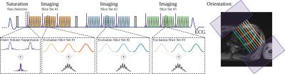
Feasibility Study: 2-D Self-Navigation using Compressed Sensing Reconstruction for Respiratory Gating in Free-breathing 3-D CINE Imaging
Ivo Prochaska, Jens Wetzl, Christoph Forman, Armin Nagel, Andreas Maier
We investigate the feasibility of  using 2-D self-navigation for respiratory gating for free-breathing whole-heart 3-D CINE imaging, where respiration-induced cardiac motion may be more easily detected than in commonly used 1-D self-navigation methods. We compare self-navigation images, derived gating signals and resulting 3-D CINE images of the 1-D and 2-D methods and find that respiratory motion can be well visualized with the 2-D method; both methods show a good overlap of gating signals and little difference in resulting image quality. 2-D self-gating may thus be considered a promising alternative to 1-D self-navigation as it allows easier detection of respiratory motion.


3162.   
71 Accelerated 2D Cine MRI Featuring Compressed Sensing and ECG-triggered Retro-gating - permission withheld
Christoph Forman, Randall Kroeker, Michaela Schmidt

We present the combination of ECG-triggered retrospective gating and compressed sensing for segmented 2D Cine imaging at high spatiotemporal resolution. This enables capturing the complete cardiac cycle in segmented acquisitions, while significantly reducing the total acquisition time with compressed sensing and auto-calibration. The method was evaluated in 8 healthy volunteers and ventricular function parameters were compared to reference 2D Cine acquisitions featuring ECG-triggered retro-gating. Both methods resulted in comparable image quality and equivalent quantitative values for ventricular function parameters.


3163.   
72 
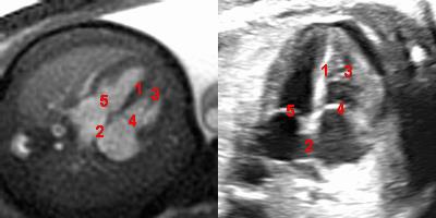
Identification and Measurement of Anatomical Landmarks Using Fetal Cardiac Cine MRI in Comparison with the Clinical Gold Standard Echocardiography
Jerome Yerly, Jerome Chaptinel, Yvan Mivelaz, Milan Prsa, Leonor Alamo, Yvan Vial , Gregoire Berchier, Jean-Baptiste Ledoux, Chantal Rohner, Matthias Stuber
The recent development of a self-gated framework to reconstruct cardiac cine MR images without the need for an external ECG signal has opened the door to prenatal cardiac examination with MRI. This study investigates the potential of this technique for fetal cardiac MRI and compares its performance with the clinical gold standard echocardiography. Standard views from clinical fetal echocardiographic examinations were acquired with both imaging modalities and two experienced readers independently compared them qualitatively and quantitatively. The results showed good agreement between the two modalities and validate the use of MRI for prenatal evaluation of the heart.
Velocity & Flow
Electronic Poster
Cardiovascular

 
Monday, 24 April 2017
Exhibition Hall  09:15 - 10:15


    Computer #

 
3212.   
49 Fast self-gated 4D flow measurements in the murine aortic arch with retrospective radial sampling
Patrick Winter, Kristina Andelovic, Thomas Kampf, Peter Jakob, Wolfgang Bauer, Volker Herold
A self-navigated radial 4D-PC sequence is presented for accelerated ECG-free 4D flow measurements in the murine aortic arch. Self-navigation signals were extracted from the radial DC signal and used for retrospective motion synchronization. 3D-Cines with 30 frames were reconstructed with a spatial resolution of 100 µm. The volume flow was determined at 4 2D slices extracted from the 3D dataset and the 3D flow was visualized with streamlines. The results are in good accordance with results reported for ECG-triggered measurements. The new method yields high potential for preclinical studies of hemodynamics and can also be transferred to applications in humans.


3213.   
50 Fast self-navigated PC-MRI for human cardiac flow measurements using retrospective radial sampling - permission withheld
Patrick Winter, Kristina Andelovic, Peter Jakob, Wolfgang Bauer, Volker Herold
A self-navigated radial PC-MRI sequence is presented for fast ECG-free 2D-Cine measurements with 3-dimensional flow encoding. The radial DC signal was used for retrospective self-navigation. Reconstructions were performed with an iterative CG-Sense algorithm. For each flow encoding step 2D-Cines with 30 frames, respectively, and a spatial resolution of 2.34 mm were reconstructed.  The time-dependent volume flow through the pulmonary artery was quantified and the blood flow through the ventricles was visualized. The proposed method does not rely on ECG signals and is immune to distortions often observed with conventional triggering. It hence yields high potential for clinical applications.


3214.   
51 Cardiac 4D phase-contrast MRI at 9.4 T using self-gated ultra-short echo time (UTE) imaging
Martin Krämer, Abdallah Motaal, Karl-Heinz Herrmann, Bettina Löffler, Jürgen Reichenbach, Gustav Strijkers, Verena Hoerr
Time resolved 4D phase contrast (PC) MRI in mice is challenging and often susceptible to artifacts due to long scan times, ECG-gating and the rapid blood flow and cardiac motion of small rodents. To overcome several of these technical challenges we implemented a retrospectively self-gated 4D-PC radial UTE acquisition scheme and assessed its performance in healthy mice by comparing the results with those obtained with an ECG-triggered 4D-PC fast low angle shot (FLASH) sequence. 


3215.   
52 Effect of field strength and spatial resolution on quantifying intracranial hemodynamics with PEAK-GRAPPA accelerated Dual-Venc 4D flow MRI
Susanne Schnell, Maria Aristova, Can Wu, Pierre-Francois Van de Moortele, Bharathi Dasan Jagadeesan, Kamil Ugurbil, Michael Markl, Sebastian Schmitter
Dual-venc 4D flow MRI was applied in 5 healthy volunteers at two different field strengths of 3T and 7T and at two different isotropic spatial image resolutions, approximately (1.2mm)3 and (0.8mm)3. The aim of this study was to systematically investigate the effect of field strength as well as the impact of resolution for intracranial 4D flow MRI with respect to image quality (vessel sharpness and depiction of small intracranial vessels) and quantification of intracranial flow parameters (net flow, peak velocity).


3216.   
53 An MRI Phantom Study to Assess the Effects of Localized Stiffness on Aortic Hemodynamics
Khalil Rachid, Dima Rodriguez
Cardiovascular complications have been highly associated with arterial stiffness which results from aging and/or vascular disease. Reduced arterial elasticity, particularly at the aorta level, increases the left ventricle load. In this work, we studied the effects of localized stiffness on an aortic phantom with respect to its severity and position relative to an upstream proximal site. As expected, our results showed that the aortic hemodynamics were altered: the pulse pressure was increased, and the flow rate decreased. Moreover, the local proximal compliance was reduced as the stiffness was brought closer to the heart. However, the pulse wave velocity remained unchanged suggesting that global stiffness measuring approaches might not detect regional wall alterations 


3217.   
54 Interobserver reproducibility of blood flow measurements with an abdominal 4D flow MRI sequence with spiral sampling and compressed sensing
Octavia Bane, Steven Peti, Mathilde Wagner, Stefanie Hectors, Hadrien Dyvorne, Bachir Taouli
Our study evaluated the interobserver reproducibility of flow quantification in abdominal vessels using a novel 4D flow phase-contrast (PC) MRI sequence. The effect of liver fibrosis on blood flow metrics was also examined in 20 patients. Time-averaged vessel cross-section area, through-plane velocity and volume flow measured in 14 abdominal vessels in a subset of 10 patients were found to have acceptable interobserver agreement (Cohen’s kappa 0.762). A significant increase in the hepatic artery velocity and flow, splenic vein area and flow, middle hepatic vein velocity and flow, and right hepatic vein velocity was observed in patients with advanced fibrosis/cirrhosis.  


3218.   
55 Comparison of SENSE, GRAPPA, SPIRiT and ESPIRiT for accelerated 4D flow MRI Imaging
Xiaole Wang, Aiqi Sun, Jianwen Luo, Rui Li
Parallel imaging is a promising method to shorten the scanning time of 4D flow MRI. We applied SENSE, GRAPPA, SPIRiT and ESPIRiT in 4D flow imaging on six healthy volunteers and compared the accuracy of four algorithm. We found that ESPIRiT and SPIRiT showed the better velocity maps than SENSE and Grappa. The temporal fidelity of ESPIRIT can be the best among four methods, while SENSE and GRAPPA resulted in overestimates of peak flow. In conclusion, we validate the accuracy of four widely-used parallel imaging methods for the reconstruction of velocity map in 4D flow MR Imaging.


3219.   
56 UNFOLDed Spiral SPiRIT Phase Contrast Velocity Mapping for Scan Time Reduction of Coronary Blood Flow Measurements
Axel Krafft, Simon Reiss, Jürgen Hennig, Marius Menza
Coronary flow measurements can provide important information, e.g. to assess coronary stenosis, but remain challenging due to the need for high spatio-temporal resolution in the presence of heart and respiratory motion. Progress has been made by using spiral acquisitions enabling data acquisition within a breath hold. We combined spiral SPiRIT flow measurements with UNFOLD, a technique that allows for further data undersampling and scan time shortening (here by a factor of 2), to achieve coronary flow measurements with a spatio-temporal resolution of 1×1×8mm³/<15.5msec. The combined UNFOLD-spiral approach was implemented into different spiral sampling patterns and evaluated in healthy volunteers.


3232.   
69 Blood flow velocity and pulsatility measurement of lenticulostriate arteries with 3T 2D Qflow MRI
Lennart Geurts, Jeroen Hendrikse, Jaco Zwanenburg
The lenticulostriate arteries are involved in small vessel disease and their blood flow velocity and pulsatility has been measured with 7T 2D Qflow. To make this measurement more widely available we aimed to translate it to 3T by increasing blood SNR with a contrast agent. In this study we show that the 2D Qflow acquisition can measure blood flow velocity and its pulsatility in lenticulostriate arteries at 3T, even without the use of a contrast agent.


3220.   
57 Respiratory changes in pulmonary flow distribution in Fontan circulation using “5-D” flow MRI
David Rutkowski, Christopher François, Alejandro Roldán-Alzate
The purpose of this study was to evaluate the respiratory effects on particle trace distribution from the vena cava to the pulmonary arteries in single ventricle patients with TCPC. Six patients were imaged using PC-VIPR and a scheme that allows for double gating to the ECG and respiratory cycles, providing flow data for separate respiratory phases. Results displayed a non-equal flow distribution to the pulmonary arteries from the vena cava and a significant difference in pulmonary flow distribution between inspiration and expiration.  This may indicate the importance accounting for respiratory cycle variations when interpreting clinical TCPC results.


3221.   
58 Bootstrapped Estimates of Velocity Uncertainty for 4D Flow PC-MRI
Michael Loecher, Daniel Ennis
We implement and analyze a bootstrapping method to estimate VNR measurements with a limited number of 4D Flow acquisitions.  Bootstrapped estimates were created from sets of 2 acquisitions and sets of 5 acquisitions.  The VNR maps were compared to a ground truth VNR measurement generated from 50 acquisitions in a flow phantom.  The method was also demonstrated in vivo to generate VNR maps of the carotid arteries.


3222.   
59 Evaluate right ventricle-pulmonary artery interaction in patients with repaired tetralogy of Fallot by 4D flow-derived kinetic energy
Meng-Chu Chang, Ming-Ting Wu, Ken-Pen Weng, Mao-Yuan Su, Marius Menza, Hung-Chieh Huang, Hsu-Hsia Peng
We quantified the kinetic energy (KE) of intraventricular RV flow (KERV,flow), PA forward flow (KEPA) and PA retrograde flow (KEPA,regur). The purpose is to establish 4D flow-derived KE-related indices for better understanding of the RV-PA interaction. We observed the interaction between RV and PA by assessing the correlation between diastolic KEPA,regur and late diastolic KERV,flowas well as between systolic KEPA and RV mean pressure. In conclusion, we verified that altered hemodynamic conditions of RV interacted with severe PR, which may lead to RV overloading pathology and adverse outcome in rTOF patients. 


3223.   
60 Energy-loss in Fontan Circulation Develops Heterogeneous Pulmonary Perfusion: Hybrid Analysis of 2D Vortex Flow Map and 4D Flow MRI
Michinobu Nagao, Umiko Ishizaki, Yumi Shiina, Kenji Fukushima, Yuka Matsuo, Tatsunori Takahashi, Kei Inai, In-Sam Park, Yasuhiro Goto, Yamato Shimomiya, Yuzo Yamasaki, Ichiro Sakamoto, Kenichiro Yamamura, Atsushi Takemura, Masami Yoneyama, Shuji Sakai
Patients with Fontan-operation, single-ventricle heart frequent have systematic collaterals that increase pulmonary blood flow. The competitive flow elevates pulmonary artery pressure, a process leading to erosion of flow energy. We developed a novel post-processing of 2D cine MRI named for “vortex flow map” to evaluate the energy-loss in Fontan-circuit. Vortex flow map shows energy-loss as small magnitude of vortex flow (MVF) and reveals the relation to heterogeneous pulmonary perfusion. In total cavopulmonary conversion, 4D flow can visualize energy-loss caused by a meandering extra-cardiac conduit and the conflict flow, leading to reduce unilateral lung perfusion.


3224.   
61 Accurate Flow MRI: the importance of velocity distribution asymmetry
Antoine Vallatos, Haitham Al-Mubarak, James Mullin, William Holmes
This work proposes a theoretical and experimental investigation into the unexplored effect of asymmetric distribution of intra-voxel velocities on the accuracy of Flow MRI. Our experimental results show that asymmetric velocity distributions can compromise the linearity of measured phase against applied gradient, leading to important velocimetry errors. A theoretical expression of the observed phase measurement errors is introduced, relating them to velocity distribution properties such as variance, skewness and kurtosis. This enables to explain previously reported velocimetry errors and propose solutions so as to increase the accuracy of velocity measurements.


3225.   
62 Comparison of Doppler Echocardiography Mean Transvalvular Aortic Pressure Gradient to 4D Flow MRI
Michael Rose, Kelly Jarvis, Susanne Schnell, James Thomas, Joshua Robinson, Cynthia Rigsby, Michael Markl, Alex Barker
Mean and peak pressure gradients are an important measure of aortic stenosis severity. In this study, we present a novel method for measuring mean pressure gradients using 4D flow MRI. 4D flow MRI peak and mean pressure gradients were measured in 23 pediatric BAV patients and compared to Doppler echocardiography (echo). There was no significant difference between 4D flow MRI and echo in peak or mean pressure gradients. 4D flow MRI mean pressure gradients correlated better with echo than peak pressure gradients (R2= 0.78 vs. R2=0.27), suggesting 4D flow to be better suited for measuring mean pressure gradients.


3226.   
63 On Partial Fourier Acquisition in 4D Flow MRI of Mean Velocities and Turbulent Kinetic Energy
Jonas Walheim, Sebastian Kozerke
Scan time reduction with partial Fourier was investigated for 4D flow MRI of mean velocity and turbulent kinetic energy. It was shown analytically, that PF leads to a loss in resolution for phase images. According to experiments with in-vivo data, the precondition of slowly varying phases is not met for PF reconstructions of 4D flow MRI, making homodyne reconstruction and POCS fail. Therefore, it is concluded, that PF cannot recover missing k-space samples in 4D flow MRI and does not offer a benefit over symmetric k-space sampling with the same number of phase encodes.


3227.   
64 Comparison of 4D Flow MRI and Tomographic Particle Image Velocimetry
Rafael Medero, Alejandro Roldán-Alzate
4D flow MRI has shown promising results, assessing hemodynamics in different vascular territories. In order to keep expanding its clinical use, reliable validation of 4D flow MRI is needed for improvement of its accuracy and precision while reducing scan time. The purpose of this study was to compare velocity measurements through an in vitro carotid artery bifurcation model using 4D Flow MRI and particle image velocimetry. PIV system provides great insight into the velocity field within the model, using an excellent acquisition quality.   


3228.   
65 Combining Phase Information from Phased Array Coils without Phase Unwrapping in Phase Contrast Imaging
Yi Wang, Xingxian Shou, Hai Luo, Xiang Zhou, Bin Wang, Leping Zha
In phase contrast flow quantitation, high spatial/temporal resolution and parallel imaging often entail the high density multiple phased array receive coils.  To overcome ambiguities in coil signal combination introduced by motion, phase wrapping, image noise, and the unknown phase-offset among the coils, we used the k-space complex division of the same coil element signals from both flow compensated (FC) and flow encoded (FE) scans to eliminate the coil sensitivity phase associated with that coil element and then add all phase difference of each elements in complex domain to avoid phase unwrapping for a fast and accurate flow velocity mapping.


3229.   
66 Computational Fluid Dynamics of Pulmonary Circulation Before and After Induced Pulmonary Hypertension: 2D Flow versus 4D Flow MRI-Based Boundary Conditions
Sylvana García-Rodríguez, James Leschke, Alejandro Roldán-Alzate, Christopher François

Acute pulmonary hypertension was induced in dogs by micro embolization. RHC was performed, as well as 4D flow MRI pre and post-embolization. MRI data was used to develop a CFD model of the proximal pulmonary circulation while comparing two inlet boundary conditions: velocity definition as defined from 2D flow MRI and from 4D flow MRI, which takes into account the directionality of the velocity. WSS tended to decrease post-embolization. Pressure gradient distribution shows that during peak systole, a constant normal inlet condition might be sufficient for CFD simulation; however, differences might be present during diastole.


3230.   
67 Improving visualization of cardiac QFlow by acquiring bSSFP cine images within the same breath hold
Matthew Lanier, Ryan Moore, Michael Taylor, Charles Dumoulin, Hui Wang
In this study, we propose a method to acquire QFlow and bSSFP cine images in a single breath-hold that eliminates the need to have these scans acquired separately; thereby overcoming the need for inter-breath hold cross-registration.  The approach provides improved registration of flow fields with high-resolution anatomic cine imaging.


3231.   
68 Improved blood flow velocity measurement in superficial perforating arteries of the white matter at 7 tesla MRI
Lennart Geurts, Geert Jan Biessels, Jaco Zwanenburg
7T 2D Qflow is capable of measuring blood flow velocity in the superficial perforating arteries. These arteries run though the semi oval center of the white matter in the brain and have diameters smaller than 200 µm. Due to the small diameters, partial volume effects of perforators with surrounding tissue cause velocity underestimation. With simulations and experiments we show that tilt optimized nonsaturated excitation (TONE) significantly increases SNR and decreases velocity underestimation in superficial perforating arteries.


3233.   
70 Respiratory Controlled Adaptive K-space Reordering (ReCAR) Improves 4D Flow Image Quality
Ning Jin, Andreas Greiser, Kelvin Chow, Susanne Schnell, Alex Barker, Michael Markl

4D flow normally uses respiratory gating to mitigate breathing artifacts for thoracic and abdominal applications. Respiratory Controlled Adaptive k-space Reordering (ReCAR) employs k-space reordering based on the current respiratory position. It has been reported to increase respiratory gating efficiency for 4D flow applications. The purpose of this study was to systematically compare the image quality of 4D flow data acquired with and without adaptive k-space reordering.


3234.   
71 Respiratory self-gated golden-angle spiral 4D flow MRI - permission withheld
Rene Bastkowski, Kilian Weiss, David Maintz, Daniel Giese
A spiral self-gated 4D Flow sequence is presented that operates at a predictable scan time. The respiratory self-navigation signal is extracted from the FID during the fat saturation pulse. The sequence allows to reconstruct 4D flow data in multiple respiratory states. This technique potentially allows to analyze respiratory dependent flow.


3235.   
72 The importance of correcting for through-plane heart motion in the assessment of aortic regurgitation using PC-MRI as estimated by feature tracking cine-MRI - permission withheld
Frida Svensson, Alexander Johansson, Åse Johnsson, Kerstin Lagerstrand
Phase contrast measurements are prone to velocity offsets due to through-plane motion of the heart. Here we examine the impact of these velocity offsets on the assessment of aortic regurgitation using a promising method for quantification of the through-plane heart motion. Without correction for through-plane heart motion, the phase contrast measurements significantly underestimated the severity of the regurgitation and the underestimation varied highly between individuals. This calls for development of reliable and robust methods, such as the proposed tracking method, for individual correction of through-plane heart motion in the phase contrast measurements.
Myocardial Ischemia Imaging
Electronic Poster
Cardiovascular

 
Monday, 24 April 2017
Exhibition Hall  09:15 - 10:15


    Computer #

 
3236.   
1 Coronary flow velocity reserve by 3T-MRI  fast velocity-encoded cine can detect patients with high-risk coronary artery disease
Masanao Naya, Yasuka Kikuchi, Noriko Oyama-Manabe, Masahiko Obara, Tadao Aikawa, Osamu Manabe, Hiroyuki Sugimori, Nagara Tamaki
Low coronary flow velocity reserve (CFVR) on left main coronary trunk can predict downstream coronary organic stenosis and myocardial scar, suggesting that CFVR derived by flow velocity by MRI is a simple and reliable index to detect patients with high-risk coronary artery disease.


3237.   
2 Cardiac ASL using Single-Shot EPI at 3T
Ahsan Javed, Krishna Nayak
Arterial spin labeling (ASL) is a non-contrast method for measuring tissue perfusion, and can be applied to the measurement of myocardial blood flow and myocardial perfusion reserve. Current cardiac ASL methods predominantly use balanced steady state free precession (bSSFP) imaging. In this work, we revisit one of the original cardiac ASL imaging schemes, single-shot EPI (Poncelet et al., MRM 1999), and experimentally demonstrate its potential advantages and its current limitations at 3T. 


3238.   
3 Quantitative dynamic MRI in the phase domain: Characterization of coronary arterial morphology using a simple Fourier method to accurately estimate bolus arrival times in DCE-MRI perfusion imaging
Karl Kunze, Teresa Vitadello, Christoph Rischpler, Markus Schwaiger, Stephan Nekolla
This abstract proposes a simple method to extract the bolus arrival time (BAT) from myocardial DCE-MRI perfusion data using a low-frequency phase reconstruction of the deconvolution of arterial input functions with myocardial tissue curves. A simulation study is performed to test the proposed technique with respect to accuracy, robustness and superiority to existing approaches. A clinical PET/MRI example and a control group are examined to show the robustness of BAT estimation even without post-processing steps like surface coil intensity correction or saturation correction. The interpretation of the BAT as a surrogate for coronary path lengths is supported by coronary angiography.


3239.   
4 Validation of the Intravoxel Incoherent Motion Model in the Healthy Human Heart using Black-Blood Preparation
Georg Spinner, Constantin von Deuster, Christian Stoeck, Sebastian Kozerke
In vivo cardiac Intravoxel Incoherent Motion Imaging (IVIM) offers the potential to estimate myocardial perfusion without the need for contrast agents. The IVIM parameter estimates, however, suffer from low signal-to-noise ratio, patient motion and are depending on imaging settings as well as on diffusion gradient shapes. In the present work, further evidence is presented that estimation of perfusion using a second-order motion compensated diffusion weighted sequence in the in vivo human heart is possible. 


3240.   
5 Accelerated Cardiac Perfusion MRI with Radial k-space Sampling, Compressed Sensing, and KWIC filtering to Enable Qualitative and Quantitative Analyses of Perfusion.
Nivedita Naresh, Hassan Valizadeh, KyungPyo Hong, Amir Rahsepar, Daniele Procissi, Jeremy Collins, James Carr, Daniel Lee, Daniel Kim
First-pass cardiac perfusion MRI is widely used as an important diagnostic tool for cardiovascular disease and extensive efforts are focused on improving spatial coverage, minimizing dark rim artifacts and quantifying absolute myocardial blood flow. In this study, we used a combination of radial k-space sampling, compressed sensing, and KWIC filtering to address these issues. Compared to the conventional perfusion technique, the accelerated method improved spatial coverage, minimized dark rim artifact and enabled quantification of myocardial blood flow.


3241.   
6 Heart-Rate Independent T2 Mapping for Overcoming Loss of BOLD Sensitivity in Conventional Cardiac T2 MRI Acquired Under Vasodilator Stress
Hsin-Jung yang, Damini Dey, Jane Sykes, John Butler, Xiaoming Bi, Behzad Sharif, Ivan Cokic, Sotirios Tsaftaris, Debiao Li, Piotr Slomka, Frank Prato, Rohan Dharmakumar
Despite the advances to date, myocardial BOLD MRI continues to be plagued by imaging confounders, which limit its reliability. We hypothesized that (a) the loss in BOLD sensitivity is dependent on the magnitude of the change in heart rate (HR) between rest and vasodilator stress; and (b) HR-insensitive T2 maps can enable BOLD changes to be accurately captured. We tested our hypothesis by examining the BOLD response to a HR-insensitive T2 mapping approach and conventional T2 mapping. Our results show that reliability of T2-based myocardial BOLD MRI could be markedly improved through heart-rate-insensitive T2 acquisitions.


3242.   
7 Spiral simultaneous multi-slice first-pass myocardial perfusion imaging
Yang Yang, Matthew Van Houten, Patrick Norton, Klaus Hagspiel, Christopher Kramer, John Mugler, Michael Salerno
First-pass contrast-enhanced myocardial perfusion imaging is an important tool to assess patients with coronary artery disease, but current techniques are still limited by spatial-temporal resolution and ventricular coverage. Simultaneous multi-slice (SMS) utilizes multi-band (MB) RF pulses which can greatly improve sampling efficiency. We develop an SMS-spiral perfusion pulse sequence by modulating the phase of excitation RF pulses of multiple slices with a Hadamard matrix to achieve significant signal cancellation and an incoherent aliasing pattern. The pulse sequence is evaluated in 23 patients with MB factors of 2, 3 and 4 resulting in high image quality.


3243.   
8 Quantitative 3D myocardial perfusion at systole and diastole with a dual echo arterial input function
Jason Mendes, Ganesh Adluru, Devavrat Likhite, Apoorva Pedgaonkar, Merlin Fair, Peter Gatehouse, Brent Wilson, Edward DiBella
Quantitative 3D myocardial perfusion is a clinically realistic goal and this work further develops and combines several innovative sequence designs to achieve this goal. These developments include an optimized 3D stack of stars readout (150ms per beat), acquisition and T2* correction of an arterial input function, tailored saturation pulse design and potential whole heart coverage at both systole and diastole at heart rates up to 109 bpm. Implementation and quantitative perfusion results are shown for healthy volunteers and a patient with known coronary disease.


3244.   
9 10-fold Spatial-Only Acceleration For High-Resolution Myocardial Perfusion Using Multi-Band Imaging and Multi-Band Outer Volume Suppression
Sebastian Weingärtner, Steen Moeller, Chetan Shenoy, Mehmet Akçakaya
Myocardial perfusion imaging is clinically established for the detection of myocardial ischemia and requires rapid imaging to monitor the uptake of a contrast-agent in the heart. Spatial resolution or coverage is commonly increased by exploiting temporal correlations, at the risk of inducing temporal blurring. Here, we investigate the use of simulteaneous multi-slice imaging for high spatial-only acceleration. Outer-volume-suppression using multi-band saturation-slabs (MB-OVS) were used to facilitate high multi-band factors. Phantom results, show through signal suppression outside region-of-interest with MB-OVS. In-vivo results show robust image quality throughout the contrast uptake and washout with 9-slice LV coverage at a temporal resolution <550ms.


3245.   
10 Estimating extraction fraction and blood flow by combining first-pass myocardial perfusion and T1 mapping results
Devavrat Likhite, Promporn Suksaranjit, Ganesh Adluru, Chris McGann, Brent Wilson, Edward DiBella
Dynamic contrast enhanced MRI is maturing as a tool in contemporary cardiovascular medicine. However, there are challenging areas that have not been fully understood, such as modeling extraction of the contrast agent from the vasculature to the extravascular space. We present a technique that exploits information overlap between two different cardiac MRI techniques, namely, DCE-MRI and T1 mapping, in order to estimate extraction and flow. Our study shows that extraction fraction and myocardial blood flow can be estimated by fixing extracellular volume (ECV) to values obtained from T1 mapping. 


3246.   
11 Optimized 2D radial CAIPIRINHA for cardiac perfusion MRI
Ye Tian, Ganesh Adluru, Jason Mendes, Edward DiBella
We propose an optimized offset golden angle trajectory of 3-slice radial CAIPI for more uniform distribution of phase modulated rays. Both computer simulation and phantom scan demonstrate that our proposed method has less artifact. Simulation studies show a 41% reduction in aliasing energy when compared to conventional golden angle trajectory. As well, optimization with an asymmetric echo readout resulted in a reduction of acquisition time by 15% with little sacrifice of image quality. These two methods can benefit simultaneous multi-slice perfusion acquisitions.


3247.   
12 Analysis of Coronary Contrast Agent Transport in Bolus Based Quantitative Myocardial Perfusion MRI Measurements with Computational Fluid Dynamics (CFD) Simulations in High Performance Computing (HPC) Environments
Johannes Martens, Sabine Panzer, Jeroen van den Wijngaard, Maria Siebes, Laura Schreiber
Aim of the project is the execution of computationally extremely challenging CFD simulations of blood flow and contrast agent (CA) transport in coronary arteries on HPC clusters. Therefore scalability testing is performed to assess the suitability of applied software codes in HPC environments. Cardiovascular 3D-models are extracted from high-resolution cryomicrotome imaging data and meshed with computational grids. Navier-Stokes-equations for blood flow and the advection-diffusion-equation for CA transport are solved to obtain CA bolus dispersion in the epicardial vessels. We find indications of asymptotically decreasing dispersion effects with increasing vessel generation.


3248.   
13 An analysis of radial sequence parameters in myocardial first-pass perfusion for optimised imaging
Merlin Fair, Peter Gatehouse, Edward DiBella, David Firmin
A simulation framework is devised to analyse sequence parameters in radial first-pass perfusion for optimised imaging. A model containing left-ventricular myocardium, bloodpool and defect was radially sampled in 2D and 3D under multiple sequence parameters variations, tested with differing imaging environments. Automatic measures of sharpness, signal variation and CNR were made on the N=1016 simulation reconstructed images and results of interest are presented.


3249.   
14 Quantitative MRI measurement of the interplay between myocardial function, perfusion, structure and metabolism during acute and chronic remodeling in a porcine model of myocardial infarction
Smita Sampath, Sarayu Parimal, Wei Huang, Ibrahim Mazlan, Grace Croft, Teresa Totman, Yvonne Tay, Elaine Manigbas, Miko Chang, Anqi Qiu, Michael Klimas, Jeffrey Evelhoch, Dominique de Kleijn, Chih-Liang Chin
We present a comprehensive characterization of LV structure, function, perfusion and metabolism in a porcine animal model with myocardial infarction with view to develop a translational platform for future testing of safety and efficacy of novel heart failure therapeutics.  Dynamic contrast enhanced MRI and tagging MRI are employed to obtain non-invasive quantitative magnetic resonance imaging (MRI) biomarkers for characterization.  Results demonstrate an acute decrease in function and perfusion in the infarct region and functional compensation in the remote region with associated increased metabolic activity.  Chronic remodeling is associated with decreased metabolic activity in the infarct region along with increased fibrosis.         


3250.   
15 Exenatide decreases ectopic fat accumulation but have no impact on myocardial function and perfusion in patients with obesity and type 2 diabetes
Ines Abdesselam, Anne Dutour, Alexis Jacquier, Frank Kober, Patricia Ancel, Oliver Rider, Monique Bernard, Benedicte Gaborit
The objective of the study is to assess the impact of Exenatide on endothelial reactivity, and change in ectopic fat and cardiac function. This study included 44 patients (mean 52 years) randomized to Exenatide or reference treatment. Magnetic resonance imaging was used to assess ectopic fat accumulation, coronary vasoreactivity and cardiac function. 16-weeks of Exenatide treatement resulted in a significant improvement in glycemic control and a significant reduction of both epicardial fat and hepatic steatosis. However, we found no effect of Exenatide on myocardial function.  In addition, one-week of exenatide treatment had only a modest effect on vascular reactivity, albeit non-significant.


3251.   
16 BOLD Effect of Oxygen-Inhalation T2-star MRI Surrogates Systemic and Myocardial Oxygen Consumption in Heart Failure and Myocardial Infarction
Michinobu Nagao, Kenji Fukushima, Umiko Ishizaki, Yuka Matsuo, Yuzo Yamasaki, Tetsuya Matoba, Tomomi Ide, Atsushi Takemura, Mitsuru Momose, Shuji Sakai
Measurement of oxygen consumption (OC) has been less than satisfactory in patients with heart failure. This has necessitated the invasive techniques to measure the total oxygen demand with cardiopulmonary exercise (CPX). The present study proposes a novel method to quantify OC using BOLD effect of oxygen-inhalation T2* MRI. Difference between oxygen-T2* and room-air-T2* (ΔT2*) was identified as an estimate of OC. ΔT2* was significantly correlated with systematic and myocardial OC expressed as the results of CPX and myocardial fatty acid scintigraphy. Our method allows assessing non-invasively OC in vivo. ΔT2* could be used as an imaging biomarker for heart failure.


3252.   
17 Analytically-derived Parameter Scouting for Dark-Blood Late Gadolinium Enhancement (DB-LGE) Imaging
Ahmed Fahmy, Tamer Basha, Reza Nezafat
In Dark Blood Late Gadolinium Enhanced (DB-LGE) imaging, simultaneous signal nulling of the healthy myocardium and the blood and thus yields superior contrast-to-noise ratio of the scared tissues. The method employs a T2-preparation pulse applied after the inversion recovery pulse to rapidly damp the myocardium magnetization relative to the blood. Accurate timing of the inversion pulse, T2-preparation pulse, and echo acquisition is essential for the success of the technique. In this work, we present a simple method for accurately estimating these parameters through fast low-resolution scouting scans played prior to DB-LGE scans.


3253.   
18 Myocardial Edema Imaging using Single-shot T2STIR Prepared bSSFP
Yanjie Zhu, Dan Yang, Yucheng Chen, Dong Liang, Xin Liu, Yiu-Cho Chung
STIR-TSE is useful in imaging myocardial edema. However, it is sensitivity to cardiac motion, resulting in myocardial inhomogeneity. T2p-bSSFP overcomes this issue, but averaging is commonly used to improve its contrast. We propose a novel single-shot imaging technique that generates "STIR like" contrast and improves contrast between edema and normal myocardium compared to T2p-bSSFP. The performance of the new sequence (T2STIR-bSSFP) was evaluated by phantom study and in patients with acute myocardial infraction. The results show that the average CNR of T2STIR-bSSFP between edema and myocardium was 1.9 times better than T2p-bSSFP.


3254.   
19 Ungated free-breathing late gadolinium enhancement imaging with a radial simultaneous multi-slice acquisition
Ganesh Adluru, Jason Mendes, Brent Wilson, Edward DiBella
Late Gadolinium Enhancement imaging is the gold standard for identifying infarcted myocardium.  Existing data acquisition methods rely on good ECG-gating signal and breathholding to acquire images in quiescent diastolic cardiac phase.  However, in patients with arrhythmias and when imaging at higher field strengths, R-R interval is inconsistent and a good ECG signal can be challenging.  This can lead to increased acquisition time and inconsistent recovery of longitudinal magnetization affecting signal-to-noise ratio and optimal myocardial nulling.  Here we propose an ungated free-breathing scheme that acquires multiple slices at the same time with consistent nulling of healthy myocardium at systole and diastole.


3255.   
20 High Resolution, Single-shot LGE MRI with Compressed Sensing and Radial k-space Sampling
Hassan Haji-Valizadeh, Jeremy Collins , Daniel Lee, James Carr, Daniel Kim
We sought to develop a high resolution single-shot LGE pulse sequence using Radial Compressed Sensing. We then evaluated in patients the performance of our acquisition scheme with respect to segmented clinical LGE.


3256.   
21 Quantitative Inversion Time Prescription for Late Gadolinium Enhancement Using T1-based Synthetic Inversion Recovery Imaging – Eliminating the Subjective Estimation of Inversion Time
Akos Varga-Szemes, Rob van der Geest, Carlo De Cecco, Taylor Duguay, U. Joseph Schoepf, Pal Suranyi
Conventional Look-Locker (LL)-based inversion time (TI) estimation prior to late gadolinium enhancement (LGE) imaging has multiple limitations, including: the long breath-hold, the collected images are in different cardiac phases, and the subjective TI estimation. In this study we aimed to develop a quantitative T1 mapping-based synthetic inversion recovery (IRsynth) approach allowing for the quantitative determination of the optimal TI for LGE imaging. We showed in 40 patients that the IRsynth method provides better quality of myocardial signal nulling, retrospective TI selection, higher TI resolution, no need for further LL correction or TI adjustment, and less operator dependence.


3257.   
22 Validation of Contrast Enhanced Cine Steady-State Free Precession and T2-Weigthed CMR for Assessment of Ischemic Myocardial Area-At-Risk
Esben Hansen, Steen Pedersen, Steen Pedersen, Hans Erik Bøtker, Won Yong Kim
Measuring myocardial salvage is important to evaluate the possible cardioprotective effects of adjunctive cardioprotective intervention in patients with myocardial infarction undergoing primary percutaneous intervention. Contrast-enhanced steady-state free precession magnetic resonance imaging (CE-CINE) has recently been used to quantify AAR and validated against myocardial perfusion SPECT. In this study we sought to determine how well T2-STIR and CE-CINE depicts AAR in an experimental porcine model of myocardial ischemia-reperfusion injury using histopathology as the reference for infarct size and AAR.


3258.   
23 Effects of a Gadolinium Based Contrast Agent on the Myocardial R2* Relaxation Rate in Patients with Chronic Myocardial Infarction
James Goldfarb, Nathaniel Reichek, Jie Cao, Dana Peters
In this proof-of-concept study, we studied the effects of a contrast agent on R2*/T2* relaxation in dense myocardial fibrosis.  A significant change in myocardial R2* was found after contrast and an R2* difference existed after contrast agent administration between fibrosis and remote myocardium. On average, viable myocardial R2* increased by 4.5 Hz while infarcted increased by 9.3 Hz at 1.5T with a clinical contrast agent.
New Methods
Electronic Poster
Cardiovascular

 
Monday, 24 April 2017
Exhibition Hall  09:15 - 10:15


    Computer #

 
3259.   
25 QISS FISS (Quiescent-Interval Slice-Selective Fast Interrupted Steady-State):  A Best-of-Both-Worlds Solution to Nonenhanced MR Angiography at 3 Tesla
Robert Edelman, Shivraman Giri, Ioannis Koktzoglou
QISS is a robust method for nonenhanced MRA that conventionally uses a TrueFISP readout.  At 3 Tesla, use of TrueFISP is challenging due to high RF power deposition and sensitivity to off-resonance artifacts.  In order to overcome these limitations, we implemented a QISS technique that incorporates a new type of readout that is a hybrid of TrueFISP and FLASH pulse sequences, which we call “Fast Interrupted Steady-State” (FISS).  A pilot study was conducted to evaluate the potential benefits of QISS FISS at 3 Tesla for evaluation of the peripheral arteries and great vessels of the chest.


3260.   
26 Characterizing the Cardiac Phase Dependence of Motion Compensated CODE cDTI
Eric Aliotta, Kevin Moulin, Patrick Magrath, Daniel Ennis
First and second order motion compensated convex optimized diffusion encoding (CODE-M1M2) enables robust, high resolution cardiac diffusion tensor imaging (cDTI). However, timing of the diffusion encoding relative to the cardiac cycle still requires careful evaluation to achieve precise and accurate measurements. In this study, CODE-M1M2 cDTI was acquired in healthy volunteers at both mid-systole and diastole to identify differences in fiber orientation, fiber cone of uncertainty (CoU), mean diffusivity (MD) and fractional anisotropy (FA). While fiber orientations were equivalent, lower CoU, lower MD, and higher FA were observed in mid-systole than in diastasis indicating improved performance.


3261.   
27 Insights from comprehensive fetal cardiovascular MRI assessment using 3D motion-correction and metric-optimised gated phase contrast in cases of suspected coarctation of the aorta
David Lloyd, Joshua van Amerom, Maria Murgasova, Bernard Kainz, Kuberan Pushparajah, John Simpson, Mary Rutherford, Jo Hajnal, Reza Razavi
The antenatal diagnosis of coarctation of the aorta can be life saving; however, it is notoriously difficult to predict using ultrasound alone. We present 16 fetal cases with suspected coarctation of the aorta between 30-36 weeks, assessed using a combination of novel prenatal MRI techniques: three-dimensional motion corrected slice-volume registration and metric-optimised gated phase contrast flow measurements. Detailed 3D analysis revealed significant correlation between the size and position of the aortic isthmus and the need for neonatal surgical repair. Retrograde flow at the aortic isthmus and the angle of its insertion relative to the arterial duct may also be important variables. The comprehensive physiological data generated by combining novel fetal MRI techniques may offer powerful insights into traditionally challenging antenatal diagnoses.


3262.   
28 Contact-free Cardiac Motion Estimation using the Scatter of a Parallel Transmit Coil at 7T MRI
Sven Jaeschke, Caitlin O'Brien, Aaron Hess, Matthew Robson
Cardiovascular MRI at ultra-high field strength is a growing field of research and as such there is a need for reliable, accurate, and user-independent triggering and gating. We propose a novel approach that uses directional couplers from the SAR monitoring system and an 8-channel pTx coil to measure the reflection coefficients simultaneously to determine cardiac motion with an independent component analysis and Gaussian shaped RF-monitoring pulses. This approach has the potential to improve cardiac imaging at ultra-high field CMR by providing robust triggering in periods of breath-hold and free-breathing, additional gating information and an optimised workflow with no additional set-up.


3263.   
29 Fetal cardiac volume reconstruction from motion-corrected multi-slice dynamic MRI
Joshua van Amerom, David Lloyd, Maria Kuklisova Murgasova, Anthony Price, Shaihan Malik, Paul Aljabar, Joanna Allsop, Ana Gomes, Mary Rutherford, Kuberan Pushparajah, Reza Razavi, Joseph Hajnal
Motion is a significant challenge in fetal cardiac MRI. Multi-slice 2D dynamic MR data was used to reconstruct high-temporal resolution real-time images to be combined in time as 2D cine images and then in space as volumes with full coverage of the heart. Retrospective, image-based post-processing (motion correction, outlier rejection) was used to improve image quality in the presence of intra- and inter-slice motion. Reconstructed multi-slice cine images depict the complex anatomy of the fetal heart across the cardiac cycle and preliminary volume reconstructions allow multiple views to be visualised.


3264.   
30 Time-Resolved Contrast-Enhanced MR Angiography with Single-Echo Dixon Background Suppression
Eric Stinson, Joshua Trzasko, Norbert Campeau, John Huston III, Phillip Young, Stephen Riederer
Contrast-enhanced MR angiography (CE-MRA) typically relies on a time-subtraction technique to suppress the background signal and emphasize the contrast-enhanced blood signal. However, Dixon-based background suppression has been reported to reduce motion sensitivity and improve signal-to-noise ratio in CE-MRA studies. Dual-echo Dixon techniques suffer a time penalty from acquiring an image at a second echo time, but single-echo Dixon techniques require an image at only one echo time and can reduce the time penalty. Here, time-resolved 3D single-echo Dixon CE-MRA at 3.0T with image update times of under 5 seconds is reported with results in the calves, hands, and brain.


3265.   
31 Free-breathing navigator 3D Cardiac Quantitative Susceptibility Mapping (QSM): initial experience at 1.5T and 3T.
Yan Wen, Thanh Nguyen, Pascal Spincemaille, Jiwon Kim, Jonathan W. Weinsaft, Yi Wang
To improve signal-to-noise ratio (SNR) in cardiac QSM (cQSM), we introduce here 3D acquisition that is more SNR efficient than previous 2D acquisition. Respiratory motion artifacts during the long 3D acquisition are compensated using navigator gating. We also investigated potential SNR gain at 3T over previous 1.5T. Our initial results seem to suggest possible issues at 3T that prevent the realization of potential cQSM improvement at from 1.5T to 3T in terms of SNR and contrast.


3266.   
32 Tomoelastography of the abdominal aorta and inferior vena cava in different hydration states
Thomas Elgeti, Jing Guo, Heiko Tzschätzsch, Florian Dittmann, Eric Barnhill, Jürgen Braun, Ingolf Sack
Tomoelastography was recently introduced for high-resolution stiffness mapping in abdominal organs. The method is based on multifrequency MR elastography and noise-robust reconstruction of shear wave speed maps. This study demonstrates the feasibility of tomoelastography for measurement of the mechanical properties of the abdominal aorta (AA) and inferior vena cava (IVC) in healthy volunteers. AA was measured stiffer than IVC with increasing disparity after drinking one liter of water. Our results demonstrate the sensitivity of MRE-measured effective stiffness values in AA and IVC to physiological alteration of the hydration state and offer a new perspective for MRE of abdominal vessels.


3267.   
33 Contact- free diaphragm navigation using the scatter of a parallel transmit coil at 7T
Aaron Hess, Matthew Robson
A method is presented to measure respiratory motion in mm units based on the scatter of a parallel transmit coil. The respiratory changes in scatter are observed by the local SAR monitor and converted into a respiratory measure through a calibration phase. The newly described measure was compared to the diaphragm position measured in MR images. The standard deviation of the difference between the two was 1.1±0.4 mm across six volunteers. The method was implemented into a free breathing cardiac cine which showed no respiratory artefact when used to gate. 


3268.   
34 Interleaved 31P-MR spectroscopy and cine 1H-MR imaging of the human heart at 3 Tesla - permission withheld
Madeleine Daemen, Aart Nederveen, Jeroen Jeneson, Gustav Strijkers, Adrianus Bakermans
Changes in the myocardial energy homeostasis have been linked to decreases in cardiac pumping performance. Current MR practice requires two consecutive recordings of 31P-MRS and 1H-MRI to assess both aspects of heart physiology, with consequently long scan times, considerable patient burden, and potentially a mismatch between data from separate sessions. By efficiently interleaving 3D ISIS acquisitions for localized 31P-MRS with short-axis cine 1H-MR imaging, both myocardial energy status as well as left-ventricular ejection fraction could be quantified from MR data that were acquired essentially simultaneously.


3269.   
35 Computational modeling of arterial wall strain in a rabbit model of atherosclerosis using an elastin-specific MR contrast agent
Andrew Comerford, Rene Botnar, Alkystis Phinikaridou
Vulnerable plaque rupture is a major cause of common clinical events such as heart attacks and strokes. Plaques form in the arterial wall due to atherosclerosis, however not all plaques that do develop are at risk from rupture. Risk stratification is a major clinical need. This study combines elastin-specific MR contrast agent and CINE MR data from a rabbit model of atherosclerosis with finite element modeling. This combination allows the biomechanics of the arterial wall to be compared with morphological and biological indicators. Wall strains were shown to vary depending on the wall thickness and elastin content.


3279.   
45 4D Ferumoxytol Enhanced MUSIC: Value-Based MRI in Neonates and Infants with Congenital Heart Disease
Kim-Lien Nguyen, Fei Han, Ziwu Zhou, Daniel Brunengraber, Ihab Ayad, Daniel Levi, Gary Satou, Brian Reemtsen, Peng Hu, J. Paul Finn
Conventional cardiovascular magnetic resonance imaging (MRI) in congenital heart disease (CHD) enables dynamic morphologic and vascular assessment. However, current techniques require multi-slice cine imaging and angiography with supplemental phase-contrast. We demonstrate that 4D multiphase steady state imaging with contrast (MUSIC) can provide high-resolution dynamic images of all relevant cardiovascular anatomy without breath-holding or operator input in a cohort of neonates and infants with CHD. Further, 4D MUSIC has high clinical impact on the care of small children.


3270.   
36 Dynamic PET/MR Imaging of Glucose Utilization and Contractile Function Under Hypoxic Stress
Gregory Barton, Kara Goss, Niti Aggarwal, Marlowe Eldridge, Alan McMillan
The purpose of this study was to establish a methodology that could be used to study the dynamic relationship between cardiac contractile function and metabolism in a serial rest-stress model using dynamic cardiac PET/MR. To assess dynamic changes in cardiac glucose metabolism we evaluated the use of a continuous infusion of 18F-FDG. We found that increases in cardiac contractility were coupled with an increase in glucose utilization in the myocardium under hypoxic stress conditions. These results demonstrate the feasibility of performing simultaneous measures of contractile function and metabolism in the heart.


3271.   
37 Statistical paradigm for composite MR Angiography generated from multi-contrast MRI
Dahan Kim, Patrick Turski, Oliver Wieben, Kevin Johnson
We present an innovative paradigm to overcome artifacts of individual MR angiography techniques by utilizing complimentary information existing across multi-contrast MR images. This technique applies Bayesian statistics to extract vessel likelihoods from each image type and generates a single ‘composite’ angiogram. Composite angiograms are computed utilizing black blood (BB), contrast enhanced MRA (CE-MRA), and phase contrast MRA (PC-MRA) images acquired in subjects with known neurovascular disease.  The composite angiogram is demonstrated to improve vessel lumen depiction overcoming artifacts in individual source images from background enhancement, air cavities, and flow in CE-MRA, BB, and PC-MRA, respectively. 


3272.   
38 Novel Ultrasound Real-time Feedback Slice Tracking with Spatially Resolved MR-compatible Ultrasound for Cardio-vascular MRI
Lindsey Crowe, Francesco Santini, Gibran Manasseh, Matthieu Destrade, Iris Friedli, Oliver Bieri, Rares Salomir, Jean-Paul Vallée
Spatially resolved MR-compatible ultrasound was used to detect a respiratory-mimicking motion for prospective correction of cine MR data acquisitions. A scaling factor relating the motion between the two modalities was derived in real-time and was chosen to compare motion-corrected MR images to a static phantom ‘breath-hold’ and an uncorrected cine image series.


3273.   
39 The impact of kinetic isotope effects in using deuterated glucose for metabolic experiments
Alexander Funk, Brian Anderson, Xiaodong Wen, Chalermchai Khemtong, Dean Sherry, Craig Malloy
Deuteration of 13C-enriched glucose has been used to prolong T1 of hyperpolarized 13C with the objective of monitoring glucose metabolism to lactate. However, the effect of deuteration on the flux in glycolysis in vivo has not been investigated extensively. Deuterated glucose was studied in perfused hearts and extracts were analyzed for a deuterium kinetic isotope effect using 1H,13C and 2H NMR spectroscopy. Unexpectedly, there was extensive exchange of methyl deuterons in lactate with aqueous medium.  Perdeuteration slows metabolism of glucose to lactate.


3274.   
40 Improving Shear Wave Signal-to-Noise Ratio in 3D High Frequency Cardiac Magnetic Resonance Elastography with Subjects in Prone Position
Shivaram Poigai Arunachalam, Arvin Arani, Ian Chang, Yi Sui, Phillip Rossman, Kevin Glaser, Joshua Trzasko, Kiaran McGee, Armando Manduca, Richard Ehman, Philip Araoz
Myocardial stiffness is a novel biomarker for diagnosing a variety of cardiac diseases. Our recent work demonstrated the feasibility of measuring in-vivo myocardial stiffness using 3D high frequency cardiac MR elastography (MRE) in normal volunteers using octahedral signal-to-noise ratio (OSS-SNR) as a shear wave quality metric. The purpose of this work is to determine whether scanning subjects in prone position can improve the OSS-SNR compared to supine position. 47 healthy volunteers were enrolled and OSS-SNR in prone position (mean: 1.98) was significantly higher (p < 0.01) than the OSS-SNR in supine position (mean: 1.52) with comparable mean stiffness. 


3275.   
41 Feasibility of Cardiac bSSFP Cine MRI at 0.35T: A Comparison with 1.5T
Shams Rashid, Fei Han, Yu Gao, Yingli Yang, Peng Hu
In this study, we demonstrate the feasibility of cardiac cine MRI at 0.35T using bSSFP and compared the results with bSSFP at 1.5T.


3276.   
42 Cardiac Cine MRI with bSSFP-EPI at 0.35T
Shams Rashid, Fei Han, Yu Gao, Yingli Yang, Peng Hu
We have developed a bSSFP-EPI cine sequence and implemented it on a 0.35T MR system. The bSSFP-EPI cine sequence has reduced acquisition duration compared to conventional bSSFP cine.


3277.   
43 Optimized 2D-PACE for aorta blood flow gating and its application in T1W morphological cMRI
Fang Dong, Kun Zhou, Shi Cheng
Electrocardiogram gating is commonly used in clinical cardiac MRI. However, it takes time to attach ECG electrodes and gating signals can be easily affected. In this abstract, the 2D-PACE technique is optimized for blood flow monitoring to explore its potential as an alternative to ECG gating. A new protocol triggered by 2D-PACE for T1W cardiac morphology was also proposed. Our results showed that the optimized 2D-PACE was robust for blood flow peak detection. And combining with our new protocol, it provided comparable image quality to those from ECG gated conventional protocol, which showed a great potential for cardiac triggering. 


3278.   
44 Post-Contrast Black-blood HASTE: Initial Results with Ferumoxytol
Takegawa Yoshida, Eun-ah Park, Peng Hu, Kim-Lien Nguyen, J. Paul Finn
Black blood imaging is helpful in cardiovascular evaluation, but is challenging due to its dependence on through-plane blood flow. We explored ferumoxytol-enhanced black-blood Half-Fourier Acquisition Single-shot Turbo Spin-echo (HASTE) imaging for cardiovascular morphologic evaluation and compared its effectiveness with pre-contrast double inversion (DIR) HASTE. Post-ferumoxytol HASTE showed complete and homogeneous blood suppression and better qualitative image scores (all, p<0.001) for all cardiovascular regions. The technique holds promise for confident definition of thrombus or plaque, independently of blood flow.


3280.   
46 Ferumoxytol Enhanced Cardiac Cine MRI in Patients with Implanted Cardiac Devices
Kim-Lien Nguyen, Takegawa Yoshida, Peng Hu, J. Paul Finn
With an aging population, more patients are receiving implantable cardiac devices (ICDs) annually.  Many will need one or more cardiac MRI exams over the course of their lifetime. ICDs can cause artifacts and obscure evaluation of intra-cardiac morphology. While researchers have dealt with artifacts during late gadolinium enhancement imaging by using wideband inversion recovery, off resonance artifacts preclude reliable SSFP cine and SGE cine is degraded by blood saturation. We explored the use of ferumoxytol to mitigate device-related artifacts and flow limiting artifacts during cine MRI in patients with ICDs.


3281.   
47 Vascular Access Mapping with Ferumoxytol MRA for TAVR Planning in Patients with Renal Impairment: A Step Closer Towards Patient-Specific Care
Kim-Lien Nguyen, Takegawa Yoshida, Adam Plotnick, John Moriarty, Peng Hu, J. Paul Finn
Transcatheter aortic valve replacement (TAVR) has transformed the treatment of critical aortic stenosis, particularly for those patients whose surgical operative risks are high.  However, pre-TAVR vascular mapping with CT requires 40-120 mls of iodinated contrast, which in older patients with renal impairment increases the risk of acute renal injury that portends poor prognosis. We demonstrated that ferumoxytol MRA can provide reliable vascular mapping in patients with renal impairment undergoing TAVR evaluation, without exposure to iodinated or gadolinium contrast agents.


3282.   
48 Transpedal MR-lymphangiography - A new imaging tool for pre-interventional planning of lymphatic interventions in patients with chylous effusions - permission withheld
Claus Pieper, Hans Schild
Lymphatic interventions are increasingly performed for the treatment of chylous leakages. Pre-interventional evaluation of the central lymphatic system is important for adequate patient selection and planning of the interventional technique. We describe a new transpedal approach for MR-lymphangiography (tMRL) to evaluate the anatomy of central lymphatics including anatomical variations, as well as for detection of leakages and analysis of a potential interventional access site. Our results demonstrate that central lymphatic anatomy can be readily evaluated using tMRL in the majority of patients scheduled for lymphatic interventions. It is a well-tolerated, valuable diagnostic tool in the pre-interventional work-up of lymphatic interventions.
 

The International Society for Magnetic Resonance in Medicine is accredited by the Accreditation Council for
Continuing Medical Education to provide continuing medical education for physicians.