Diffusion: Validation
Electronic Poster
Diffusion
Monday, 24 April 2017
| Exhibition Hall |
14:45 - 15:45 |
| |
|
Computer # |
|
3451.
 |
1 |
Quantitation of DTI changes associated with muscle injury using a 3D printed phantom 
David Berry, Shangting You, John Warner, Lawrence Frank, Shaochen Chen, Samuel Ward
Diffusion tensor imaging has been proposed as a tool to non-invasively assess skeletal muscle microstructure, which would be of significant clinical value. However, its application to the assessment of changes in muscle microstructure associated with injury, pathology, or age remain poorly defined because it is difficult to precisely control muscle microstructural features in vivo. Recent advances in bottom up fabrication technologies allow precision-engineered diffusion phantoms with histology informed skeletal muscle geometry to be manufactured. Therefore, the goal of this study was to develop skeletal muscle phantoms at relevant size scales in order to relate microstructural features to MRI-based diffusion measurements.
|
|
3452.
 |
2 |
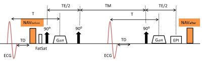
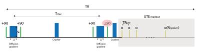
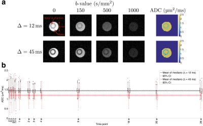
Accuracy of ADC measurements with an Ultrashort Echo Time Diffusion Weighted stimulated echo 3D Cones sequence (DW-STEAM 3D Cones UTE)
Paul Baron, Dirk Poot, Piotr Wielopolski, Edwin Oei, Juan Hernandez-Tamames
Diffusion weighted STEAM with UTE readout is a promising acquisition method to measure diffusion in tissues with short T2 and T2*, such as tendons, ligaments and menisci. However, the accuracy of the ADC obtained with this method has not been studied before. Using experiments and Bloch simulations we show that the ADC can be biased, especially when a short TR is used, and that this bias depends on T1 and T2. Randomization of the diffusion gradient direction reduces the bias, providing clear suggestions to improve acquisition and/or post processing that reduces the ADC bias.
|
|
3453.
 |
3 |
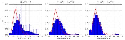
A biomimetic tumour tissue phantom for validating diffusion-weighted MRI measurements
Damien McHugh, Fenglei Zhou, Penny Hubbard Cristinacce, Josephine Naish, Geoffrey Parker
This work investigates the stability of a water-based biomimetic tumour tissue phantom, and evaluates its potential as a tool for validating diffusion-weighted (DW) MRI measurements. As with biological tissue, and unlike most previous phantoms for tumour DW-MRI, the phantom’s apparent diffusion coefficient depends on diffusion time, with values stable over six months. DW-MRI-based estimates of microstructural parameters exhibited bias, possibly indicating limitations in the analysis model or acquisition scheme. It is envisaged that such phantoms will aid investigation of DW-MRI tumour microstructural models, and more generally will act as realistic test objects for comparing DW-MRI-derived biomarkers obtained from different scanners/sites.
|
|
3454.
 |
4 |
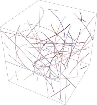
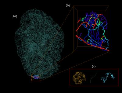
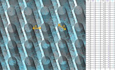
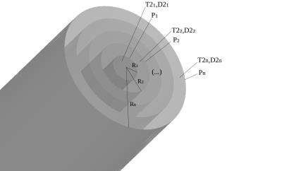
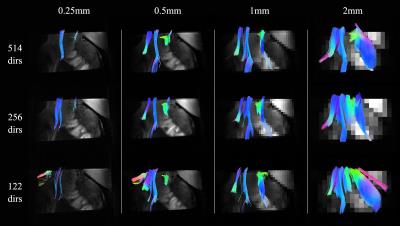
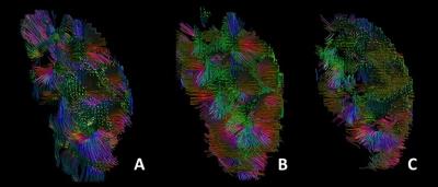
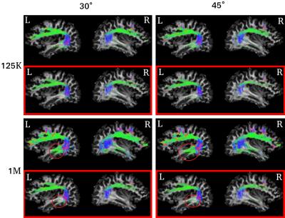
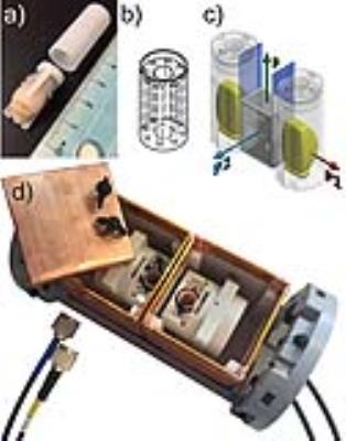
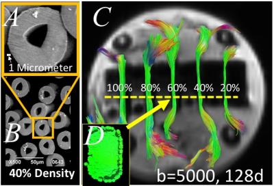
Providing Ground Truth Quantification of Anisotropic Diffusion MRI Imaging with a Hollow Textile Phantom
Sudhir Pathak, Catherine Fissell, David Okonkwo, Walter Schneider
A novel Textile Anisotropic Brain Imaging Phantom incorporating textile hollow fibers (taxons with inner/outer diameter 12/34 micron) is used to validate diffusion MRI imaging (dMRI). The taxon intra and extra spaces can be filled with water or deuterium. A taxon crossing pattern and a packing density pattern is manufactured to test orientation and the amount of taxons. NODDI based Intra-cellular volume fraction is correlated with the amount of taxons (r2=0.96) as compared to FA which is poor predictor with r2=0.11. Fiber crossings are estimated using Constrained Spherical Deconvolution techniques and can be resolved for angles greater then 45 degrees.
|
|
3455.
 |
5 |
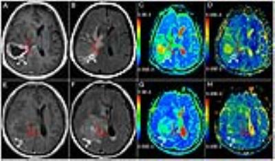
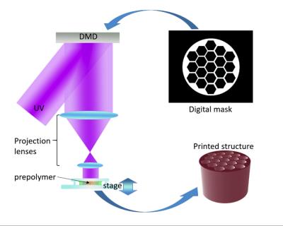
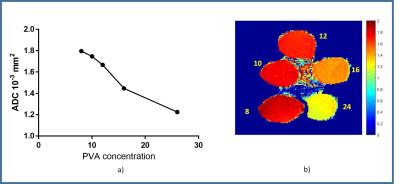
Cost effective 3D printed brain phantom for diffusion MRI
Arush Honnedevasthana Arun, Shivaprasad Chikop, Nithin Vajuvalli, Rashmi Rao, Sairam Geethanath
Diffusion weighted MRI is used to measure diffusion properties in the brain. In this paper, the method for the creation of an anatomically and mechanically realistic brain phantom from polyvinyl alcohol cryogel (PVA-C) and a 3D printable brain phantom using Poly Lactic Acid (PLA) PLA is proposed. PVA-C is material widely used in medical imaging phantoms because of its mechanical similarities to soft tissues. This brain phantom will allow testing and optimization of diffusion based MR methods.
|
|
3456.
 |
6 |
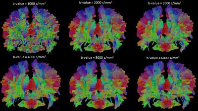
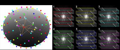
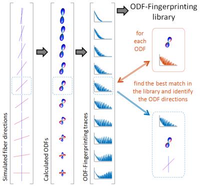
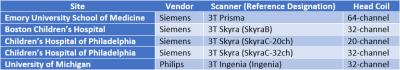
Robustness of Local Connectome Fingerprint Explored: Using a Multi-center and Multi-vendor Study
Vincent Lee, Ashok Panigrahy, Vincent Schmithorst, Thomas Chenevert, Borjan Gagoski, Deqiang Qiu, Peter LaViolette, Jeffrey Berman, Timothy Verstynen, Fang-Cheng Yeh
In this study we explore whether scanner-related-variabilities contribute to an individual’s distinct fingerprint – and whether the fingerprint specificity would be robust as a biomarker by scanning the same subject across multiple vendors and multiple scanner in four institutions. Both Diffusion Tensor Imaging and Multi-shell Multi-band diffusion imaging (MSMBDI) was tested, and differences within acquisition type (using fractional anisotropy and normalized quantitative anisotropy) and between acquisition types comparisons (using q-space diffeomorphic reconstruction) analysis were examined. We found that scanner may contribute partly to the fingerprint patter, but the fingerprint was robust at maintaining pattern, especially in MSMBDI to warrant further studies.
|
|
3458.
 |
8 |
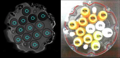
Quantitative evaluation of apparent diffusion coefficient in a large multi-unit institution using QIBA diffusion phantom
Joshua Yung, Yao Ding, Ken-Pin Hwang, Carlos Cardenas, Hua Ai, Michael Boss, Thomas Chenevert, Clifton Fuller, R Stafford
The purpose of this study was to determine the quantitative variability of apparent diffusion coefficient values across a large fleet of MR systems. Using a NIST traceable magnetic resonance imaging diffusion phantom, imaging was reproducible and the measurements were quantitatively compared to known values. Significant differences in identical phantoms were not observed, but uncertainty in the measurements was seen at low apparent diffusion coefficient values. The same trend was observed when the diffusion phantoms were imaged in 20 different MR systems. The characterization of ADC variability for these systems provides an improved quality control for quantitative diffusion weighted imaging.
|
|
3459.
 |
9 |
Reproducibility of DTI Metrics and the Influence of SNR on DTI metrics in a Longitudinal Multicenter Clinical Trial
Xiaopeng Zhou, Ken Sakaie, Josef Debbins, Robert Fox, Mark Lowe
SPRINT-MS is a large-scale longitudinal phase II trial including 27 3T MRI scanners to evaluate the treatment of progressive multiple sclerosis with Ibudilast. DTI measures showed high reproducibility in a longitudinal multicenter study, making it appropriate for use as a biomarker. The longitudinal design of the SPRINT-MS trial is expected to mitigate the systematic influence of SNR on DTI, but the observed trend may be useful as a correction factor in cross-sectional studies.
|
|
3460.
 |
10 |
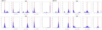
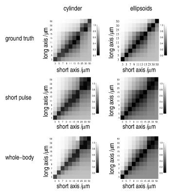
A Novel Yeast Cells- and Microcapillaries-Based Phantom for Validation of Diffusion MRI Models
Shir Levy, Darya Morozov, Inbar Seroussi, Leah Bar, Nir Sochen, Yoram Cohen
Numerous different models provide detailed microstructure information from diffusion MRI data. In order to challenge them, there is a need for complex phantoms with known structural characteristics. For this purpose, we present a novel phantom, consists of spherical fixed yeast cells and cylindrical microcapillaries. Despite of its complexity, arising from the different size, geometry and size distribution of the restricted compartments, there is a good correlation between the known ground truth and the features that were extracted from fitting single diffusion encoding (SDE) MRI experimental data, assuming continued or discrete weight of size distribution.
|
|
3461.
 |
11 |
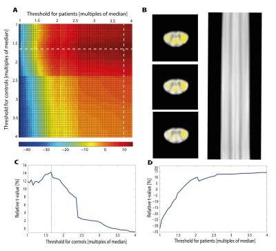
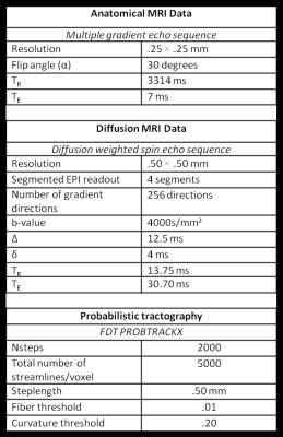
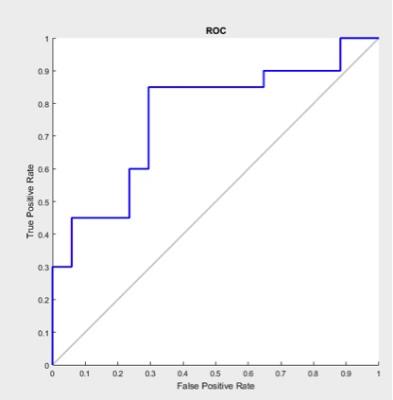
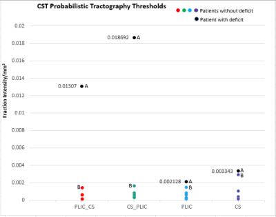
The use of novel validation methods to investigate optimal ROI location and probabilistic thresholds in tractography for presurgical planning in brain tumour patients.
Gideon Oluniran, Marcelo Lemos, Jeorg Ederle, Jozef Jarosz, Gareth Barker, Jonathan Ashmore
Although advanced tractography techniques exists and are well documented, many validation methods are not applicable to brain tumour cases and do not examine the possibility of proposing standard probabilistic thresholds. Using a novel approach, we investigate certain thresholds (‘safe’ thresholds) which can be applied to probabilistic tractography to reduce false representation of tracts, aid maximal tumour resection, and limit neurofunctional deficit from surgical treatments. We also examine optimal region of interest (ROI) location. ‘Safe’ thresholds can be determined, and with a wider confidence when tractography is defined with inclusion regions.
|
|
3462.
 |
12 |
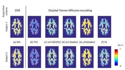
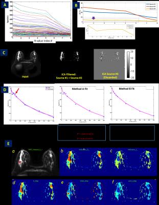
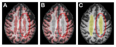
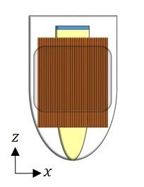
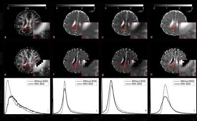
Theory, validation and application of blind source separation to diffusion MRI for tissue characterisation and partial volume correction
Miguel Molina-Romero, Pedro Gómez, Jonathan Sperl, Andrew Stewart, Derek Jones, Marion Menzel, Bjoern Menze
Here we present blind source separation (BSS) as a new tool to analyse multi-echo diffusion data. This technique is designed to separate mixed signals and is widely used in audio and image processing. Interestingly, when it is applied to diffusion MRI, we obtain the diffusion signal from each water compartment, what makes BSS optimal for partial volume effects correction. Besides, tissue characteristic parameters are also estimated. Here, we first state the theoretical framework; second, we optimise the acquisition protocol; third, we validate the method with a two compartments phantom; and finally, show an in-vivo application of partial volume correction.
|
|
3463.
 |
13 |
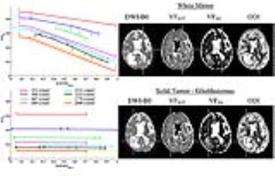
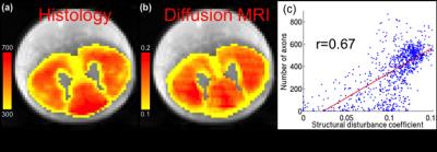
Histological validation of microstructural measures derived from the PICASO model
Lipeng Ning, Tanguy Duval, Julien Cohen-Adad, Yogesh Rathi
We propose to validate the PICASO (Precise Inference and Characterization of Structural Organization)1 biophysical model of tissue microstructure using a full-slice histology of cat spinal cord. The PICASO model was fit to high resolution diffusion MRI (dMRI) of a cat spinal cord to estimate microstructural measures of diffusion disturbance, which is directly related to axonal packing and density. We found that the structural disturbance coefficient (SDC) in the direction orthogonal to the fiber orientation from the PICASO model was strongly correlated with the axonal density obtained from histology2, with a correlation coefficient of r=0.67. Thus, the SDC could provide very precise information about the microscopic arrangement of axons or cells in biological tissue.
|
|
3464.
 |
14 |
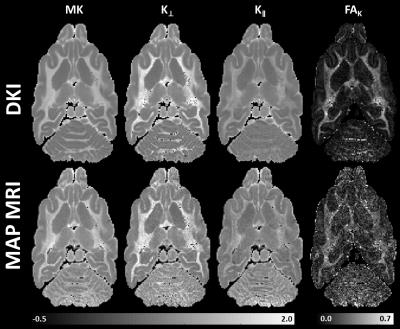
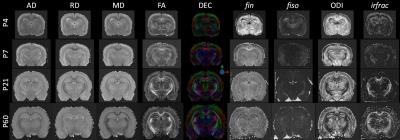
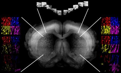
The histological validation analysis of diffusional kurtosis imaging with a cleared mouse brain.
Ryusuke Irie, Koji Kamagata, Aurelien Kerever, Suguru Yokosawa, Yosuke Otake, Hisaaki Ochi, Kazuhiko Tagawa, Hitoshi Okazawa, Ryo Ueda, Kohske Takahashi, Kanako Sato, Masaaki Hori, Eri Hirasawa, Shigeki Aoki
Diffusional kurtosis imaging (DKI) is a sensitive technique to analyze brain microstructure but that has little histological foundation. In this study, we evaluated a relationship between DKI parameters with neurite density measured by a confocal microscopy of the cleared mouse brain. There was a strongly positive correlation between neurite density and DKI parameters in the caudate nucleus and putamen, whereas the correlation between neurite density and fractional anisotropy was moderate. DKI reflect neurite density in an area with crossing fibers, so that can evaluate more complex microstructures than diffusion tensor imaging.
|
|
3465.
 |
15 |
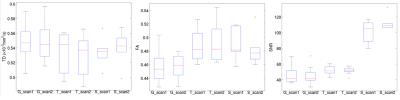
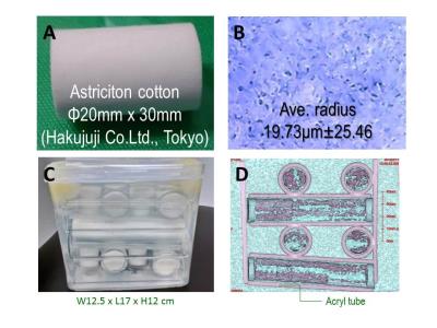
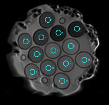
An affordable phantom for ADC/FA; a device for multi-site studies
Koji Sakai, Toshiaki Nakagawa, Ryusuke Nakai, Hiroyasu Ikeno, Seiji Yamaguchi, Hiroaki Takadama, Kei Yamada
Multi-site study gives large statistical power for the results. Some intrinsic differences among scanners are recognizing as an inevitable property. Isotropic diffusion has already been discussing. In contrast, anisotropic diffusion has been evading the endeavor because of the absence of an easily available anisotropic diffusion phantom. We compared DTI measures among five different MR scanners at three different sites using commercially and easily available astriction cotton. The averaged coefficient of variation for longitudinal repeated acquisitions of DTI on five different MR scanners were stable. The scan-rescan properties of five different MR scanners can be comparable by anisotropic diffusion phantom.
|
|
3457.
 |
7 |
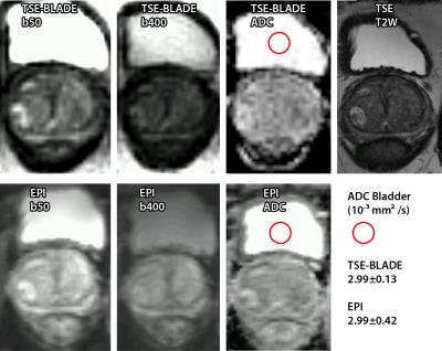
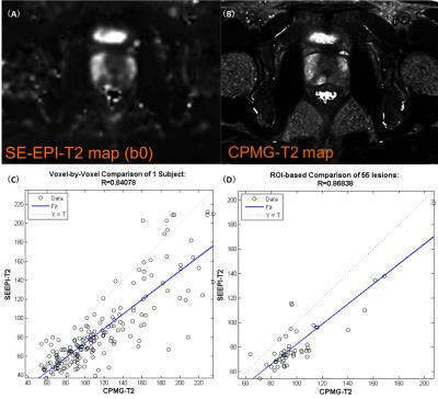
Quantitative evaluation of PROPELLER DWI using QIBA diffusion phantom
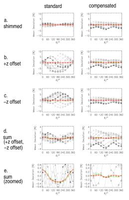
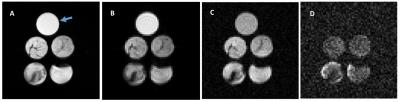
Joshua Yung, Hua Ai, Ho-Ling Liu, R Stafford
The purpose of this study was to characterize ADC values when varying imaging parameters in a diffusion-weighted (DW) FSE sequence with Periodically Rotated Overlapping ParallEL Lines with Enhanced Reconstruction (PROPELLER) k-space trajectory. The QIBA diffusion phantom was used to quantitatively evaluate the different pulse sequences. The DW PROPELLER sequence showed good agreement with the QIBA SE EPI sequence with the ETL=20 and b-value of 0 and 2000 s/mm2 sequence having with a r2=0.9034. The DW PROPELLER sequence is promising for quantitative evaluation of ADC values and this study may help improve clinical protocols using diffusion weighted imaging.
|
|
3466.
 |
16 |
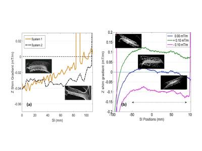
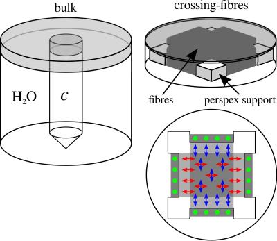
Reduction of susceptibility-induced field gradients in multi-fibre diffusion phantoms via susceptibility matching
Ezequiel Farrher, Johannes Lindemeyer, Farida Grinberg, Ana-Maria Oros-Peusquens, N. Jon Shah
Studies performing diffusion-weighted MRI on anisotropic fibre phantoms suffer from microscopic background field gradients induced by differences in the magnetic susceptibility of the employed materials. We present a particularly promising approach that makes use of a matched magnesium chloride solution to eliminate these effects. The method is thoroughly studied and successfully validated on a crossing-fibre phantom containing two perpendicularly crossing fibre populations. The obtained results are no longer subject to any orientation-dependence with respect to B0.
|
|
3467.
 |
17 |
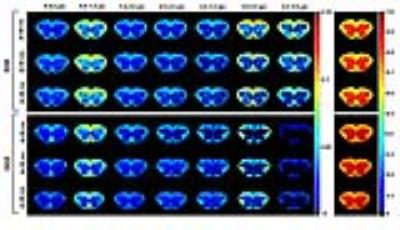
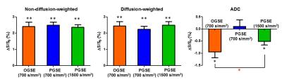
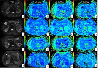
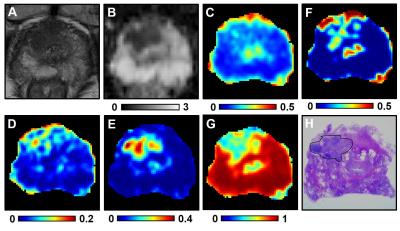
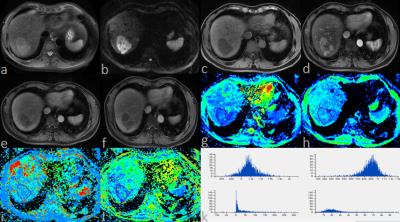
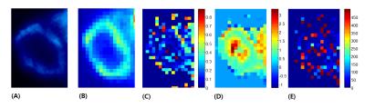
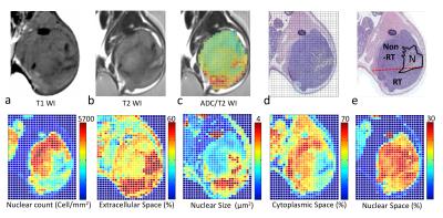
Diffusion radiomics analysis in intratumoral heterogeneity of murine prostate cancer following radiotherapy: pixel-wise correlation with histology
Yu-Chun Lin, Gigin Lin, Chun-Chieh Wang
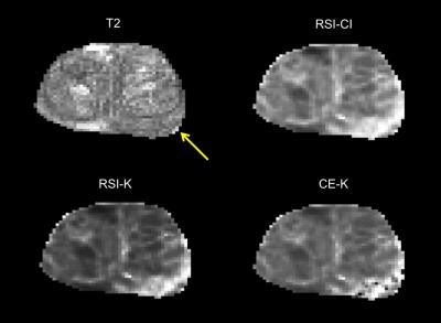
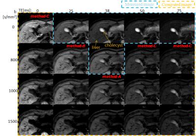
To investigate the biological meaning of apparent diffusion coefficient (ADC) in tumors following radiotherapy. Five mice bearing TRAMP-C1 tumor were half-irradiated. Diffusion-weighted images were acquired using multiple b-values of up 0 to 3000 s/mm2. The pixelwise ADC positively correlated with extracellular space and nuclear size, and negatively correlated with nuclear count, cytoplasmic space and nuclear space. Optimal ADC was achieved at b-value of 800 s/mm2 in determining the treatment response. Pixelwise ADC values correlate with histology metrics might be a means of in vivo radiomics biomarkers for evaluating tumor heterogeneity and responses to radiotherapy.
|
|
3468.
 |
18 |
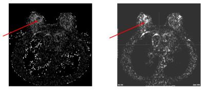
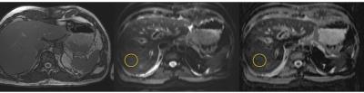
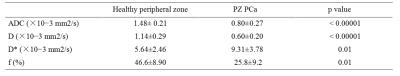
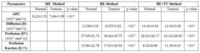
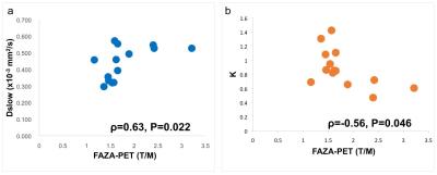
Hypoxia imaging of head and neck carcinoma: Correlation between DWI parameters and FAZA-PET activity
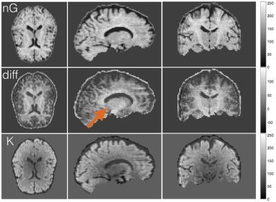
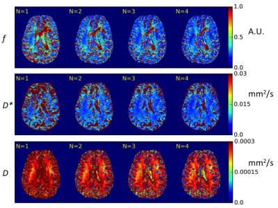
Akiko Imaizumi, Takayuki Obata, Yasuhiko Tachibana, Masayuki Inubushi, Mitsuru Koizumi, Kyosan Yoshikawa, Ming-Rong Zhang, Katsuyuki Tanimoto, Rintaro Harada, Takashi Uno, Tsuneo Saga
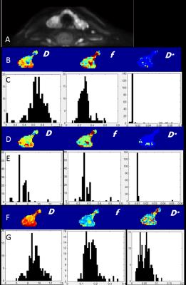
To investigate the usefulness of diffusion-weighted imaging (DWI) for visualizing hypoxia of head and neck carcinoma, the correlation between DWI parameter estimates and 18F-fluoroazomycin arabinoside (FAZA) positron emission tomography (PET) activity was evaluated. The diffusion coefficients and fractions of the fast and slow compartments according to the 2-compartment model (Dfast, Dslow and Ffast, Fslow) were estimated. The diffusional kurtosis (K) and the corrected diffusion coefficient (D) were also obtained according to the diffusional kurtosis imaging (DKI) method. Amongst the DWI estimates, Dslow and K were significantly correlated with FAZA-PET activity, which suggests they might be useful as indicators of hypoxia.
|
|
3469.
 |
19 |
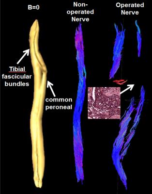
7.0 T Diffusion Tensor Imaging Evaluation of Rabbit Sciatic Nerve Microstructure with Histologic Correlation
Tina Jeon, Emil Vutescu, Eric Aronowitz, Henning Voss, Jonathan Dyke, Darryl Sneag
High-resolution DTI is a promising tool to evaluate peripheral nerve regeneration following surgical repair of nerve injury. The spatial resolution achieved with 7.0 T allowed us to more confidently interrogate the nerve for measuring fractional anisotropy (FA) and diffusivity and to perform fiber tracking as compared to 3.0 T. Among DTI metrics, FA correlated the greatest with axonal density and diameter. These findings support that DTI has the potential to measure axonal regeneration in the peripheral nerves at preclinical and clinical field strengths.
|
|
3470.
 |
20 |
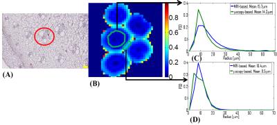
Diffusion-weighted MRI of node tissue: correlation of mean diffusivities and cellularity.
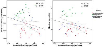
Mariaulpa Sahalan, Aritrick Chatterjee, Nyoman Kurniawan, Gary Cowin, Laurence Gluch, Carl Power, Geoffrey Watson, Kevin Tay, Julie Fletcher, David Taylor, Roger Bourne
Improvement of sensitivity and specificity in DWI-based assessment of nodal diseases is dependent on a better understanding of how nodal microstructures affect the water diffusivity in tissue. In this abstract we report the first diffusion microimaging investigation of formalin fixed node tissue with the aim of assessing any correlation between mean diffusivity and cellularity. Mean diffusivity was calculated in ROI corresponding to distinct node sub-structures. Nuclei were segmented semi-automatically to measure the cellularity metrics: nuclear count and nuclear area. The results showed there is no significant correlation between mean diffusivity with cellularity metrics in the nodal tissues.
|
|
3471.
 |
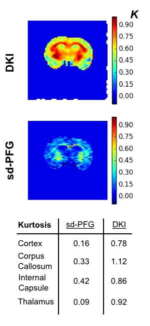
21 |
Reproducibility of SMT-Based Microscopic Diffusion Anisotropy Imaging on a Clinical MRI System
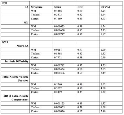
Cara Foley, Enrico Kaden, Kiran Seunarine, Matt Hall, David Carmichael, Jonathan Clayden, Chris Clark
Diffusion Tensor Imaging (DTI) is a widely used neuroimaging technique, but it lacks specificity. Advanced diffusion models aim to yield greater biophysical information than DTI. This information is redundant if the parameters cannot be accurately reproduced across time-points. Multi-shell diffusion images were acquired on a single 3T scanner at two time-points for ten healthy adults. Mean parameter values from SMT-based microscopic diffusion anisotropy imaging and DTI were obtained in white matter, cortex and thalami. The advanced model performed similarly well to DTI in white matter, but was less consistent in the cortex and thalami, potentially due to its increased complexity.
|
|
3472.
 |
22 |
Scan-rescan of AxCaliber, macromolecular tissue volume and g-ratio in the spinal cord
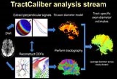
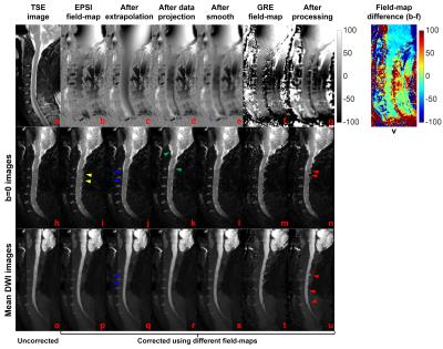
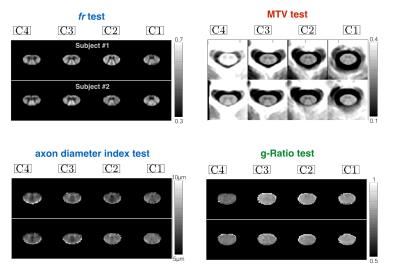
Tanguy Duval, Victoria Smith, Eric Klawiter, Nikola Stikov, Julien Cohen-Adad
Translating quantitative MRI to clinical research raises many challenges in term of acquisition strategy, modeling of the MRI signal, artifact corrections (sensitivity to motion and distortion) and metric extraction (template registration and partial volume effects). In this work, we wanted to validate the repeatability of this entire framework, from the acquisition to the extraction of the metrics using a template-based approach. We took advantage of the 300 mT/m gradients from the connectome scanner for estimating robustly AxCaliber metrics, MTV, and g-ratio in the spinal cord of eight healthy subjects, scanned and rescanned in two different sessions. Our results show good scan-rescan repeatability (r>0.7, small deviations <5%), and demonstrate the capability of these metrics to detect inter-subjects (through ICC) and inter-fiber-pathways differences (through ANOVA analysis).
|
|
3473.
 |
23 |
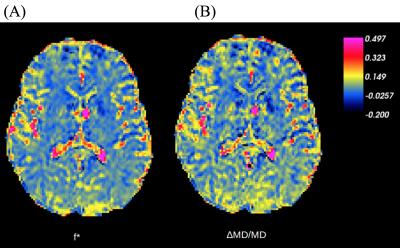
Reducing complexity of functional imaging: free-breathing imaging based ADC and IVIM measurements are as accurate as breath-hold measurements in renal cell carcinoma
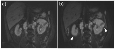
Neil Jerome, Matthew Orton, James d'Arcy, David Collins, Martin Leach, Dow-Mu Koh
Respiratory motion represents a serious confounding factor for abdominal imaging; for more complex diffusion models such as IVIM, acquisition of diffusion-weighted images in successive breath-holds offers control of motion for sharper images. In this patient study, DWI was performed in free-breathing and consecutive breath-holds, without registration, on successive days without intervention to determine repeatability. Derived tumour ROI parameters from ADC and IVIM models were not significantly affected between breathing regimes, but observed coefficients of variation for free-breathing were smaller for all pseudo-diffusion related parameters. Breath-holding is time inefficient, and free-breathing allows more data collection for development of robust DWI markers.
|
|