MS: Longitudinal Studies
Electronic Poster
Neuro
Tuesday, 25 April 2017
| Exhibition Hall |
13:45 - 14:45 |
| |
|
Computer # |
|
4046.
 |
1 |
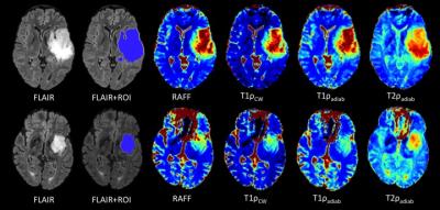
Longitudinal Study of MS lesions using Multi-contrast Ultra-high Field (7Tesla) MRI 
SANJEEV CHAWLA, Ilya Kister, Tim Sinnecker, Jens Wuerfel, Friedemann Paul, Yulin Ge
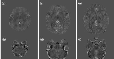
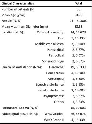
To track evolution of multiple sclerosis (MS) lesions, 9 patients underwent gradient-echo-T2* and quantitative susceptibility mapping on 7T MR system at baseline and at follow-up period (mean duration=2.4years). Majority of lesions were non-iron laden at baseline and most of them remained unchanged in size, morphology and susceptibility patterns. Some of these lesions accumulated iron deposition on follow-up. A minority of iron-laden lesions underwent redistribution of iron content. Small increase in lesion count was observed at follow-up. Interestingly, majority of these lesions were iron-enriched. This study may provide insights into pathophysiological features of MS lesions during the course of disease evolution.
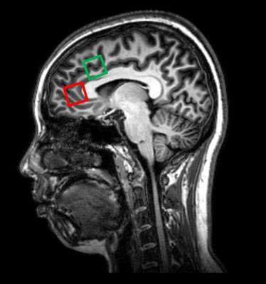
|
|
4047.
 |
2 |
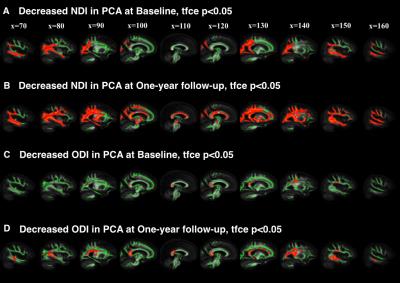
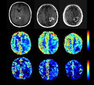
A longitudinal study of neurite orientation dispersion and density imaging in relapsing-remitting multiple sclerosis
Elda Fischi-Gomez, Guillaume Bonnier, Pavel Falkowskiy, David Romascano, Myriam Schluep, Renaud Du Pasquier, Alessandro Daducci, Jean-Philippe Thiran, Gunnar Kruger, Cristina Granziera
We explored the sensitivity of a novel diffusion MRI method i.e. “Neurite orientation dispersion and density imaging”, to detect and characterize brain microstructure alterations in relapsing-remitting multiple sclerosis patients that we followed up over 2 years. Cross-sectionally, NODDI revealed that an increase in orientation dispersion and a decrease in neurite density in NAWM and in lesions of RRMS patients compared to healthy subjects. Longitudinally, NODDI measured a decreased dispersion and an increased neurite density in MS lesions at 2 years follow-up. Also, NODDI metrics at baseline were highly related to cognition at both baseline and follow-up.
|
|
4048.
 |
3 |
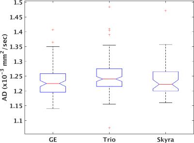
Systematic Differences in High Angular Resolution Diffusion Imaging at Baseline in a Multicenter Longitudinal Clinical Trial
Ken Sakaie, Xiapeng Zhou, Josef Debbins, Mark Lowe, Robert Fox
The lack of imaging biomarkers is a key obstacle to the development of treatment for progressive multiple sclerosis. While diffusion MRI is a promising biomarker, variability among scanners may limit its use. We examine site-related variability among multiple sclerosis patients to inform the design of multicenter trials.
|
|
4049.
 |
4 |
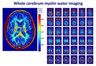
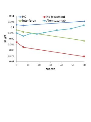
Myelin water imaging provides evidence of long-term remyelination and neuroprotection in Alemtuzumab treated multiple sclerosis patients
Irene Vavasour, Cornelia Laule, Shannon Kolind, Roger Tam, David Li, Alex MacKay, Anthony Traboulsee
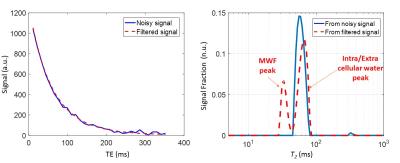
To test the potential neuroprotective and reparative properties of alemtuzumab (a highly effective disease modifying therapy for relapsing remitting MS), we used myelin water imaging to measure myelination in MS patients treated with either alemtuzumab, interferon, or no treatment. NAWM MWF showed a steady 4% increase in alemtuzumab-treated subjects whereas MWF in subjects treated with interferon or without treatment decreased by 10% over 5 years. Myelin recovery following treatment with alemtuzumab supports previous clinical trial findings, provides understanding of the biological mechanisms underlying observed clinical improvement and demonstrates that MWF is a powerful biomarker for neuroprotection and repair in MS.
|
|
4050.
 |
5 |
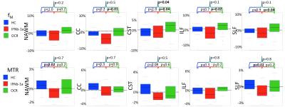
Myelin-Related MRI Metrics Demonstrate Longitudinal Differences for Relapsing MS Patients Treated with Ocrelizumab or Inteferon Beta-1a Over 96 Weeks
Shannon Kolind, Irene Vavasour, Roger Tam, Lisa Tang, Alexander Rauscher, Robert Carruthers, Rick White, Victoria Levesque, Hideki Garren, David Clayton, David Li, Anthony Traboulsee
Conventional MRI scans cannot evaluate disease-related changes in normal-appearing white matter, and have limited sensitivity for detecting changes in chronic lesions. In this work, we employed 2 quantitative MRI measures related to myelin content, myelin water fraction and magnetization transfer ratio, to evaluate the effects a potential novel therapy for multiple sclerosis (ocrelizumab) compared to a commonly-used therapy (interferon beta-1a). Over 2 years, these myelin-related measurements increased or remained stable in all regions for patients taking ocrelizumab, while they decreased for interferon beta-1a. These results support the use of quantitative MRI measures for more efficient, biologically specific clinical trial outcomes.
|
|
4051.
 |
6 |
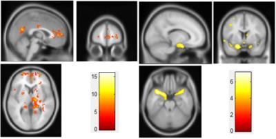
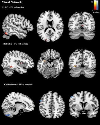
LONGITUDINAL ASSESSMENT OF LARGE-SCALE BRAIN FUNCTIONAL NETWORKS IN PATIENTS WITH MS: RELATIONSHIP WITH CLINICAL DISABILITY AND COGNITIVE IMPAIRMENT
Paola Valsasina, Maria Rocca, Fiammetta Pirro, Annalisa Colombi, Elisabetta Pagani, Ermelinda De Meo, Bruno Colombo, Paolo Preziosa, Vittorio Martinelli, Giancarlo Comi, Andrea Falini, Massimo Filippi
Aim of this study was to investigate the temporal evolution of resting state (RS) functional connectivity (FC) in patients with multiple sclerosis (MS) and its correlation with clinical and cognitive worsening. The predictive value of baseline functional network measures on the worsening of clinical disability/cognitive impairment was also explored. No significant RS FC changes were detected in healthy controls, while MS patients showed a complex pattern of longitudinal changes in the different networks, with a trend towards an increase (or stability) of RS FC in clinically stable MS patients, and a decrease of RS FC in clinically worsened MS patients.
|
|
4053.
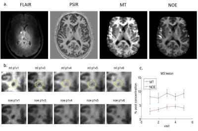 |
8 |
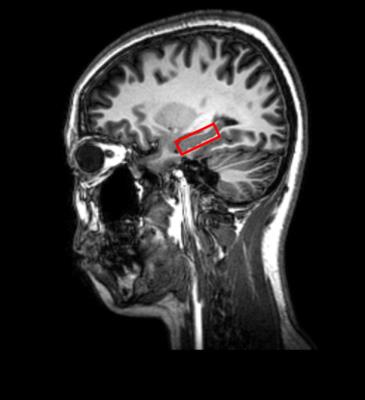
A longitudinal study of the effect of Multiple Sclerosis on surrounding white matter using z-spectrum imaging at 7T
Kingkarn Aphiwatthanasumet, Olivier Mougin, Nick Geades, Nikos Evangelou, Richard Bowtell, Penny Gowland
MS lesions are known to evolve in time, many showing signs of remyelination. To investigate this, we considered the changes in the regions around existing lesions of varying ages, to test the hypothesis that there is ongoing tissue damage in the regions around white matter lesions in MS. We found that the quantitative MT measured at 7T values in the region surrounding MS lesions decreased over the period of a longitudinal 6 month study. No systematic trend was found for the lesion core. This supports the hypothesis that an MS lesion causes ongoing damage in the region surrounding the lesion.
|
|
4054.
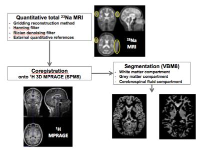 |
9 |
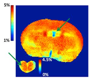
Brain sodium concentrations in healthy subjects are constant over time: a 3-year longitudinal 23Na MRI study at 3T
Adil Maarouf, Soraya Gherib, Elisabeth Soulier, Sylviane Confort-Gouny, Maxime Guye, Jean Pelletier, Jean-Philippe Ranjeva, Wafaa Zaaraoui
Longitudinal evaluation of brain sodium concentration in physiological conditions
|
|
4052.
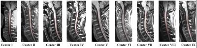 |
7 |
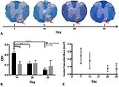
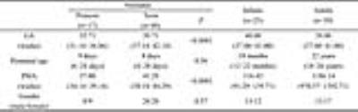
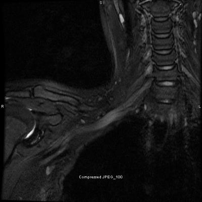
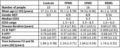
Longitudinal assessment of cervical cord atrophy across MS clinical phenotypes: a multicenter study
Paola Valsasina, Maria Rocca, Mohammad Aboulwafa, Paolo Preziosa, Frederik Barkhof, Hugo Vrenken, Claudio Gobbi, Chiara Zecca, Alex Rovira, Xavier Montalban, Hugh Kearney, Olga Ciccarelli, Lucy Matthews, Jacqueline Palace, Antonio Gallo, Alvino Bisecco, Achim Gass, Philipp Eisele, Carsten Lukas, Barbara Bellenberg, Giancarlo Comi, Massimo Filippi
Aims of this large, multicenter study were to characterize baseline cervical cord atrophy in patients with multiple sclerosis (MS) compared with healthy controls, and to evaluate the modification of cervical cord cross-sectional area (CSA) over one-year of follow-up in such patients. Results indicated that baseline cord atrophy was present in MS patients vs controls, with a differential effect across phenotypes and a greater severity of atrophy in the progressive forms of the disease. Significant CSA decrease over time was found in relapsing remitting, primary progressive MS and in clinically worsened patients.
|
|
4055.
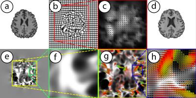 |
10 |
Manifold valued statistical models for longitudinal analysis of MRI data
Nagesh Adluru, Hyunwoo Kim, Richard Davidson, Andrew Alexander, Sterling Johnson, Vikas Singh
This work presents novel statistical image analysis methods to characterize complex morphological brain changes using MRI data. Specifically, our procedure utilizes the fundamental representations of "longitudinal change" -- voxel-wise Jacobian matrices obtained from image registration. Currently their univariate summaries (for example determinants) are ubiquitously used in neuroimaging studies. Operating directly with representations of Jacobians namely Cauchy deformation tensors, which are elements of an abstract mathematical manifold of symmetric positive definite matrices, yields promising improvements in statistical power in detecting subtle but statistically significant effects. The key technical contributions are computational algorithms for estimating multivariate general linear models with manifold-valued response variables.
|
|
4056.
 |
11 |
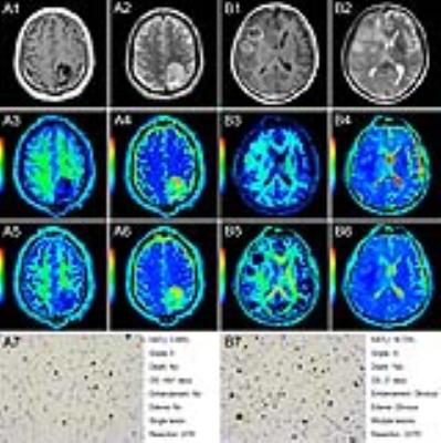
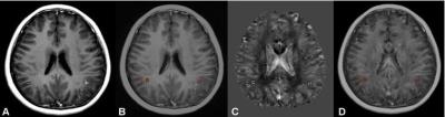
A preliminary study: the Values of Quantitative Susceptibility Mapping (QSM) in CIS and MS in Children - permission withheld
Hua CHENG, Hong ZHANG, Yang FAN, TongLi HAN, Yue LIU, Yun PENG
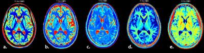
Quantitative Susceptibility Mapping (QSM) has been well used in evaluating the iron quantity changes in adult patients with multiple sclerosis (MS). However, it has not been tested in pediatric MS patients. In the present study, QSM was applied to assess difference of iron quantity in clinically isolated syndrome (CIS) and MS in children. It is shown that QSM provides a superior sensitivity method in the detection iron of changes of MS-related tissue in children, which suggests that QSM may serve as a potential sensitive biomarker in pediatric MS.
|
|
4057.
 |
12 |
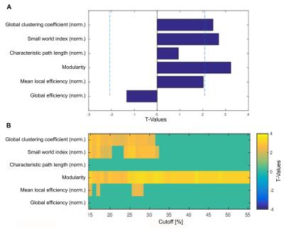
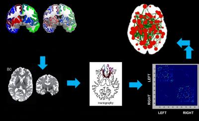
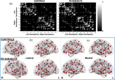
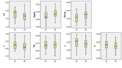
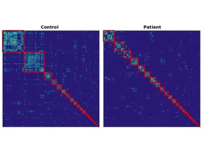
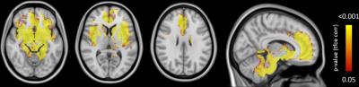
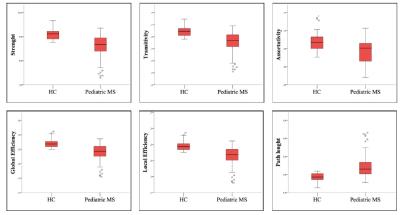
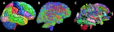
STRUCTURAL CONNECTIVITY ABNORMALITIES UNDERLYING COGNITIVE IMPAIRMENT IN PEDIATRIC MULTIPLE SCLEROSIS
Loredana Storelli, Maria Rocca, Ermelinda De Meo, Elisabetta Pagani, Lucia Moiola, Angelo Ghezzi, Pierangelo Veggiotti, Ruggero Capra, Maria Pia Amato, Agnese Fiorino, Lorena Pippolo, Maria Pera, Giancarlo Comi, Andrea Falini, Massimo Filippi
In this study, diffusion tensor (DT) magnetic resonance imaging (MRI) was applied to describe brain structural network architecture and connectivity abnormalities underlying cognitive dysfunction in 53 pediatric multiple sclerosis (MS) patients in comparison to 26 age- and gender-matched healthy controls (HC). Global and local network analyses were performed to assess between-group differences of connectivity metrics and cortical hubs. Cognitive impairment in pediatric MS patients seemed to be mainly associated to a reduced strength of connections of structural hubs and loss of efficiency in information transmission.
|
|
4058.
 |
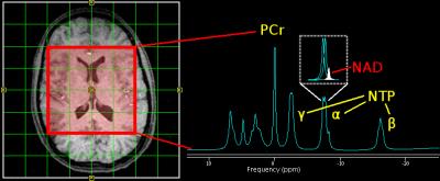
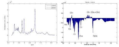
13 |
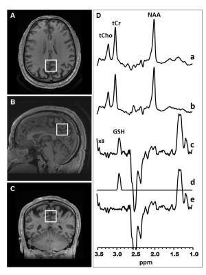
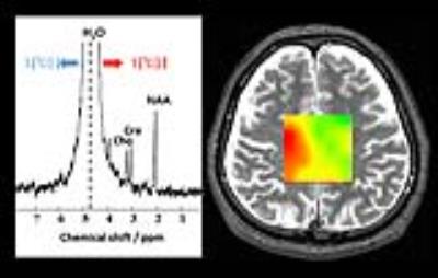
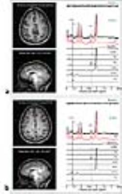
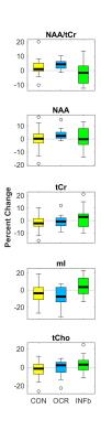
Longitudinal Changes in Magnetic Resonance Spectroscopy Over 96 Weeks in Relapsing MS Treated with Ocrelizumab versus Interferon Beta-1a
Erin MacMillan, Julia Schubert, Irene Vavasour, Roger Tam, Alexander Rauscher, Rick White, Hideki Garren, David Clayton, Victoria Levesque, David Li, Anthony Traboulsee, Shannon Kolind
Single voxel magnetic resonance spectroscopy (MRS) was performed in thirty-seven relapsing multiple sclerosis (MS) patients, who were enrolled in a phase III clinical trial of ocrelizumab versus interferon beta-1a, at baseline, 24, 48, and 96 weeks follow-up. 24 healthy controls were also scanned. MRS demonstrated a significant interaction between visit and treatment group in the NAA/tCr ratio. The change in absolute metabolite concentrations over 96 weeks revealed that this interaction was primarily driven by increased NAA and reduced inflammation in the ocrelizumab group, while the interferon beta-1a group exhibited a smaller increase in NAA and ongoing inflammation.
|
|
4059.
 |
14 |
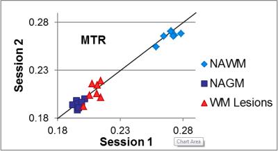
Toward a standardized quantitative imaging protocol for multiple sclerosis: a multisite study of magnetization transfer and quantitative T1 imaging techniques
Ian Tagge, Daniel Schwartz, Katherine Powers, Rohit Bakshi, Peter Calabresi, Todd Constable, John Grinstead, Roland Henry, Govind Nair, Jiwon Oh, Li Pan, Nico Papinutto, Daniel Pelletier, Daniel Reich, Nancy Sicotte, Jack Simon, William Stern, William Rooney
The current lack of standardization in MRI protocols leads to increased variability, particularly in semi-quantitative techniques such as MTR, and makes comparisons between studies almost impossible. A single subject with clinically stable RRMS travelled to seven North American sites and underwent two distinct 3T MRI sessions following a standardized MTR and qT1 protocol at each site. Both MTR and qT1 mapping have been shown to have potential in elucidating tissue characteristics and underlying pathology. This work demonstrated that use of carefully standardized protocols produces consistent quantitative and semi-quantitative measurements across sites in MS brain tissue in-vivo.
|
|
4060.
 |
15 |
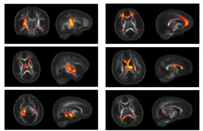
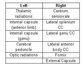
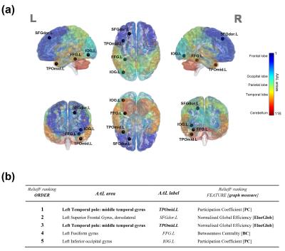
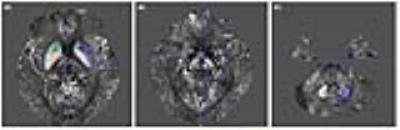
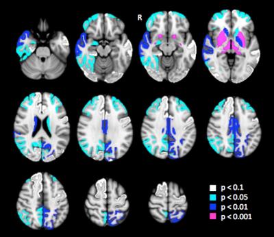
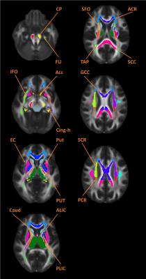
Graph theoretical measures predict volumetric changes in multiple sclerosis
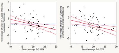
Thalis Charalambous, Carmen Tur, Ferran Prados, Steven Pavert, Declan Chard, David Miller, Sebastien Ourselin, Jonathan Clayden, Claudia Gandini Wheeler-Kingshott, Alan Thompson , Ahmed Toosy
Numerous studies demonstrated structural network changes in patients with multiple sclerosis (MS). However, the predictive nature of the graph-derived metrics is not yet examined. In this longitudinal study, we constructed baseline diffusion-based structural networks and we used multiple linear regression analysis to assess the ability of the network measures to predict follow-up increased lesion load and brain atrophy in MS (n=49). Our results suggest that edge density, global and local efficiency can predict follow-up brain atrophy after adjusting for the nuisance variables, signifying that network analysis can provide new insights into disease trajectories and offer potential biomarkers for MS progression.
|
|
4061.
 |
16 |
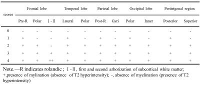
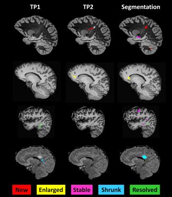
THE EVOLUTION OF CORTICAL AND SUB-CORTICAL LESION SIZE AND NUMBER CORRELATES WITH CHANGES IN COGNITION IN EARLY-STAGE RELAPSING-REMITTING MULTIPLE SCLEROSIS PATIENTS
Alexandra Sorega, Mário João Fartaria , Guillaume Bonnier, Tobias Kober, Renaud Du Pasquier, Myriam Schluep, Gunnar Krueger, Meritxell Bach Cuadra, Cristina Granziera
Lesion load and activity in multiple sclerosis (MS) patients, as identified by conventional magnetic resonance imaging (MRI), correlate only moderately with patients clinical status and evolution. Cortical lesion number and volume measured with advanced MRI may provide better correlates to cognitive dysfunction and disability. In this work, we studied the clinical impact of advanced MRI metrics of cortical and subcortical lesion evolution in a cohort of early relapsing-remitting MS patients. The number and volume of lesions that “shrunk”, disappeared or remained stable over time were strong determinants of changes in cognition in our patients cohort.
|
|
4062.
 |
17 |
Longitudinal outer and inner cortical MTR abnormalities in different MS clinical phenotypes
Rebecca Samson, Manuel Cardoso, Nils Muhlert, Varun Sethi, Özgür Yaldizli, Maria Ron, Ferran Prados , Sebastian Ourselin, David Miller, Claudia Gandini Wheeler-Kingshott, Declan Chard
Outer cortical magnetisation transfer ratio (cMTR) is potentially a sensitive measure of pathology linked to clinical disease progression in multiple sclerosis (MS). Here we aimed to investigate longitudinal outer and inner cMTR changes in healthy controls (HC) and people with MS of different clinical subtypes. Follow-up (FU) outer cMTR showed larger reductions in secondary progressive (SP)MS than other subtypes, and inner cMTR was also reduced more than in HC and relapsing-remitting (RR)MS, although cMTR was reduced in all MS patients at FU. This supports histopathological findings and suggests that cMTR measurement may be relevant to clinical disease progression in MS.
|
|
4063.
 |
18 |
Monitoring Disease Progression in Experimental Autoimmune Encephalitis using VDMP-CEST MRI
Aline Thomas, Jiadi Xu, Peter Van Zijl, Jeff Bulte
VDMP-CEST MRI can spatiotemporally monitor disease progression in a mouse model of multiple sclerosis. Lower VDMP-CEST signals corresponded to a decreased lipid and metabolite content in the peri-ventricular region of the brain, characteristic of the disease, with magnetic resonance spectroscopy used as validation.
|
|
4064.
 |
19 |
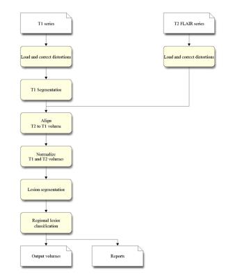
ACCURACY AND REPRODUCIBILITY OF AN AUTOMATED LESION SEGMENTATION TOOL-LESIONQUANT
Weidong Luo, Kelly Leyden, Aziz Ulug, Sebastian Magda, Julia Albright, Robert Haxton, Chris Airriess
Quantitative measures such as lesion volume and distribution have significant value for clinicians evaluating disease progression. Clinical standards for lesion evaluation include visual inspection of MRI images, or expert manual segmentation of lesions. These subjective measurements are often vulnerable to inter- and intra-rater variability, resulting in low reproducibility. CorTechs Labs’ LesionQuant is a fully-automated lesion segmentation tool for clinical use designed to provide accurate and reproducible lesion segmentations. This study objectively evaluates the segmentation results of LesionQuant compared to expert manual segmentation.
|
|
4065.
 |
20 |
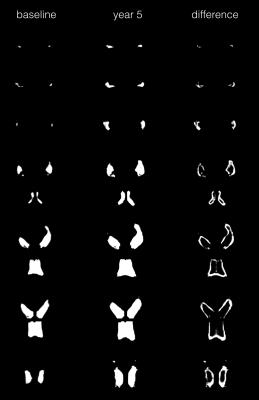
Automated Lateral Ventricle Segmentation in Multiple Sclerosis – Assessing Reliability and Clinical Impact in a 5-Years Follow-Up Cohort Study
Esther Ruberte, Tim Sinnecker, Michael Amann, Yvonne Naegelin, Iris-Katharina Penner, Matteo Pardini, Jens Kuhle, Tobias Derfuss, Christoph Stippich, Ludwig Kappos, Jens Wuerfel, Özgür Yaldizli
As pars pro toto, lateral ventricle enlargement might give an indirect estimate of brain atrophy. In contrast to whole brain atrophy, ventricle enlargement is, however, clinically easy to assess and its quantification is robust to MR images of less than perfect quality. Here we investigate i) the applicability of an automatic lateral ventricle delineation algorithm (ALVIN) in multiple sclerosis (MS), and ii) the association between of lateral ventricle enlargement and clinical disability in MS longitudinally. We found that ALVIN reliably estimates the lateral ventricle volume in MS that is associated with whole brain atrophy and neurological as well as cognitive disability.
|
|
4066.
 |
21 |
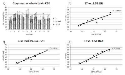
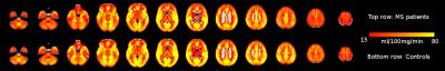
Using multi-inversion time ASL to explore gray matter perfusion in patients with multiple sclerosis: repeatability and relationship to disease characteristics
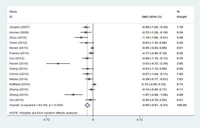
Ilona Lipp, Catherine Foster, Rachael Stickland, Alison Davidson, Richard Wise, Valentina Tomassini
The first aim of this study was to investigate retest-reliability of gray matter perfusion estimates in patients with multiple sclerosis. Using multi-inversion time pulsed ASL, we demonstrate good repeatability of global and local perfusion estimates over a four-week interval. The perfusion estimates were consistently lower in the patients than in a group of healthy controls. The second aim was to relate GM perfusion to disease characteristics. We could not find an effect of disease duration or stage (relapsing-remitting vs progressive) on perfusion, but we did observe a weak correlation with cognition, supporting previous studies.
|
|
4067.
 |
22 |
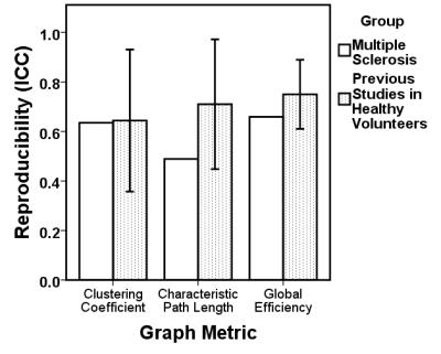
Reproducibility of Brain Network Metrics in People with Multiple Sclerosis
Thomas Welton, Dorothee Auer, Cris Constantinescu, Rob Dineen
Knowledge of reproducibility of graph-theoretic brain network metrics in disease populations is critically important for applications in clinical studies but non-existent in published literature. We compared reproducibility of graph metrics over time in a cohort of MS patients to reported values in published studies of reproducibility in healthy volunteers. We found that reproducibility was good in MS patients but slightly lower than in healthy people. Reproducibility of graph-theoretic brain network metrics in MS patients is not greatly dissimilar to that in healthy populations, which supports their use in clinical studies.
|
|
4068.
 |
23 |
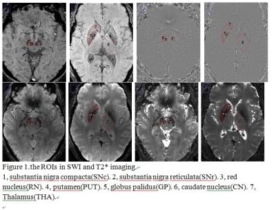
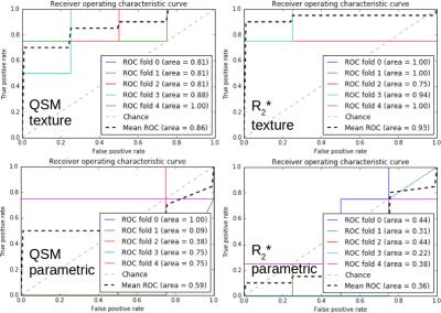
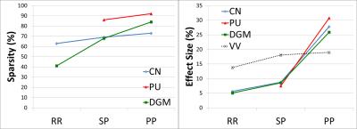
Progressive Iron Accumulation in Multiple Sclerosis Phenotypes Revealed by Sparse Classification of Deep Gray Matter
Ahmed Elkady, Dana Cobzas, Hongfu Sun, Gregg Blevins, Alan Wilman
Purpose: To create an anatomically-interpretable framework for localized analysis of brain iron accumulation/demyelination, and apply this framework to Multiple Sclerosis (MS) Deep Gray Matter (DGM). Materials and Methods: Quantitative Susceptibility and R2* maps were computed for 110 MS and 75 control subjects.
Results: Significant iron accumulation and insignificant demyelination were detected in MS DGM. Common MS pathological volumes and their pathological effect size progressively increased with advanced phenotypes. The developed framework offered improved statistical power and iron specificity compared to whole structure and singular analysis.
Conclusion: Using a novel localized analysis pipeline, we demonstrated the progressive iron accumulation in MS DGM.
|
|
4069.
 |
24 |
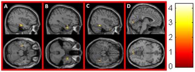
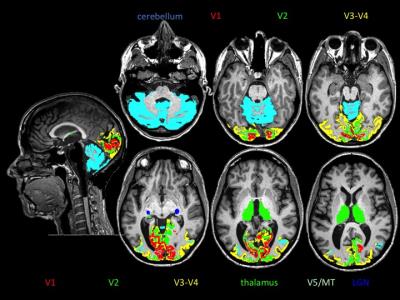
Predicting visual function clinical outcome in MS: a MRI and OCT metrics study.
Eduardo Caverzasi, Christian Cordano, Alyssa Zhu, Antje Bischof, Gina Kirkish, Nico Papinutto, Michael Devereux, Nicholas Baker, Sam Arnow, Justin Inman, Hao Yiu, Carolyn Bevan, Jeffrey Gelfand, Bruce Cree, Stephen Hauser, Roland Henry, Ari Green
Fifty Multiple Sclerosis subjects were evaluated by optical coherence tomography and MRI, including multi-shell and putative myelin content imaging focused on primary visual area, thalamus and cerebellum. Predictive models of visual function performance, measured by visual evoked potentials and low contrast visual acuity were tested using a partial least square regression analysis. Combination of MRI and OCT metrics appears to strongly describe the visual function. Myelin content imaging, in particular, has a strong predictive value once there is history of optic neuritis. These preliminary results may improve the understanding of the pathological mechanisms underlying clinical dysfunction in multiple sclerosis.
|
|