ISMRM 25th Annual Meeting & Exhibition • 22-27 April 2017 • Honolulu, HI, USA

Electronic Poster Session: General Cancer Imaging
4351 -4374 Cancer Treatment Response
4375 -4398 Perfusion, Permeability & Diffusion in Cancer
4465 -4488 Preclinical Imaging in Cancer
4489 -4512 General Cancer Including Preclinical
 
Cancer Treatment Response
Electronic Poster
General Cancer Imaging

 
Tuesday, 25 April 2017
Exhibition Hall  16:15 - 17:15


    Computer #

 
4351.   
 
1 A multi-parametric MRI-based radiomics approach to predict the high level of microsatellite instability in colorectal cancer
Zaiyi Liu, Yanqi Huang, Xin Chen, Zhongping Zhang, Lan He, Xiaomei Huang
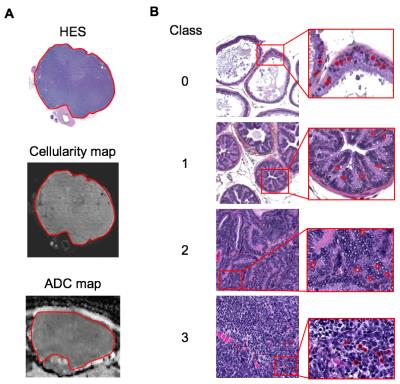
Microsatellite instability (MSI) is the condition of genetic hypermutability that results from impaired DNA mismatch repair (MMR). High levels of microsatellite instability (MSI-H) is regarded as a prognostic marker and predictor of the response to chemotherapy in colorectal cancer (CRC). This study presented a multi-parametric radionics classifier for preoperative and individualized prediction of MSC-H status in CRC patients. The potential application of this radiomics approach may aid the prognostic evaluation and decision-making in CRC patients.


4352.   
2 
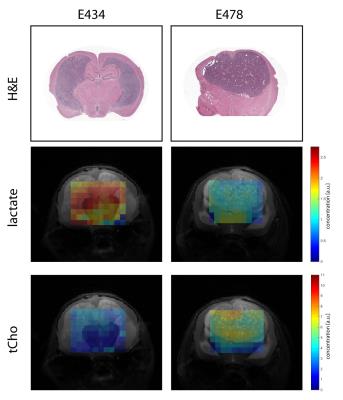
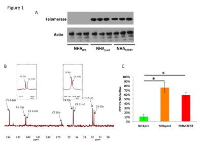
Magnetic Resonance Spectroscopic Early Predictors of 2-Year Progression Free Survival in IDH Mutated WHO II and III Gliomas
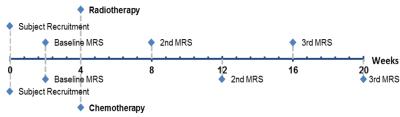
Min Zhou, Raymond Huang, Huijun Liao, Benjamin Rowland, Yue Zhou, Nils Arvold, Alexander Lin
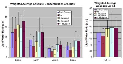
Most MR spectroscopy studies of patients with IDH-mutated gliomas have focused on the sensitivity and specificity of the 2-hydroxyglutarate measures but none that have examined the predictive value of 2HG in comparison to other brain metabolites for treatment outcome.  This prospective longitudinal study measured MRS at baseline and two time points after radio/chemotherapy of IDH-mutated gliomas.    Results showed that Cho/NAA shows the greatest predictive value followed by Cho/Cr, NAA/Cr, and Lac/Cr ratios but that all three time points 2HG levels were not predictive of outcome demonstrating that it is likely reflective of pathways distinct from membrane proliferation.


4353.   
3 
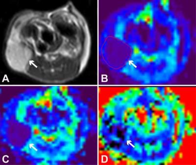
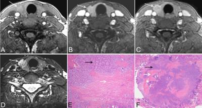
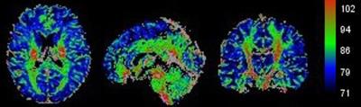
Radiation-induced changes in normal-appearing brain in patients with nasopharyngeal carcinoma: a MR T1? imaging study
Xiang Xiao, Yikai Xu, Yuankui Wu, Yingjie Mei, Queenie Chan
Radiation encephalopathy is the primary complication in patients with nasopharyngeal carcinoma (NPC) following radiotherapy (RT). In order to detect early radiation-induced alterations in the brain of NPC patients after RT, we recruited NPC patients before RT and after RT with normal-appearing brain for MR T1ρ examinations. We found abnormal microstructural changes of gray matter and white matter in NPC patients after RT can be detected by MR T1ρ even when routine MRI findings are negative. MR T1ρ may be used to predict early radiation-induced alterations of the brain following RT for NPC patients.


4354.   
4 
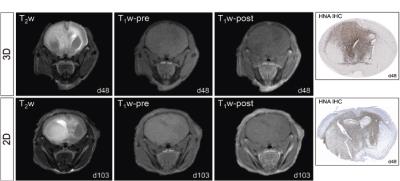
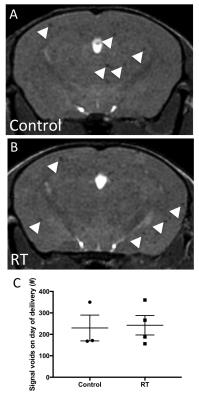
Using MRI to investigate the impact of radiation-induced damage on promoting tumor growth in a mouse model of brain metastasis
Amanda Hamilton, Eugene Wong, Paula Foster
Whole brain radiotherapy (RT) is the standard of care for breast cancer patients with multiple brain metastases but there are multiple negative consequences associated with the irradiation of normal brain tissue. In our study we investigated the influence that RT-induced damage of healthy brain has on the arrest and growth of metastatic breast cancer cells in a mouse model of breast cancer brain metastasis. We observed that irradiated but otherwise healthy neural tissue had an increased propensity to support metastatic growth compared to control. Elucidating the impact of RT on normal neural tissue could have implications in clinical patient management.   


4355.   
5 
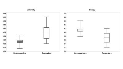
Quantitative assessment of water diffusivity in bladder tumors: can response be predicted prior to neoadjuvant chemotherapy?
Huyen Nguyen, Amir Mortazavi, Kamal Pohar, Lai Wei, Zarine Shah, Debra Zynger, Guang Jia, Michael Knopp
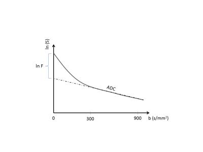
This study is to correlate the degree of tumor heterogeneity in Apparent Diffusion Coefficient (ADC) at baseline with chemotherapeutic response in bladder cancer patients. MRIs of twenty muscle-invasive bladder cancer patients were performed with Diffusion weighted MRI (DWI). Freehand ROIs were placed on the whole tumor volume on ADC maps to obtain a dataset of voxel-wise ADC values for each patient. Histogram analysis was performed on each patient’s ADC dataset to calculate uniformity (U) and entropy (E) at baseline. These quantities were then correlated with the patient’s chemotherapeutic response. Our data showed that there was a strong correlation of tumor heterogeneity, which is characterized by U and E, and the patient’s chemotherapeutic response. While U was significantly higher, E was significantly lower in responders (both P<0.01) compared to non-responders. In conclusion, quantification of tumor ADC heterogeneity can provide useful information that enables the ability to predict chemotherapeutic response prior to the treatment, to improve the patient outcomes. 


4356.   
6 
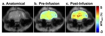
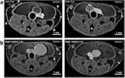
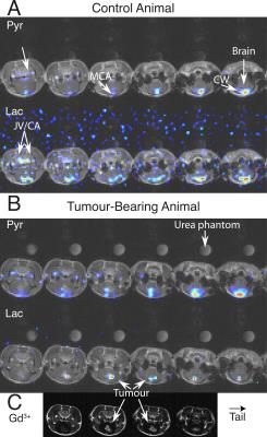
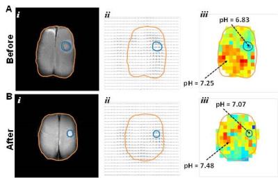
Simultaneous imaging of tumor size with contrast-enhanced MRI and response to therapy with extracellular pH readout from BIRDS
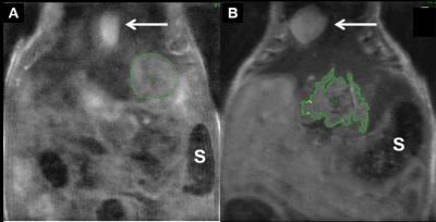
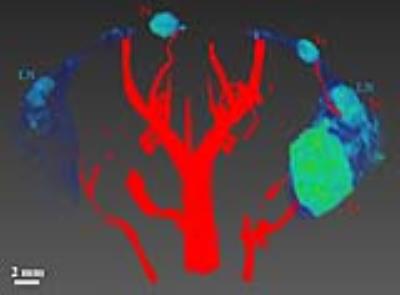
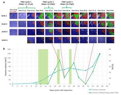
Jyotsna Upendra Rao, John Walsh, Maxime Parent, Yuegao Huang, Meser Ali, Daniel Coman, Fahmeed Hyder
Acidic extracellular pH (pHe) of gliomas promotes tumor growth and builds resistance to therapy. Thus, to monitor therapeutic response, we used Biosensor Imaging of Redundant Deviation in Shifts (BIRDS), to generate pHe maps of rat brains bearing U251 tumors. Upon TmDOTP5- infusion, MRI identified tumor boundary and BIRDS imaged the intratumoral and peritumoral pHe gradient (DpHe). Two weeks post implantation of U251 glioma cells, animals were either treated with temozolomide (40 mg/kg) or were left untreated. The results of both terminal and longitudinal studies suggest that temozolomide therapy hinders tumor growth and normalizes intratumoral pHe.


4357.   
7 
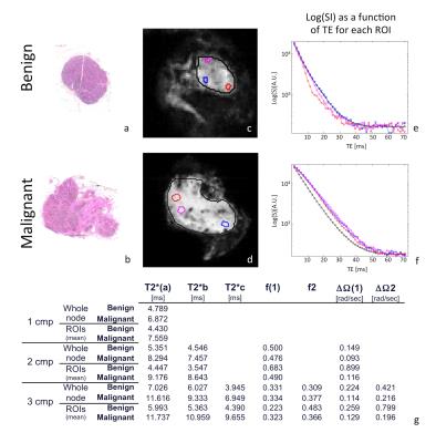
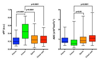
Anatomical sites’ dependency of 3.0 T whole-body MRI’s signal fat fraction and apparent diffusion coefficient in multiple myeloma focal lesions
Arash Latifoltojar, Margaret Hall-Craggs, Alan Bainbridge, Neil Rabin, Rakesh Popat, Ali Rismani, Kwee Yong, Shonit Punwani
The increasing utility of MRI's quantitative imaging biomarkers for disease characterisation and response monitoring necessitates a better understanding of underlying pathophysiological changes.

This might be more pertinent when heterogeneous organ such as skeletal system is being investigated.  In this work, we carry out a study into the heterogeneity of multiple myeloma's focal lesions on the basis of the various anatomical locations in skeleton.


4358.   
8 
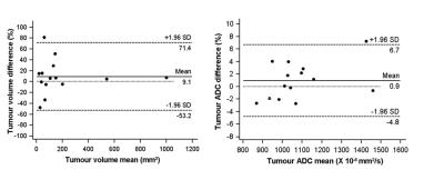
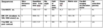
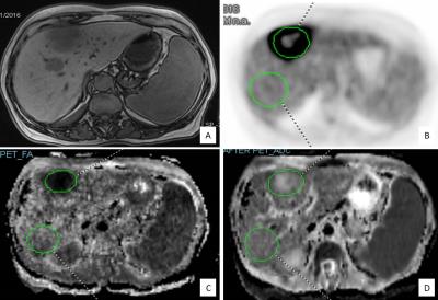
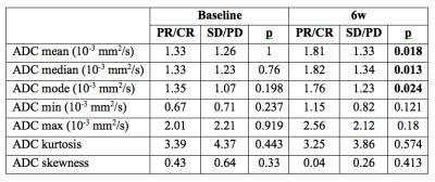
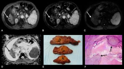
HCC treated with 90Yttrium radioembolization: can pre-treatment and 6week post-treatment volumetric ADC histogram measurements predict subsequent tumor response?
Sonja Gordic, Mathilde Wagner, Riccardo Zanato, Stefanie Hectors, Cecilia Besa, Edward Kim, Bachir Taouli
We evaluated the potential of volumetric ADC histogram measurements (vADC) obtained pre- and 6 weeks (6w) post-treatment for prediction of HCC response to 90Yttrium radioembolization (RE). 22 patients underwent MRI at baseline and 6w after RE using a routine liver MRI protocol including DWI. Tumor response was assessed by mRECIST at 6 months post treatment. vADC mean, median and mode obtained at 6w post-treatment were significantly different between patients with partial/complete response vs. those with stable/progressive disease, and were a significant predictor of complete tumor response at 6 months, with vADC mode performing best. Pre-treatment vADC did not have any predictive value for response. 


4359.   
9 Evaluation of HCC Response to Loco-regional Therapy: Validation of Response Criteria with MRI using Explant as a Reference
Sonja Gordic, Idoia Corcuera-Solano, Ashley Stueck, Pamela Argiriadi, Preethi Guniganti, Michael King, Edward Kim, Swan Thung, Bachir Taouli
We assessed the performance of various imaging response criteria for the prediction of complete pathologic necrosis (CPN) of hepatocellular carcinoma post locoregional therapy on liver explant. Patients who underwent liver transplantation after locoregional therapy were included in this retrospective study. All patients underwent MRI using routine liver protocol within 90 days of liver transplant. RECIST, mRECIST, EASL, percentage of necrosis on subtraction images, and DWI (signal intensity and ADC) were assessed. CPN was retrospectively assessed in all tumors at histopathology. mRECIST, EASL, percentage of necrosis and signal intensity on DWI were all significant predictors of CPN while RECIST and ADC criteria were not. 


4361.   
11 
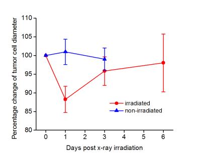
in vivo detection of tumor response to radiotherapy using Imaging Micro-structural Parameters Using Limited Spectrally Edited Diffsuion
xiaoyu jiang, hua li, zou yue, junzhong xu, john Gore
The changes that occur over a cell cycle play a vital role in mediating a cell’s sensitivity towards radiation therapy. Radiation exposure is expected to arrest cells at a particular cell cycle phase which improves the effectiveness of subsequent doses of radiation/chemotherapy. Cells in different phases have different sizes that can be detected by diffusion MRI with appropriate diffusion times. In this study, we evaluate the hypothesis in a rat glioma model that measurements of mean tumor cell size provides a means to quantify changes of cell phase distributions, and hence is capable of monitoring tumor response to radiotherapy.


4362.   
12 
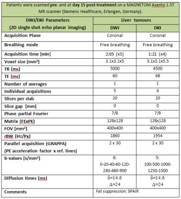
ADC and Kurtosis parameters show early response to anti-angiogenic therapy in patients with liver metastases - permission withheld
Mihaela Rata, Khurum Khan, David Collins, Matthew Orton, James d'Arcy, Nina Tunariu, Maria Bali, Ian Chau, Nicola Valeri, David Cunningham, Martin Leach, Dow-Mu Koh
Diffusion Weighted Imaging (DWI) is a valuable method of characterising tumour cellularity and assessing tumour response to therapy. Diffusion Kurtosis Imaging (DKI) is now being investigated outside of the brain, as the diffusional kurtosis metrics are strongly linked to cellular microstructure and heterogeneity in tissues. This work assesses tumour response to anti-angiogenic therapies as derived from DWI and DKI data in a cohort of patients with liver metastases enrolled on an early phase clinical trial. Our results demonstrate a significant cohort response to treatment of the ADC parameter, but also observed significant non-Gaussian diffusion behaviour.


4363.   
13 
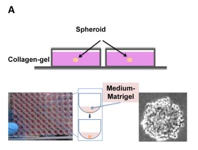
Impacting cancer cells via mechanical waves: can we change cellular behaviour?
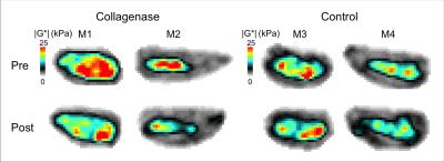
Marlies Hoelzl, Frederic Festy, Gilbert Fruhwirth, Ralph Sinkus
90% of cancer related deaths are caused by cancer metastasis, a process where cells leave the primary tumour, disseminate and form outgrowth at the secondary metastatic site. The tumour microenvironment provides crucial signals ((bio)chemical, mechanical) to regulate tumour formation, progression, and cell spread throughout the body. Translation of mechanical forces, displacements and deformations into biochemical signals (i.e. mechanotransduction) affects their cell behaviour (adhesion, spread, survival).1,2 We show here that multiple treatment of tumour spheroids (solid tumour model in vitro) with focussed shear waves operating at specific frequency and amplitude results in reduced growth and reduced invasive behaviour of cancer cells.


4365.   
15 
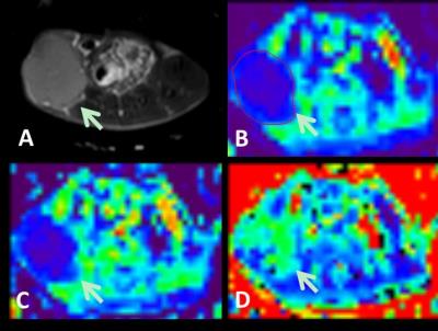
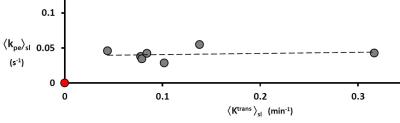
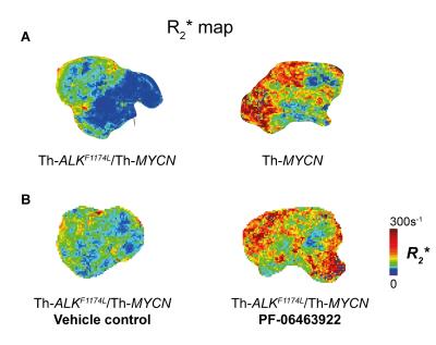
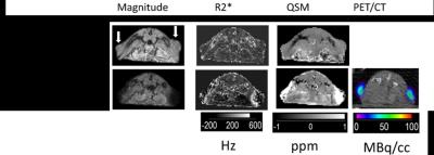
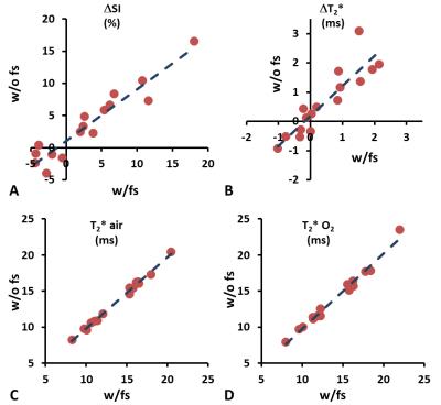
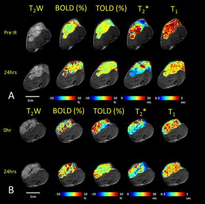
Multiparametric MRI assessment of tumor physiological changes during hypofractionated SABR
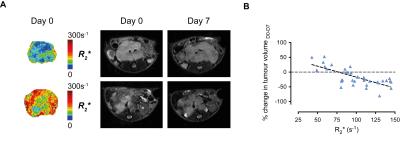
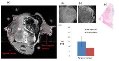
Heling Zhou, Zhang Zhang, Rebecca Denney, Jessica Williams, Jeni Gerberich, Strahinja Stojadinovic, Debabrata Saha, John Shelton, Ralph Mason
Hypofractionated stereotactic body radiation therapy, a new radiation treatment scheme, may be particularly susceptible to tumor hypoxia. We applied oxygen enhanced MRI together with DCE MRI to observe tumor physiological changes induced by a radiation fraction in a human lung cancer xenograft rat model. This study showed reduced vascular oxygenation 24 hours after the first fraction of 12 Gy, as indicated by the significant decrease in T2* compared to baseline, but no significant response in dynamic contrast enhanced MRI. DCE parametric maps revealed a multinodular structure in the tumor as confirmed by histology.


4366.   
16 
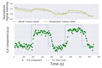
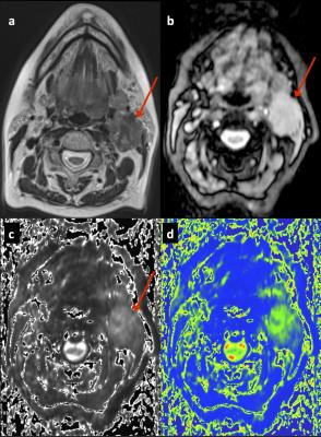
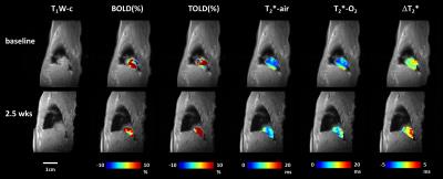
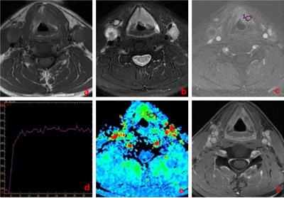
Absence of oxygen enhanced changes in T2* within head and neck cancer metastatic cervical lymph nodes is associated with local disease recurrence within 2-years following chemoradiotherapy
Harbir Sidhu, Chiara Tudisca, David Price, Sola Adeleke, Marianthi-Vasiliki Papoutsaki, Martin Forster, Ruheena Mendes, Stuart Taylor, Shonit Punwani
Hypoxia within head and neck squamous cell cancer metastatic lymph nodes is associated with poorer outcomes following chemoradiotherapy when measured directly using polarographic probes.

The utility of non-invasive pretreatment T2* measurement in prediction of chemoradiotherapeutic response was investigated. Our data suggest, however,  that nodes demonstrating sustained post-therapy complete local response based on two-year follow-up are significantly more hypoxic compared with relapsing-nodes and paradoxically demonstrate a significant increase in hypoxia on breathing 100%-oxygen.

Following further work to ascertain the mechanisms of these observed changes, the differential response to oxygen and lower baseline oxygenation in responding-nodes could be exploited in risk stratification.


4367.   
17 Prediction of Individual Breast Tumor Therapeutic Response
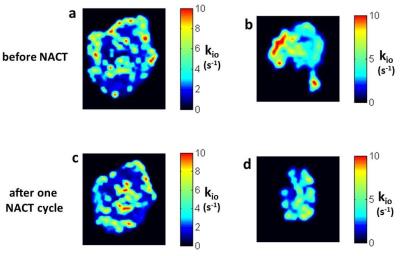
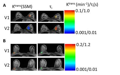
Charles Springer, Jr., Xin Li, Megan Troxell, Karen Oh, Arpana Naik, Kathleen Kemmer, Aneela Afzal, May Mishal, Alina Tudorica, Wei Huang
Synopsis:  A kio parametric image maps Na+,K+ATPase activity with intra-tumor resolution.  For breast tumors, the kio hot spot fraction decreases after one NACT cycle if the tumor goes on to be cancer-free after NACT completion, but not if it maintains residual cancer.  Also, though kio hot spots are reduced after one NACT cycle, new ones appear in different loci.  This is consistent with metabolic competition between different cancer cell populations within the tumor.  


4364.   
14 Evaluation of tumor oxygenation following radiation and PS-targeting antibody therapy in an orthotopic lung cancer model
Heling Zhou, Olivier Belzile, Zhang Zhang, Debabrata Saha, Jo Wagner, Brock Sishc, Strahinja Stojadinovic, Rolf Brekken, Ralph Mason
Combining phosphatidylserine (PS)-targeting monoclonal antibodies with radiation therapy can potentially enhance treatment efficacy. Oxygenation is important in radiation therapy response and could influence future treatment design. Oxygen enhanced MRI was used to examine changes in oxygenation in orthotopic lung tumors in rats treated by radiation or radiation plus a PS-targeting antibody. Orthotopic tumors were well oxygenated and responsive to oxygen breathing challenges before and after treatment. Combination therapy appeared to be more effective in tumor control than radiation alone. 


4368.   
18 
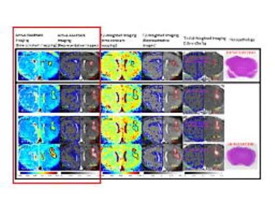
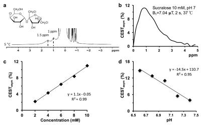
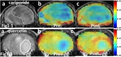
In-vivo Detection of Acute Intracellular Acidification in Glioblastoma Multiforme by AACID CEST MRI Following a Single Dose of Cariporide and Quercetin
Mohammed Albatany, Alex Li, Susan Meakin, Robert Bartha
Identification of tumor boundaries is challenging due to the infiltrative nature of the cancer. Cariporide and quercetin are drugs approved for human use that target different pH regulatory mechanisms in cancer. The goal of the current study is to determine whether chemical exchange saturation transfer (CEST) MRI is sensitive to tumor acidification after cariporide or quercetin injection. In mice with U87 glioblastoma brain tumors, we found both drugs significantly reduced tumor pH within two hours of treatment measured by CEST MRI. The physiological change induced by cariporide or quercetin could help localize brain cancer and monitor tumor response to chemotherapy. This unique approach to cancer detection does not require injection of an imaging contrast agent.


4360.   
10 
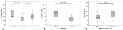
Value of Tumor Stiffness Measured with MR Elastography for Assessment of Response of Hepatocellular Carcinoma to Locoregional Therapy
Sonja Gordic, Jad Bou Ayache, Paul Kennedy , Cecilia Besa, Mathilde Wagner, Octavia Bane, Richard Ehman, Edward Kim, Bachir Taouli
We correlated tumor stiffness (TS) measured with MR elastography (MRE) to degree of tumor enhancement and necrosis on contrast-enhanced T1-weighted imaging (CE-T1WI) in hepatocellular carcinomas (HCC) treated with locoregional therapy.  Patients with HCC who underwent locoregional treatment and controls with newly diagnosed untreated HCC were included. TS values were obtained by placing regions of interest (ROIs) over HCCs on stiffness maps. Visual assessment of tumor necrosis on subtraction images and calculation of enhancement ratios by placing ROIs over tumors on CE-T1WI was performed. TS measured with MRE showed a significant correlation with tumor enhancement and necrosis. 


4369.   
19 The use of DCE MRI in predicting early chemo-radiotherapy treatment response for Larynx and hypopharynx carcinoma - permission withheld
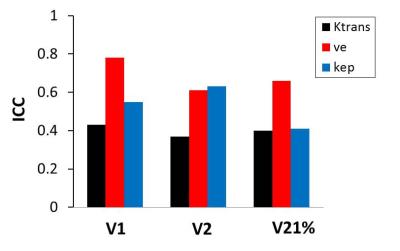
Wei Guo, Dehong Luo, Xinyi Chen, Bing Wu, Meng Lin, Lin Li, Yanfeng Zhao, Xinming Zhao, Chunwu Zhou
In this work, we evaluated the utility of pretreatment semi-quantitative dynamic contrast-enhanced magnetic resonance imaging (MRI) in predicting early response to CRT (chemo-radiotherapy) in patients with larynx and hypopharynx carcinoma from primary tumors. To our knowledge, few studies correlate DCE-MRI semi-quantitative parameters on larynx and hypopharynx carcinoma. These quantitative methods do not require high computational power and were very suitable for clinical application.


4370.   
20 DCE-MRI Assessment of Breast Cancer Response to Neoadjuvant Chemotherapy: Early Prediction of Response and Evaluation of Residual Disease
Alina Tudorica, Karen Oh, Kathleen Kemmer, Megan Troxell, Arpana Naik, Neda Jafarian, Yiyi Chen, Stephen Chui, Eric Goranson, Nicole Roy, Aneela Afzal, May Mishal, Megan Holtorf, Charles Springer, Xin Li, Wei Huang
DCE-MRI was performed on 47 breast cancer patients (49 primary tumors) before, during, and after neoadjuvant chemotherapy (NACT).  DCE-MRI data were subjected to Tofts model (TM) and Shutter-Speed model (SSM) pharmacokinetic (PK) analysis.  Imaging metrics and the corresponding percent changes were correlated with binary pathologic complete response (pCR) and non-pCR endpoints, as well as residual cancer burden (RCB) index values.  By NACT midpoint, several DCE-MRI PK parameters and percent changes are good early predictors of pCR vs. non-pCR, while tumor size is a poor predictor.  Both PK parameters and tumor size after NACT completion are good markers of RCB.  TM and SSM parameters perform equally well for prediction of NACT response and evaluation of RCB.


4371.   
21 
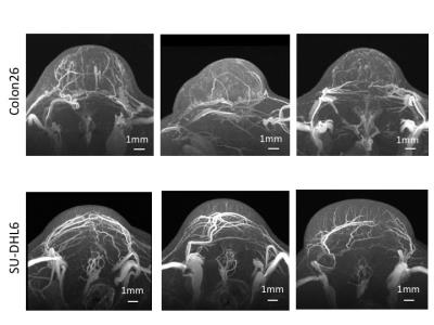
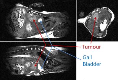
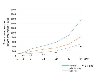
Near infrared photoimmunotherapy for lung cancer in a transgenic mouse model evaluated by MRI - permission withheld
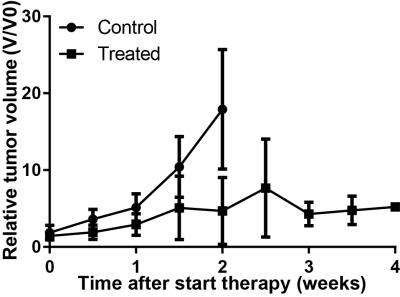
Yuko Nakamura, Marcelino Bernardo, Zoe Ohler, Tadanobu Nagaya, Shuhei Okuyama, Fusa Ogata, Peter Choyke, Hisataka Kobayashi
Near infrared photoimmunotherapy (NIR-PIT) is a new cancer treatment that combines the specificity of antibodies for targeting tumors with the toxicity induced by photoabsorbers after irradiation with NIR light. The purpose of this study was to determine whether MRI can monitor the therapeutic effect of NIR-PIT in spontaneously occurring lung cancers that express epidermal growth factor receptor. Tumor volume ratio was inhibited significantly in the NIR-PIT group compared with control group. Thus, MRI can be a useful imaging modality for monitoring the therapeutic effects of NIR-PIT for cancer.


4372.   
22 Rapid MR Pancreatic and Ovarian Screening Imaging for Patients with BRCA Mutation Undergoing Screening Breast MRI – Pilot Study
Sandra Huicochea Castellanos, Mitchell C. Raeside, Andrea Agostini, Richard K.G. Do, Amita Amita Shukla-Dava, David Aramburu Nunez, Ramesh Ramesh , Olga Smelianskaia, Monika Khan, Yuliya Lakhman, Evis Sala, Lorenzo Mannelli
The purpose of this study was to develop and optimize a rapid MR pancreas and ovarian screening protocol to be performed in conjunction with breast MRI screening in BRCA mutation carriers. Images were acquired with the patient in the prone position, with the breast coil still in place, but using the built-in body coil on a 3T magnet, and evaluated for image quality and detection of lesions. 30 women had research MR for pancreatic screening and 5 of them also underwent for rapid ovarian MR screening which provided diagnostic quality images within a short time of acquisition.


4373.   
23 
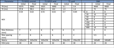
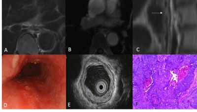
Preoperative T Staging of Potentially Resectable Esophageal Cancer: 3T MRI based on T2-TSE-BLADE and contrast-enhanced free-breathing radial VIBE (StarVIBE) vs endoscopic ultrasound
Jinrong Qu, Hongkai Zhang, Hui Liu, Xu Yan, Zhaoqi Wang, Hailiang Li, Kiefer Berthold, Nickel Marcel Dominik, Ihab R. Kamel
 This study compared MRI based on the combination of T2-TSE-BLADE and contrast-enhanced T1 radial VIBE (StarVIBE) against endoscopic ultrasound (EUS) for T staging of potentially resectable esophageal cancer (EC). The histology confirmation of the T stage was used as reference. The results showed that the combination of T2-TSE-BLADE and StarVIBE is comparable to EUS in T staging of potentially resectable EC with lesions of T1/T2 stage, and is superior to EUS with lesions of T3/T4 stage.


4374.   
24 
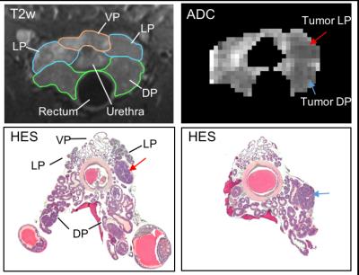
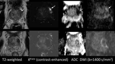
Appearance of Changes From Focal Therapy on Multiparametric Prostate Magnetic Resonance Imaging
Daniel Margolis, Ely Felker, Shyam Natarajan, Chris Alabastro, Leonard Marks
Understanding the changes corresponding to focal therapy of prostate cancer on MRI is paramount to appropriate management, as serum tests may fail to accurately monitor these patients.
 
Perfusion, Permeability & Diffusion in Cancer
Electronic Poster
General Cancer Imaging

 
Tuesday, 25 April 2017
Exhibition Hall  16:15 - 17:15


    Computer #

 
4375.   
25 The Effects of AIF Quantification Variations on DCE-MRI Prediction of Soft Tissue Sarcoma Response to Preoperative Therapy: A Preliminary Multicenter Study
Kimberly Li, Yiyi Chen, Yun Yu, Xia Li, Andriy Fedorov, Guido Jajamovich, Dariya Malyarenko, Madhava Aryal, Peter LaViolette, Matthew Oborski, Finbarr O'Sullivan, Richard Abramson, Kourosh Jafari-Khouzani, Aneela Afzal, Alina Tudorica, Brendan Moloney, Sandeep Gupta, Cecilia Besa, Jayashree Kalpathy-Cramer, James Mountz, Charles Laymon, Mark Muzi, Paul Kinahan, Kathleen Schmainda, Yue Cao, Thomas Chenevert, Bachir Taouli, Fiona Fennessy, Thomas Yankeelov, Xin Li, Wei Huang
Soft tissue sarcoma DCE-MRI data collected at baseline and after one chemotherapy cycle were shared among nine centers and individual arterial input functions (AIFs) were quantified with center-specific methods.  Pharmacokinetic (PK) modeling of the data was performed with these AIFs and the Tofts model.  Considerable variations in estimated PK parameters and the corresponding percent changes were observed due to AIF variations.  kep is less susceptible to AIF variation than Ktrans and may be a more robust imaging biomarker of microvasculature.  kep percent change correlates in a uniformly negative relationship with necrosis percentage of resection specimen across all individually measured AIFs.


4376.   
26 Predicting MGMT expression levels in glioma patients using multi-inversion-time PASL - video not available
Yuchao Liang, Yinyan Wang, Tianyi Qian, Josef Pfeuffer, Shaowu Li, Tao Jiang, Lei Wang
To evaluate the differences in cerebral blood flow between glioma patients with high MGMT expression and those with low MGMT expression, we retrospectively compared cerebral blood flow in pre-operative MR images acquired using multi-inversion-time PASL from glioma patients with histopathologically confirmed high MGMT expression and those with histopathologically confirmed low MGMT expression. The results demonstrate that MGMT expression status significantly correlates with both quantitative CBF and relative CBF.


4377.   
27 Utilizing correlations between Ktrans and plasma volume fraction estimates improves extended Kety modelling of DCE-MRI data
Matthew Orton, Mihaela Rata, David Collins, James d'Arcy, Martin Leach
The extended Kety model is widely used for deriving quantitative perfusion and vascularity measures from DCE-MRI. In practice the signal contribution from the plasma space can be low compared to the signal-to-noise ratio (SNR) of the data which leads to cases where Ktrans is non-zero (indicating delivery of contrast to the tissues), but where the plasma volume fraction is estimated as zero: a logical contradiction.  This work describes a model and fitting methodology to overcome this that makes use of a relationship between Ktrans and the plasma volume fraction to ensure both are positive or both are zero.


4378.   
28 Application of dynamic contrast-enhanced MRI in differential diagnosis of benign and malignant lung lesions
Zhi-yun Jiao, Fang Du, Jianxun Qu, Ling He, Chao Xu, Xia Ye, Jiangfen Wu
Dynamic contrast enhancement (DCE) MRI is rarely used in lung lesions in previous studies, because of the influence of heart rate and respiratory motion. With the development of 3D non-rigid registration algorithm, high resolution dynamic enhanced MRI application in lung disease is increasing. After registration, the image is more clearer and the quantitative parameters are more accurate. The quantitative parameters Ktrans and Kep are capable of differentiating benign and malignant lung lesions, which has an important value in clinical work.


4379.   
29 Differential diagnosis of hepatocellular carcinoma and hepatic metastases using Radiomics Features from Quantitative DCE-MRI
Ye Li, Lian Liu, Ning Huang, Yan Guo
To evaluate the value of Ktrans and HPI (hepatic arterial perfusion index), and their radiomics features in the differential diagnosis of hepatocellular carcinoma (HCC) and hepatic metastases from colorectal cancer (HM) using dynamics contrast enhanced MRI (DCE-MRI). Exchange model with dual input-based arterial input function was used to obtain the radiomics features that can provide a comprehensive assessment of tumors’ physiologic properties including vascular permeability, microcirculation and hemodynamic information. Our results indicated that IDM (Ktrans), inertia and correlation (HPI) are of great significance to differentiate HM and HCC.


4380.   
30 R2* relaxation affects pharmacokinetic analysis of DCE-MRI at high field strength
Eugene Kim, Jana Kim, Siver Moestue
In DCE-MRI, the effect of the r2* relaxivity of gadolinium-based contrast agents is often assumed to be negligible. Here, the validity of this assumption at 7T was tested. DCE-MRI was performed on two preclinical cancer models using a spoiled multiple gradient-echo acquisition, which enabled correction for transverse relaxation. Not accounting for R2* resulted in underestimation of the Tofts pharmacokinetic parameters, with Ktrans being the most affected and vp the least. Simulations showed that the R2* effect can be significant even at 3T in highly perfused regions but can be mitigated by decreasing TE and TR or increasing the flip angle.


4381.   
31 Arterial Input Function Selection in DSC-MRI of Brain Tumors Using Differential Evaluation Clustering Method - video not available
Hossein Rahim Zadeh, Anahita Fathi Kazerooni, Mohammad Reza Deevband, Hamidreza Saligheh Rad
Proper arterial input function (AIF) selection is a critical step for accurate quantification of dynamic susceptibility contrast enhanced (DSC) MRI in brain tumor patients. In this study, we have employed differential evaluation (DE) clustering method on processed perfusion images for accurate AIF selection. The procedure consists of two main steps: preprocessing for eliminating non-arterial curves including tissue, noisy and those contaminated with partial volume effects; and AIF selection using DE clustering method. The performance of this clustering method was compared to K-means and Hierarchical Clustering techniques and the results show the superiority of the proposed approach for accurate AIF selection.


4382.   
32 Quantitative Perfusion Imaging in Rectal Cancer – Choice and Influence of the Arterial Input Function to Perfusion Parameters
Tanja Gaa, Sonja Sudarski, Lothar Schad, Frank Zöllner
Quantitative perfusion analysis in rectal cancer with DCE-MRI is highly dependent on the choice of the arterial input function (AIF). In nineteen patients it was investigated whether the selection affects the quantification of plasma flow (PF) applying with three different perfusion models (fast deconvolution algorithm, two-compartment uptake model, two-compartment exchange model). Results show disagreements in PF between the two AIFs for all three models with significant differences for the two-compartment exchange and uptake model.


4383.   
33 Model-based and Non-model-based Perfusion-related Predictors of Response to Chemotherapy in Intrahepatic Cholangiocarcinoma
Kristen Zakian, Aditi Iyer, Aditya Apte, Taryn Boucher, William Jarnagin, Nancy Kemeny, Richard Do
Hepatic arterial infusion of floxuridine has shown benefit in intrahepatic cholangiocarcinoma. Due to the complexity and expense of this treatment, a pre-treatment or early biomarker of efficacy would be advantageous. We employed non-model-based (NMB) analysis to maximize information from breath-motion degraded DCE-MRI in 24 patients. Model-based and NMB parameters were compared to RECIST response. The kurtosis value of pre-treatment time-to-half-maximum correlated with response, as did 1 month changes in perfusion parameters (Ktransand signal at half maximum).  NMB analysis resulted in fewer voxels being discarded due to motion and thus may be more representative of tumor physiology while not requiring modeling.


4384.   
34 Optimization of acquisition and modelling parameters for accurate and precise estimation of tumor vascular permeability using modified K-CNR
Thomas Ng, Ravi Seethamraju, Ritu Gill
Adoption of quantitative clinical DCE-MRI remains limited given the challenges in accurate and precise estimation of kinetic parameters. Simulations are being increasingly used to guide protocol optimization1, but the relative effects of altering protocol variables are seldom considered. We used a modified K-CNR  metric to quantify tumor Ktrans estimation. K-CNR provided a simple way to compare how input variables affect Ktrans output. The extended Toft’s model was shown to be robust for tumor relevant Ktrans. Lengthening baseline time can improve Ktrans estimation. Care must be taken when using nested model analysis; wrong model convergence can occur with non-optimized acquisition variables.


4385.   
35 Robust and Efficient Perfusion Parameter Estimation for DCE-MRI of the Prostate Utilizing the Variable Projection (VARPRO) Method - video not available
Soudabeh Kargar, Eric Stinson, Eric Borisch, Adam Froemming, Akira Kawashima, Lance Mynderse, Joshua Trzasko, Stephen Riederer
Dynamic contrast enhanced-MRI is essential for assessment of tissue microvasculature, and thus improved methods of parameter estimation for evaluation of tissue perfusion can be valuable. This work demonstrates how application of Variable Projection (VP) in perfusion analysis increases the robustness and computation efficiency compared to the conventional Levenberg-Marquardt (LM) method. A numeric simulation is used to validate the perfusion estimation and evaluate the robustness with respect to noise. The computation speed of VP is shown to be improved about 5× faster compared to LM method. VP-based perfusion analysis is demonstrated in eight patients with known prostate cancer.


4386.   
36 Weighted Total Least Squares for Parameter Estimation with the Two Compartment Exchange Model
Anders Garpebring, Tommy Löfstedt
When using linear least squares to estimate pharmacokinetic model parameters in dynamic contrast-enhanced MRI there is a risk of introducing bias due to noise correlations. By using a weighted total least squares (WTLS) method instead, these correlations are taking into account. The results of this study shows that the WTLS method is able to reduce the bias considerably compared to the linear least squares method for the two compartment exchange model, however, at the expense of increased computational time.  


4387.   
37 Diffusion-weighted MRI of rectal cancer: baseline tumour perfusion fraction predicts chemoradiotherapy response and survival
Kine Bakke, Knut Hole, Svein Dueland, Krystyna Grøholt, Kjersti Flatmark, Anne Ree, Therese Seierstad, Kathrine Redalen
More accurate diagnostics for prediction of treatment responses in locally advanced rectal cancer is warranted. We employed a simplified approach to the intravoxel incoherent motion imaging method to estimate the tumour perfusion fraction from diffusion-weighted MRI. The perfusion fraction was predictive of the histologic tumour response after chemoradiotherapy (p = 0.02), and in combination with tumour volume this parameter was also predictive of five-year progression-free survival of the patients (p = 0.002). This simplified approach does not require substantial extra scan time in a routine diagnostic scanning, and may offer a clinically feasible approach to stratifying patients to individualised treatment.    


4388.   
38 Anomalous diffusion in cerebral gliomas assessed using a fractional motion model
Boyan Xu, Lu Su, Zhenxiong Wang, Yaoyu Zhang, Yang Fan, Bing Wu, Gaolang Gong, Wenzhen Zhu, Peiyi Gao, Jia-Hong Gao
Several models have been proposed to explain the anomalous diffusion in biological tissues. Among them, the fractional motion (FM) model was considered more appropriate. In this study, the FM model was applied in gliomas to assess its feasibility for grading gliomas. It was found that the FM model could improve the diagnostic accuracy in differentiation low- and high-grade gliomas, indicating the potential of the FM model to facilitate future studies of neuro-pathological changes in clinical populations.


4389.   
39 Discriminatory ability of MRSI and DW-MRI in prostatic diseases in biopsy naïve men with PSA 4-10 ng/ml - permission withheld
Durgesh Dwivedi, Rajeev Kumar, Alok Dwivedi, Girdhar Bora, Sanjay Thulkar, Sanjay Sharma, Siddhartha Gupta, Naranamangalam Jagannathan
Multivariate approach of including different MR parameters offer a holistic view of entire data, which could provide better approach in differentiating various prostatic diseases. This approach may help in reducing the burden of unnecessary biopsy in men with PSA 4-10 ng/ml.  we evaluated discriminatory ability of MRSI and DW-MRI in prostatic diseases in biopsy naïve men with PSA 4-10 ng/ml. The combined model (PSA + ADC + metabolite ratio) showed highest discriminatory ability in various prostatic diseases with AUC of 89% in men with clinically challenging group of PSA.


4390.   
40 Diffusion Weighted Fast Spin Echo for tumor delineation in head-and-neck radiotherapy: a comparison with FDG-PET
Tim Schakel, Boris Peltenburg, Jan-Willem Dankbaar, Carlos Cardenas, Michalis Aristophanous, Chris Terhaard, Hans Hoogduin, Marielle Philippens
DWI shows high contrast between tumor and surrounding tissue, which shows potential for target volume delineation in head-and-neck radiotherapy treatment planning. In this study we assess the performance of a diffusion weighted fast spin echo sequence (DW-SPLICE) for target delineation in terms of interobserver agreement and spatial concordance with PET. Fifteen patients underwent both PET and DW-SPLICE. PET was segmented using a Gaussian mixture model, DW-SPLICE was delineated by 3 observers. Target volume delineation using DWI is feasible in head-and-neck radiotherapy. Using an optimized DWI sequence, target volumes could be defined with good interobserver agreement and large similarity with PET.


4391.   
41 Distortion-Free Diffusion MRI using an MRI-Guided Tri-Cobalt 60 Radiotherapy System: Sequence Validation and Preliminary Clinical Experience
Yu Gao, Minsong Cao, Tania Kaprealian, Mitchell Kamrava, Michael Wang, John Neylon, Daniel Low, Yingli Yang, Peng Hu
Limited resolution, severe distortion, and inaccurate quantification in low SNR scenarios associated with the conventional DW-ssEPI are problematic for adaptive radiotherapy based on tumor response. In this study, we sought to develop a reliable, accurate and distortion-free diffusion sequence that is practicable for longitudinal assessment of tumor response using an MRI-guided radiotherapy system. Quantitative phantom studies validated the superiority of the proposed technique over standard DW-ssEPI in terms of geometric fidelity and ADC accuracy. Excellent ADC reproducibility makes it a promising candidate for tumor response monitoring. Preliminary patient study demonstrated its feasibility to be used for tumor response assessment. 


4392.   
42 Proton Magnetic Resonance Spectroscopy of the Lipid Metabolic Response of a Murine Tumor Model to Radiation Therapy
Anthony Tessier, Atiyah Yahya, Matthew Larocque, B. Gino Fallone, Alasdair Syme
The temporal dependence of the lipid metabolic response to single fraction radiation therapy of human glioblastoma multiforme xenografts in NIH III nude mice was assessed with in vivo 1H magnetic resonance spectroscopy (MRS) on a 9.4T magnetic resonance system. Lipid MRS can measure fat polyunsaturation, and therapeutic response can be monitored. The methylene:methyl peak ratio (CH2/CH3) can assess drug treatment and disease progression. Herein, lipid parameters and their response to radiotherapy in tumors are studied, including methyl, lactate + methylene, diallylic, and allylic protons, protons in α position to the carbonyl group, CH2/CH3, and mean polyunsaturation.


4393.   
43 Longitudinal diffusion MRI for treatment assessment of sarcoma patients with pre-operative radiation therapy
Yingli Yang, Minsong Cao, Yu Gao, Mitchell Kamrava, Anusha Kalbasi, Nzhde Agazaryan, James Lamb, Ke Sheng, Daniel Low, Peng Hu
Diffusion weighted MRI (DWI) is promising for early prediction of tumor response to radiation therapy. We report our results of using longitudinal DWI approach performed on ViewRay system for predicting the response of sarcoma patient to pre-op RT. Six sarcoma patients were recruited in this study. Each patient subsequently underwent surgery. The tumor necrosis score was then used to compare to the ADC values to assess the predictive value of longitudinal DWI. Each patient successfully underwent 3–5 diffusion MRI scans. Based on the data from 6 patients, our longitudinal changes in tumor ADC matched well with pathology necrosis results. 


4394.   
44 Integrated volumetric diffusion-weighted MR imaging and HPV genotyping in outcome prediction for stage IB-IV cervical cancer patients following chemoradiation therapy
Gigin Lin, Chyong-Huey Lai, Yu-Chun Lin
We evaluated the values of integration of pretreatment volumetric ADC analysis with human papillomavirus (HPV) genotyping in prediction of survival and recurrence for women with locally advanced cervical cancer in 82 women following concurrent chemoradiation therapy (CCRT). Independent poor prognostic indicators for OS and DFS by stepwise multivariate analysis, i.e., lower ADC10, advanced T or M stage and high-risk HPV status were composed to generate a novel outcome-predicting model, in which death was found in all the high-risk group patients whiles none of the low-risk group died or recurred during the follow up.  


4395.   
45 Longitudinal assessment of tumor volume and apparent diffusion coefficient in patients on active surveillance: a good way of monitoring disease progression?
Veronica Morgan, Christopher Parker, Nandita deSouza
Tumor growth on T2-W MRI in 151 men with low-risk prostate cancer managed by active surveillance was related to tumor apparent diffusion coefficient (ADC). Volume increases greater than the 95% upper Limits of Agreement of reproducibility (~60%, n=20) were seen in 52 (34.4%) men. ADC was more reproducible (~5% variability). Baseline ADC values did not differ between those with and without measurable growth (p=0.06) but change in ADC did (-6.8±12.3% for those with measurable growth vs. 0.23±10.1% for those without, p=0.0005). A 5.6% reduction in ADC with time indicated a measurable increase in tumor volume (specificity 77.0%, sensitivity 54.9%, AUC=0.67).


4396.   
46 Textural Analysis based Segmentation of Bone tumours using Diffusion Weighted MR Image
Amit Mehndiratta, Esha Kayal, Rishabh Gupta, Abhimanyu Sahai, Jayendra Alampally, Sameer Bakhshi, Raju Sharma, Devasenathipathy Kandasamy
Diffusion weighted imaging (DWI) plays a crucial role in diagnosis and prognosis of cancerous diseases. Proper demarcation of tumour on DWI is therefore necessary for qualitative assessment as well as quantitative image analysis like ADC or IVIM. Manual demarcation of tumour on each DWI slice is time consuming, prone to error and hard to reproduce. Automated and semi-automated algorithms were implemented and tested to segment bone tumours specifically on DWI. Experimental results reveal that semi-automated Active-Contours performed tumor segmentation better than other methods with acceptable levels of accuracy with considerable less time and manual effort.


4397.   
47 MR imaging in the prediction of aggressive histological features in papillary thyroid carcinoma
Bin Song, Hao Wang
Purpose To investigate the value of magnetic resonance imaging (MRI) features for prediction of tumor aggressiveness in papillary thyroid cancer (PTC). Method A consecutive series of 119 lesions with PTC were preoperatively evaluated by MR imaging. A multivariate analysis was performed to predict tumor aggressiveness by 18 independent variables. Results Multivariate analysis demonstrated that lesion size classification and tumor margin on delayed contrast-enhanced images were the independent predictors of high aggressive PTC. Conclusion Lesion size classification and tumor margin on delayed contrast-enhanced images can be used preoperatively to estimate the risk of high aggressive of PTC.


4398.   
48 Optimization of b-values in iShim diffusion-weighted MR imaging of the thyroid gland: to improve the visualization of malignant thyroid nodules and the accuracy in differentiating malignant from benign thyroid nodules - video not available
Qingjun Wang, Qinglei Shi, Tianyi Qian, Alto Stemmer, Yong Guo
To investigate the optimal b-values of diffusion-weighted imaging (DWI), a prototype sequence with integrated slice-specific shimming (iShim) was applied for visualizing thyroid malignant nodules and making accurate differentiation between benign and malignant nodules. DWI images were acquired with five b-values including 0, 800, 1000, 2000 and 3000 sec/mm2. The results show that the iShim-DWI with b-value of 800 s/mm2 can present the clearest delineation of malignant thyroid nodules and that the images with b-value of 3000 s/mm2 had the best performances in differentiating malignant from benign thyroid nodules.
 
Preclinical Imaging in Cancer
Electronic Poster
General Cancer Imaging

 
Tuesday, 25 April 2017
Exhibition Hall  17:15 - 18:15


    Computer #

 
4465.   
1 Histology-Derived pseudo-ADC correlates with measured ADC and extranuclear space in a transgenic model of prostate cancer, identifying contribution of luminal space to measured ADC.
Matthew Blackledge, Konstantinos Zormpas-Petridis, Andreas Heindl, Siver Moestue, Yinyin Yuan, Dow Koh, David Collins, Yann Jamin, Tone Bathen, Martin Leach, Deborah Hill
In this preclinical study we investigate the utility of in-silico simulations of the pseudo-apparent diffusion coefficient (pADC) of water within extra-nuclear regions segmented on large field-of-view haematoxylin, eosin, and saffron (HES) slides from a transgenic mouse model of prostate cancer.   We demonstrate that pADC is correlated within in-vivo measurements of the apparent diffusion coefficient (ADC) measured by diffusion-weighted magnetic resonance imaging and may thus be used as a surrogate for exploring the effect of tissue structure on measured ADC values.  Furthermore, we demonstrate that ADC is correlated with fractional space occupied by lumen, derived from semi-automatic segmentation of HES slides. 


4466.   
2 Tumor vasculature differs between cell and fragment derived murine orthotopic models of hepatocellular carcinoma
Sibu Kuruvilla, Colleen Crouch, Joan Greve
We report the use of semi-quantitative dynamic contrast enhanced (DCE)-MRI to identify differences in the growth and vascular environment seen within two murine hepatocellular carcinoma (HCC) models - cellular orthotopic injection (COI) (representing a traditional tumor model) or surgical orthotopic implantation (SOI) of tumor fragments (representing a patient-derived orthotopic xenograft model, or PDOX). Uptake curves of gadolinium contrast agent indicate differences in vasculature between the two models, highlighting important considerations to be made when trying to use PDOX methods to model HCC in the clinic.


4467.   
3 Intrinsic susceptibility MRI predicts response to the vascular endothelial growth factor receptor inhibitor cediranib in the Th-MYCN model of neuroblastoma
Yann Jamin, Evon Poon, Neil Jerome, Alexander Koers, Laura Danielson, Dow-Mu Koh, Louis Chesler, Simon Robinson
In this study we demonstrate that the transverse relaxation rate R2* predicts response to the vascular endothelial growth factor receptor (VEGFR) inhibitor cediranib in the Th-MYCN genetically-engineered murine model of neuroblastoma, a childhood cancer of the developing nervous  system. 


4468.   
4 Papillary thyroid carcinoma with hobnail pattern: unique MRI features and correlated with the histopathologic findings
Lanyun Wang
Hobnail papillary thyroid carcinoma (HPTC) is a moderately differentiated PTC variant with aggressive clinical behavior and significant mortality.The purpose of this study was to recognize the unique MRI features of HPTC .61 patients with PTC confirmed histopathologically[19 lesions of HPTC and 42 NHPTC (PTC without hobnail features)lesions] undergoing MRI with T1W, T2W,DWI and contrast material–enhanced sequences prior to thyroidectomy were included retrospectively. there was a significant tendency toward T2WI significantly high signal ?T2WI linear low signal ?Lace levy ?Gyrus-like structure ?higher ADC values ?irregular shape present in HPTCs. HPTC lesions have unique MRI features.   


4469.   
5 Cell Nuclear Size and Shape are Associated with ADC, Cellularity, and Histological Class in a Transgenic Mouse Model of Prostate Cancer
Leslie Euceda, Matthew Blackledge, Andreas Heindl, Yinyin Yuan, Dow-Mu Koh, David Collins, Siver Moestue , Martin Leach, Tone Bathen, Deborah Hill
The relationship between tissue microstructure and ADC is poorly understood in cancer. We identified quantitative nuclear characteristics from haematoxylin, eosin, and saffron stained prostatic histology samples. ADC maps were calculated for transgenic prostate cancer mice and healthy controls. Matched histology images of excised prostates were used to determine cellularity and histological class. ADC and cellularity correlated with a set of nuclear features (describing texture, lightness, radius, and shape), which exhibited mirrored trends, implying an inverse relationship between ADC and cellularity. The same nuclear features correlated with histological class. This will be useful for underpinning relationships between ADC and tissue microstructure.


4470.   
6 A Genetically Engineered Mouse Model Recapitulates Radiological Features of Human Adamantinomatous Craniopharyngioma
Jessica Boult, Ciaran Hutchinson, John Apps, Gabriela Carreno, Laura Danielson, Laura Smith, Alex Virasami, Alexander Koers, Louis Chesler, Owen Arthurs, Juan Pedro Martinez-Barbera, Simon Robinson
Tumours in Hesx1Cre/+;Ctnnb1lox(ex3)/+ mice resemble human adamantinomatous craniopharyngioma (ACP) molecularly and histologically. MRI and ex vivo micro-CT were used to assess the radiology of this model for the first time. Early enlargement and heterogeneity of Hesx1Cre/+;Ctnnb1lox(ex3)/+ pituitaries was evident; enlargement of a solid tumour, and development of cysts and haemorrhage subsequently occurred. Solid components showed heterogeneous T1-weighted signal enhancement following Gd-DTPA administration, and in some animals cysts were hyperintense on FLAIR and T1-weighted images, both emulating clinical observations. Cyst calcification was not observed by micro-CT but we show that Hesx1Cre/+;Ctnnb1lox(ex3)/+ tumours faithfully recapitulate the MRI radiology of the human disease.


4471.   
7 Probing tumor metabolism using dynamic lactate chemical exchange saturation transfer MRI
Puneet Bagga, Mohammad Haris, Pranav Jain, Alekya Bheemreddy, Francesco Marincola, Hari Hariharan, Ravinder Reddy
Lactate CEST (LATEST) MRI method has been shown to be applicable in detecting and imaging changes in the lactate level in human subjects post heavy exercise and to measure the lactate in a mouse model of lymphoma. In this study, LATEST was implemented to probe the lactate dehydrogenase (LDH) activity in vivo in a rat glioma model. Following the administration of pyruvate, the LATEST contrast is elevated in the tumor region while it is unaltered in the unaffected region.


4472.   
8 Molecular effects of various chemotherapeutic agents on choline phospholipid metabolism of triple-negative breast cancer cells
Menglin Cheng , Zaver Bhujwalla, Kristine Glunde
The MRS-detected total choline (tCho) signal is a promising non-invasive surrogate marker of chemotherapy response in breast cancer patients. The molecular mechanisms by which common chemotherapeutic drugs affect the tCho signal, consisting of glycerophosphocholine (GPC), phosphocholine (PC), and free choline (Cho), are unknown. We have employed widely used cancer chemotherapeutic drugs including doxorubicin, paclitaxel, and vinorelbine to treat triple-negative human MDA-MB-231 breast cancer cells to elucidate their molecular effects on choline phospholipid metabolism using high-resolution 1H MRS to detect changes in cellular choline metabolite profiles, and quantitative RT-PCR to assess the corresponding changes in the expression levels of choline-metabolizing enzymes.


4473.   
9 The Metabolic Secretome of Cachexia Inducing Pancreatic Ductal Adenocarcinoma
Santosh K Bharti, Paul T Winnard Jr., Louis Dore-Savard, Yelena Mironchik, Marie-France Penet, Zaver M Bhujwalla
Tumor interstitial fluid (TIF) contains the secretome of cancers that can impact most phenotypic aspects of cancer.  One characteristic phenotype of pancreatic ductal carcinoma (PDAC) is its ability to induce profound weight loss or cachexia through a multifactorial syndrome that affects multiple organs.  To understand the interaction between cancer cells and body organs, here, for the first time, we have characterized the metabolomic profile of TIF from cachexia and non-cachexia inducing pancreatic cancer xenografts and identified clear differences in the metabolic secretome between cachexia inducing and the non-cachexia inducing tumors.  


4474.   
10 Metabolic reprogramming in a relevant IDH1-mutated human glioma xenograft model
Tom Peeters, Krissie Lenting, Jack van Asten, William Leenders, Arend Heerschap
Understanding metabolic aberrations in IDH-mutated gliomas requires xenograft models growing in a relevant tissue microenvironment and resembling its human genetic counterparts. We performed in vivo 1H MRSI of human-derived oligodendroglioma xenograft models to map lactate and total choline concentrations. Lactate levels were significantly lower and total choline higher in mutated tumor tissue compared to non-tumor brain in the same animal, or to its wild-type counterpart model. This outcome was correlated with expression levels of enzymes and transporters in both lactate- and phospholipid-related metabolic pathways. The findings point to a metabolic reprogramming of aerobic glycolysis and lipid synthesis by the IDH1 mutation.


4475.   
11 Intratumoral vessel evaluation using magnetic resonance micro-angiography with a vascular pool Gd-DOTA-dendron liposomal contrast agent
Nobuhiro Nitta, Daisuke Kokuryo, Sayaka Shibata, Kenji Kono, Akihiro Tomita, Jeff Kershaw, Ichio Aoki, Masafumi Harada
Evaluation of the intratumor environment, such as cell distribution and vessel structure, are important for understanding the characteristics of tumors. In particular, the vessel structure and its permeability is critical for the malignant potential of the tumor and drug-targeting. The purpose of this work was to non-invasively visualize intratumoral vessels using MR micro-angiography (MRmA) and a liposomal contrast agent having long blood half-life. In addition, we tried to identify differences in the vascular structures for Colon26 and SU-DHL6 cells. The proposed method visualized the intratumoral vessels clearly and revealed differences in the structure depending on the cell type.


4476.   
12 MR image analysis to identify habitats through comparison with histology and immunohistochemistry in breast cancer
Bruna Jardim-Perassi, William Dominguez-Viqueira, Mikalai Budzevich, Epifanio Ruiz, Suning Huang, Jan Poleszczuk, Alex Lopez, Debora Zuccari, Gary Martinez, Robert Gillies
Breast cancer shows significant heterogeneity at both inter- and intratumoral levels. In this study, a distribution clustering of multiple MRI pulse sequences was used in combination with a 3D printed approach, and showed a qualitatively comparable pattern of intratumoral heterogeneity (habitats) in MRI and histological images. This approach could potentially be used as a non-invasive imaging method for the monitoring of the intratumoral heterogeneity following the therapy in breast cancer.


4477.   
13 Electric field and current density mapping during reversible Electroporation
Munish Chauhan, Neeta Ashok Kumar, Vikram Kodibagkar, Rosalind Sadleir
Electroporation is popular in cancer therapy and gene or drug delivery. Magnetic Resonance Electrical Impedance Tomography (MREIT) was used to monitor the applied electric field and current density during electroporation. Electroporation fields were applied by novel low-susceptibility carbon fiber electrodes in a bovine liver tissue sample. Projected current density (JP) and Electric field (E) were computed from MR data collected under electroporation fields. Susceptibility artifacts around the pair of carbon fiber electrodes were greatly reduced. In this study, we demonstrated the feasibility of MREIT technique to monitor the current density and electric field distribution during ex-vivo tissue electroporation.  


4478.   
14 Diffusion Kurtosis Imaging Evaluating Epithelial–mesenchymal Transition in Colorectal Carcinoma Xenografts Model: Initial Experience - video not available
Huanhuan Liu, Caiyuan Zhang, Jinning Li, Weibo Chen, Dengbin Wang
Colorectal cancer (CRC) is one of the most common malignant tumors in gastrointestinal tract. Tumor recurrence and metastasis are still a major cause of death in rectal cancer patients. Epithelial-mesenchymal transition (EMT) is reported a critical process providing tumor cells with the ability to migrate and metastasize to distant sites, leading to a poor prognosis in CRC. The diffusion kurtosis imaging (DKI) could quantify non-Gaussian behavior of water diffusion and provide more precise information of tissue characteristics. Our study demonstrated that DKI could be used to identify EMT in CRC xenograft models.


4479.   
15 Telomerase expression enhances pentose phosphate pathway flux resulting in an MR-detectable increase in reduced glutathione levels in mutant IDH1 glioma cells - permission withheld
Pavithra Viswanath, Russell Pieper, Sabrina Ronen
Telomerase is the enzyme responsible for maintenance of telomeres, which are special capped structures that protect chromosomal ends from degradation. Telomerase activation and metabolic reprogramming have both emerged as hallmarks of cancer. Here, we investigated the link between telomerase and metabolism in mutant isocitrate dehydrogenase 1 (IDH1) glioma cells with the goal of identifying MR-detectable biomarkers of telomerase expression. Using 13C- and 1H- MRS, we show that telomerase expression is associated with elevated flux through the pentose phosphate pathway resulting in increased levels of reduced glutathione (GSH). Thus, GSH is a potential biomarker of telomerase expression in mutant IDH1 gliomas. 


4480.   
16 Dietary Fat Results in Increased Tumor Burden in a Mouse Model of Human Triple-Negative Breast Cancer Based on Magnetic Resonance Imaging and Histology
Devkumar Mustafi, Sully Fernandez, Erica Markiewicz, Xiaobing Fan, Marta Zamora, Jeffrey Mueller, Matthew Brady, Suzanne Conzen, Gregory Karczmar
Breast cancer is the most commonly diagnosed malignancy among women in the United States and the second leading cause of cancer mortality worldwide. Epidemiological studies suggest an increase in the risk of triple-negative breast cancer (TNBC) in association with a high animal fat diet. Based on previous MRI studies in SV40Tag mice, we examined the effect of pre-pubertal exposure to high dietary fat in this model of TNBC. The results reported here demonstrate that a high animal fat diet significantly increased the number of aggressive cancers detected by MRI in a mouse model of human TNBC.


4482.   
18 In vivo hyperpolarized [1-13C]pyruvate and [18F]-FDG PET/CT studies of prostate cancer metastasis xenografts in mice
Frits van Heijster, Sandra Heskamp, Andor Veltien, Tom Peeters, Tom Scheenen, Otto Boerman, Arend Heerschap
Understanding the underlying mechanisms of aggressiveness is important for better staging and treatment of prostate cancer. In this study two murine xenograft models of early and late stage prostate cancer were investigated. [1-13C]pyruvate was hyperpolarized by Dynamic Nuclear Polarization (DNP) and pyruvate metabolism is followed by 13C-MR. This is combined with [18F]FDG-PET/CT to study glucose metabolism. We found differences in pyruvate conversion and glucose uptake in vivo in the murine models.


4481.   
17 Magnetic Resonance Angiography Shows Increased Arterial Blood Supply Associated with Murine Mammary Cancer
Devkumar Mustafi, Abby Leinroth, Xiaobing Fan, Erica Markiewicz, Marta Zamora, Jeffrey Mueller, Suzanne Conzen, Gregory Karczmar
Breast cancer is a major cause of morbidity and mortality in Western women. Tumor neo-angiogenesis may be an MRI-detectable prognostic marker for cancer progression. Clinical practice uses DCE-MRI to detect cancers based on increased blood flow and capillary permeability. However, DCE-MRI requires repeated injections of contrast media; therefore we used time-of-flight MR angiography to measure the number and size of arteries feeding mammary glands with and without cancer, and demonstrated that blood vessels in and near mammary glands grew significantly as invasive cancers developed.


4483.   
19 Multi-agent dynamic contrast enhanced MRI to assess vascular changes induced by prolonged VEGFR2 inhibition in oesophageal cancer
Remy Klaassen, Anne Steins, Matthias Schabel, Igor Jacobs, Maarten Bijlsma, Aart Nederveen, Gustav Strijkers, Hanneke van Laarhoven
The effective vascular changes induced by prolonged VEGFR2 inhibition and the resulting changes in vascular function are difficult to monitor in vivo and therefore largely unexplored. In this study we show a multi-agent DCE-MRI approach, using 3 different-sized contrast agents, to quantify effects on vascularization induced by prolonged VEGFR2 inhibition in an oesophageal cancer mouse model. Effects on vascularization were predominantly observed in parameters dependent on the larger-sized contrast agents, where no effects where found for the traditional low-molecular weight contrast agent.


4484.   
20 In-vivo targeting and imaging of super-paramagnetic iron-oxide particles to subcutaneous tumour models - permission withheld
Mohammad Mohseni, John Connell, Stephen Patrick, May Zaw-Thin, Tammy Kalber, Tom Roberts, Quentin Pankhurst, Mark Lythgoe, Bernard Siow
Magnetic targeting of drug-conjugated iron oxide nanoparticles has the potential to increase the concentration of therapeutic agents to tumours whilst reducing off-target side effects of current chemotherapy methods. This preclinical work demonstrates that SPION accumulation can be increased in subcutaneous tumours using magnetic fields and can then be detected by MRI.  In addition, key physiological parameters can be measured before magnetic targeting for future optimisation of the strategy.


4485.   
21 Imaging Collagenase-Induced Changes in the Mechanical Phenotype of Orthotopic BT474 Breast Cancer Xenografts Using Magnetic Resonance Elastography
Jin Li, Jessica Boult, Craig Cummings, Jeffrey Bamber, Ralph Sinkus, Yann Jamin, Simon Robinson
We tested the hypothesis that MRE can inform on collagenase-induced matrix degradation in orthotopic BT474 breast carcinoma xenografts in vivo.  An acute reduction in  the absolute value of the complex shear modulus |G*| was detected in tumour just 5 hours after collagenase administration, mostly likely a consequence of both collagen degradation and reduction of interstitial fluid pressure.  The study highlights the utility of MRE-derived quantitation of tumour viscoelasticity for monitoring the response of stromal rich tumours to modification of the extracellular matrix. 


4486.   
22 Multiparametric MRI for optimal prostate cancer detection in TRAMP mice
Jana Kim, Eugene Kim, Deborah Hill, Dan Meyer, Karina Langseth, Frits Thorsen, Tone Bathen, Siver Moestue
Multiparametric MRI is the standard for the detection and characterization of prostate cancer. In this study we compared the diagnostic value of clinically used DW-MRI and DCE-MRI in a transgenic mouse model of prostate cancer (TRAMP).  Additionally, we investigated the potential utility of SSC-MRI using a USPIOs contrast agent for prostate cancer detection. The results of this study confirm the utility of DW-MRI and the potential value of DCE-MRI in early-stage detection and monitoring of prostate cancer. SSC-MRI appears to be less useful for this task in the TRAMP model, but further analysis is required to draw a clear conclusion. 


4487.   
23 Transport across the blood-brain-barrier may be limiting for hyperpolarized [1-13C]pyruvate neuro-oncology studies
Jack Miller, James Larkin, Katherine Fisher, Vicky Ball, Kevin Ray, Sebastien Serres, Damian Tyler, Angus Lau, Nicola Sibson
Hyperpolarized pyruvate has previously been used to probe primary brain cancer. Through imaging the delivery and metabolism of both hyperpolarized [1-13C]pyruvate and ethyl-[1-13C]pyruvate in a rodent model of cancer metastasis to the brain, we show that the transport of [1-13C]pyruvate across the blood brain barrier may be limiting until it is compromised by metastatic cell infiltration. 


4488.   
24 Validation of an efficient and robust MRI-guided radiotherapy planning approach for targeting abdominal organs and tumours in the mouse.
Veerle Kersemans, John Beech, Stuart Gilchrist, Paul Kinchesh, Philip Allen, James Thompson, Ana Gomes, Zenobia D'Costa, Luke Bird, Iain Tullis, Robert Newman, Abul Azad, Ruth Muschel, Borivoj Vojnovic, Mark Hill, Emmanouil Fokas, Sean Smart
The aim was to develop and validate a robust and accurate method of MR-IGRT delivery to abdominal targets in the mouse that takes advantage of the strengths of each of its components: MRI for soft tissue contrast and target identification, CBCT for accurate dose calculation and IGRT for accurate, collimated X-ray beam delivery. A multimodality cradle was developed and evaluated to enable transfer of the mouse between MR and the IGRT platform. Additionally, each step of the MG-IGRT process was validated, both in vitro using BANG gel dosimeters and in vivo by targeting the adrenal glands in mice.
 
General Cancer Including Preclinical
Electronic Poster
General Cancer Imaging

 
Tuesday, 25 April 2017
Exhibition Hall  17:15 - 18:15


    Computer #

 
4489.   
25 Diffusion kurtosis imaging can stratify differentiation of colorectal cancers: a preliminary study - video not available
Huanhuan Liu, Caiyuan Zhang, Jinning Li, Weibo Chen, Dengbin Wang
Colorectal cancer (CRC) is the third most common cancer in the world and its incidence is on the rise. Management is particularly challenging technically for surgeon and local recurrence is a common result of treatment failure. Tumor differentiation grade is one of the factors that influence the choice of individual management, then affecting the prognosis. Diffusion kurtosis imaging (DKI) could quantify non-Gaussian behavior of water diffusion and provide more precise information of tissue characteristics. Our study demonstrated that DKI is conducive to distinguishing the various differentiations of CRC models, and reflecting the proliferation indirectly in tumor cells.


4490.   
26 Multi-phase contrast-enhanced magnetic resonance imaging with respiratory gating in papillary thyroid carcinoma
Hao Wang, Bin Song
Purpose To investigate the role of multi-phase contrast-enhanced MR imaging in papillary thyroid carcinoma (PTC). Methods A consecutive series of 62 lesions with PTC were prospective evaluated by MR imaging. Lesions were evaluated for location, size, shape, margin, rim, degree and pattern of enhancement.  Results 52 (83.9%) lesions showed moderate enhancement. 45 (72.6%) lesions showed ring-enhancement and 42 (67.7%) showed central washout enhancement during the delayed phase. 36(58.1%) manifested ill-defined margin and irregular rim (49, 79.0%) after administration of the contrast medium. Nine nodules (14.5%) showed extrathyroidal extension.   Conclusion PTC commonly manifest moderate enhancement during the initial phase, with ill-defined margin and irregular rim. Central wash out enhancement and ring-enhancement occur during delayed contrast –enhanced phase.  


4491.   
27 Radiological comparisons of 3D and 2D-cell culture derived tumours in an orthotopic mouse xenograft model of paediatric glioblastoma
Mariama Fofana, Jessica Boult, Maria Vinci, Valeria Molinari, Sergey Popov, Alan Mackay, Angel Carcaboso, Chris Jones, Simon Robinson
Primary patient-derived paediatric glioblastoma (pGBM) cells, cultured under conditions designed to maintain stem-like phenotypes, provide valuable platforms for pre-clinical research. Culturing tumour cells in suspension (3D) or adherent on laminin (2D) may influence their growth behaviour in vivo. Longitudinal, anatomical MRI showed that orthotopic HSJD-GBM-001 xenografts derived from 3D-cultured cells grew significantly faster than those from 2D-culture. Infiltrative tumour growth, maintenance of the blood brain barrier and high ADC, T1 and T2, associated with histopathologically confirmed tumour-oedema, were observed in both cohorts. The study highlights the influence of in vitro cell culture conditions on in vivo tumour growth characteristics.


4492.   
28 Application of Cardio-Respiratory Gated High Resolution 3D Balanced SSFP to Liver Tumour Imaging in the Mouse
Ana Gomes, Paul Kinchesh, Stuart Gilchrist, Alex Gordon-Weeks, Ruth Muschel, Sean Smart
Prospective gating and automatic reacquisition of data corrupted by respiration motion were implemented in 3D balanced steady-state free precession (bSSFP) to provide contrast that enables unambiguous detection of liver tumours in the whole mouse liver with 200 µm isotropic resolution and in scan times that are routinely less than 7 minutes. The method was used for orthotopic tumour burden quantification in 8 female C57BL/6 mice at days 7, 9, and 11 post intra-hepatic injection of MC38-GFP cells, and enabled measurement of tumour volumes less than 1 mm3.


4493.   
29 Early Detection of Glioblastoma Multiforme by the Magnetic Susceptibility Effect from Deoxyhemoglobin - permission withheld
Zhao Li, Chaohsiung Hsu, Yung-Ya Lin
Early detection of high-grade malignancy, such as glioblastoma multiforme (GBM), using new contrast mechanism and enhanced MRI techniques significantly increases not only the treatment options available, but also the patients’ survival rate. For this purpose, the local magnetic-field gradient variations due to irregular water contents and deoxyhemoglobin concentration in early GBM is detected sensitively to provide the needed cancer contrast. Statistical results (N=22) for in vivo orthotopic xenografts GBM mouse models at various cancer stages validate the superior contrast and robustness of this approach (tumor time constant differs from that of the healthy brain tissue by +24%) towards early GBM detection than conventional T1-weighted (+2.6%) and T2-weighted images (-3.1%). This novel approach provides 4-8 times of improvements in early GBM tumor contrast, as measured by "tumor to normal tissue contrast", “contrast-to-noise ratio” (CNR) or “Visibility”.


4494.   
30 On Molecular Aspects of Ktrans and Angiogenesis: Cerebral Gliomas
Charles Springer, Jr., Xin Li, Seymur Gahramanov, Martin Pike, William Rooney, Edward Neuwelt
It is often thought that tumor Ktrans value differences correspond to changes in capillary contrast agent (CA) permeability.  We show this is not the case for implanted glioma in rat brain.  Over an almost eight-fold Ktrans change, the tumor CA extravasation rate constant kpe remains relatively fixed, as if regulated, but at a value greater than normal.  The Ktrans changes reflect cerebral blood volume fraction changes.  


4495.   
31 Intrinsic susceptibility MRI detects phenotypic alteration induced by a potent anaplastic lymphoma kinase inhibitor in a transgenic model of neuroblastoma
Yann Jamin, Elizabeth Tucker, Evon Poon, Alexander Koers, Laura Danielson, Louis Chesler, Simon Robinson
In this study we demonstrate that the transverse relaxation rate R2* affords a biomarker of response to a potent anaplastic lymphoma kinase inhibitor in the Th-ALKF1174L/Th-MYCN genetically engineered murine model of neuroblastoma, a childhood cancer of the nervous system


4499.   
35 Characterization of Intratumoral Heterogeneity Based on BOLD Effect: A Study in 13762NF Breast Tumor - permission withheld
Donghan Yang, Tatsuya Arai, James Campbell, Ralph Mason
Characterization of intratumoral heterogeneity is crucial for the accuracy and specificity of MRI evaluation of cancer. BOLD MRI, which is widely used for investigating tumor oxygenation, is sensitive to the structure and function of tumor vasculature. We present a method, based on BOLD effect (R2* dynamics), for differentiating subregions in rat 13762NF tumors that show different degrees of sensitivity with respect to hyperoxic and hypoxic gas breathing challenges. We further show the potential connection between the classified heterogeneity and tumor growth. 


4496.   
32 Numerical Model of Bi-Exponential T2 Decay-Based Magnetic Resonance Oximetry Imaging (MOXI).
Tatsuya Arai, Donghan Yang, James Campbell , Ralph Mason
The present work seeks to explore the accuracy and precision of proton MR oximetry imaging (MOXI) in silico. MOXI technique relies on the separation of oxygen sensitive T2Blood from the bi-exponential nature of overall T2 decay. The bi-exponential T2 decay models with Rician distribution noise were numerically generated, simulating the preclinical prostate tumor model experiments. The present in silico study showed the feasibility of the proton based MOXI technique. However, the results suggest that the MOXI technique may lack the accuracy and precision of measuring short T2Blood (< 30 ms), which is essential to measure hypoxia in a tumor. 


4497.   
33 Improved quantification of SPIO in peripheral tumor xenografts using QSM
Kofi Deh, Marjan Zaman, Pascal Spincemaille, Moonsoo Jin, Yi Wang
The use of MRI relaxometry for super-paramagnetic iron oxide (SPIO) quantification in a murine peripheral tumor xenograft, a frequently performed procedure in drug development research, may result in inaccurate estimates because of the dependence of relaxivity on tissue microenvironment. SPIO complexes conjugated to PET radiotracers have been proposed for more accurate SPIO quantification, but these have disadvantages of a cyclotron requirement, low spatial resolution and confounding tumoricidal effects. We demonstrate that SPIO quantification in peripheral tumor xenografts using new quantitative susceptibility mapping (QSM) algorithms has good agreement with quantification using PET suggesting that QSM may provide value for quantification in drug development research. 


4498.   
34 Probing Tumor Oxygenation Response to Hypoxic Gas Breathing - permission withheld
Donghan Yang, Tatsuya Arai, James Campbell, Ralph Mason
Tumor oxygenation response to hypoxic gas breathing is studied in rat 13762NF breast tumors using BOLD and TOLD MRI, with hyperoxic gas breathing as reference. Time course of R2* and R1-weighted signal is analyzed for 100%, 16%, and 14% O2 breathing challenges, respectively. Evidence for decreased blood oxygen saturation (sO2) and decreased tissue oxygen partial pressure (pO2) is observed, revealing different patterns depending on the tumor size.  


4500.   
36 Diffusion MRI can provide non-invasive early biomarkers of the outcome of irreversible electroporation tumor treatment in a mouse model.
Matteo Figini, Zhanliang Su, Tianchu Lyu, Xifu Wang, Xiaoke Huang, Daniel Procissi, Andrew Larson, Zhuoli Zhang
This study investigates diffusion MRI changes after treatment with irreversible electroporation (IRE) in a murine cancer model. The mean and standard deviation of the ADC in regions of interest covering the whole tumors increased 1 day after treatment and then returned gradually to the pre-treatment values. A strong correlation was found between the volume increase and the maximum relative change in ADC mean and standard deviation. Therefore we propose diffusion MRI as an early tool to predict the outcome of IRE treatment in tumors.


4501.   
37 Extracting oxygen-enhancing components from OE-MRI using ICA
Firas Moosvi, Jennifer Baker, Andrew Yung, Piotr Kozlowski, Stefan Reinsberg
MRI methods to assess tissue oxygenation have recently flourished. Here we explore how a common signal processing technique (independent component analysis) can be used to extract and amplify signal from oxygen stimuli in mouse tumours. With just dynamic T1-weighted images and ICA to extract the enhancing pixels, we can create rich parameter maps corresponding to a response to an oxygen challenge.


4502.   
38 Extracellular-extravascular accumulation of non-caloric sweetener (sucralose) provides CEST contrast for cancer detection
Puneet Bagga, Mohammad Haris, Kevin D'Aquilla, Francesco Marincola, Hari Hariharan, Ravinder Reddy
We show the utility of a non-nutritive sweetener, sucralose, as a contrast agent in cancer MRI studies by exploiting its chemical exchange saturation transfer (sucCEST) property. The extracellular-extravascular accumulation of sucralose in a glioma model provides localized, temporal changes in the CEST contrast, corroborating the gadolinium-enhanced MRI. These findings illustrate the potential of sucCEST for diagnosis and monitoring of the therapeutic response of cancers, including gliomas in preclinical studies.


4503.   
39 Characterization of lymph nodes in colorectal cancer using non-exponential modeling of T2* decay - permission withheld
Inês Santiago, Andrada Ianus, Celso Matos, Noam Shemesh
Lymph node staging is determinant in the management of cancer patients. Noninvasive imaging modalities have limited accuracy to distinguish malignant form benign lymph nodes. For that purpose, we investigated putative nonexponential (multicompartmental) and potentially non-monotonic decay in simple multi-gradient-echo(MGE) MRI in colorectal cancer. We find that frequency offsets arising from multicompartment models distinguish malignancy from normal tissues (as identified from histopathology), while relaxation rates or fractions do not. Such experiments are potentially suggestive of a simple yet useful tool for nodal staging.    


4504.   
40 Evaluation of PET and MR datasets in integrated 18F-FDG PET/MRI: a comparison of different MR sequences for whole-body restaging of breast cancer patients. - video not available
Johannes Grueneisen, Axel Wetter, Julian Kirchner, Sonja Kinner, Verena Ruhlmann, Michael Forsting, Lale Umutlu
Simultaneous PET/MR imaging has been successfully implemented for whole-body staging of numerous tumor entities within the last four years. However, to date, the introduced study protocols were acquainted with prolonged examination times, potentially impeding patient comfort and patient compliance. The present results demonstrate the high diagnostic capability of integrated PET/MR imaging for staging patients with suspected breast cancer recurrence. Furthermore, facing the need for an optimization of dedicated MR study protocols for whole-body PET/MR imaging on oncological purposes, the present data support the application of fast, yet morphologically adequate PET/MR protocols, leaving the application of contrast-agent and DWI debatable.


4505.   
41 A novel DTI derived heterogeneity index discriminating liver metastasis from normal appearing liver tissue: a PET-MRI study - video not available
DAN STEIN, Natalia Goldberg , Liran Domachevsky , Hanna Bernstine, Meital Nidam , Dorit Stern , Ifat Abadi-Korek, Jacob Sosna , David Groshar
This study's purpose was to compare DTI heterogeneity index with F18 fluorodeoxyglucose (FDG) standardized uptake value (SUVpeak) in liver metastases and in normal appearing liver tissue. The newly defined diffusion coefficient index (DCV) showed a stronger correlation to SUVpeak (r=0.714, p<0.001) than any other MR metrics evaluated. A threshold of DCV >0.05 and SUVpeak>2.88, correctly classified 28/35 (80%) and 32/35 (91.43%) liver metastases with AUC of 0.939 and 0.989, respectively.  DCV, may provide quantitative data of tissue heterogeneity associated with liver metastases. In addition, DCV introduces a potential biomarker index as a quantitative discriminator of liver metastases.


4506.   
42 Exploring flow effects on BOLD MRI with oxygen challenge in orthotopic lung tumor model
Heling Zhou, Zhongwei Zhang, Zhang Zhang, Jo Wagner, James Campbell, Shanrong Zhang, Debabrata Saha, Masaya Takahashi, Ralph Mason
Blood oxygen level dependent (BOLD) is sensitive to deoxyhemoglobin providing information on tumor oxygenation. However, the measurements are affected by blood flow. This study explored the extent of flow sensitivity by comparing the BOLD signal intensity and T2* values with and without flow suppression using an orthotopic lung tumor model. T2* appeared to be insensitive to flow for the tumor regions as observed in this preliminary study, while semi-quantitative ΔSI was strongly affected by flow and is a potential caveat. For well vascularized normal tissue (such as liver), flow suppression will be necessary for accurate measurements.  


4507.   
43 A probabilistic approach to automated classification of distinct pathological regions in soft tissue sarcoma using diffusion and T2 relaxation
Shu Xing, Carolyn Freeman, Sungmi Jung, Ives Levesque
In this work, we propose a novel probabilistic reference-region-based segmentation method to automatically distinguish various pathological tissue regions within soft tissue sarcoma, including high cellularity, high T2 and necrosis. The classification is based on a calculation of the probability that a tumour voxel belongs to a given class using the quantitative diffusion and T2 information when compared to a reference tissue. The probabilistic approach provides a more realistic classification of the complex tumour microenvironment compared to the previous proposed binary classification method.


4508.   
44 Microenvironmental changes in prostatic tissues in relation to tumor growth in low risk prostate cancer
Hugh Harvey, Veronica Morgan, Christopher Parker, Nandita deSouza
In 50 men with prostate cancer managed by active surveillance, changes in diffusion and perfusion within normal prostate and tumor were compared between those whose tumors did (“Growers”) or did not grow. 14% had >50% increase in tumor volume on MRI at 1-year. In these men, ADC of normal PZ was lower and more homogenous and remained unchanged over time; vascular metrics also remained stable. In TZ, no changes occurred in ADC or vascular metrics. In tumor, Kep increased with time in growers and ADC histogram metrics showed a left-shift indicating changes in tumor vascular and cellular microstructure with progression.


4509.   
45 Optimized Fast High-Resolution Whole-Body Imaging Protocols for Clincial Oncologic PET/MRI
Simone Angela Winkler, Anne Muehe, Ashok Theruvath, Maryam Aghighi, Sandra Luna-Fineman, Neyssa Marina, Ranjana Advani, Valentina Taviani, Samantha Holdsworth, Praveen Gulaka, Heike Daldrup-Link
We present a fast sequence with high diagnostic accuracy to move towards high-throughput clinical whole-body cancer staging using PET/MRI. Ferumoxytol-contrast enhanced T1-weighted LAVA-Flex is acquired at 16s/bed, at a voxel size of 3.4x1.5x1.9mm. This contrast-enhanced sequence offers superior vessel contrast and resolution to existing T1 co-registration modalities that are most commonly based on MR attenuation correction sequences or longer T1-weighted sequences. Integration of this sequence in a clinical protocol bears the promise of dramatically accelerated whole-body cancer staging with durations <20min, thus offering similar exam times to the more radiation-invasive alternative of PET/CT.


4510.   
46 Localization of Subcortical Structures with the Presence of Lesions in Clinical Brain MRI
Long Xie, Yanhui Ding, James Gee, Andreas Rauschecker, Jeffrey Rudie
In this study, we proposed a pipeline to locate subcortical structures in patients with deep gray matter lesions using clinical brain MRI images. Due to altered signal intensity profile caused by lesions and high slice thickness (~5mm), segmentation of clinical MRI images provide challenges for state-of-the-art algorithms. Our proposed pipeline generates better subcortical structure segmentations, including better lesion coverage and more reliable segmentations than other widely used algorithms. The proposed pipeline may have help in automating the diagnosis of subcortical lesions, potentially improving current clinical practice.


4511.   
47 An experimental and computational study of magnetic nanoparticle movement in response to an external magnetic force
Areej Alghamdi, Munitta Muthana, Martyn Paley
This study investigates the ability of two different magnet arrays to capture magnetic nanoparticles from a flow system located at different distances from the magnet face. The magnet fields and gradients and hence magnetic forces were simulated using FEMM software. MR Images were acquired to qualitatively assess the trapping which were compared with quantitative iron concentrations measured using inductively coupled plasma mass spectrometry. 


4512.   
48 Multi slice MRSI-based generation of nosological images of therapy response using a semi-supervised source extraction approach in preclinical glioblastoma.
Nuria Arias-Ramos, Silvia Lope-Piedrafita, Victor Mocioiu, Margarida Julià-Sapé, Carles Arús, Ana Candiota
Glioblastoma (GBM) is the most common aggressive primary brain tumour in adults and characterization of response to treatment turns out to be crucial for improving patient survival. In this work we obtained 3D-like information, from preclinical GBM under temozolomide (TMZ) treatment, with a multi-slice MRSI approach using source-based nosological images as response biomarker. A “Tumour Response Index” (TRI) was defined as the percentage of responding tumor pixels divided by the total tumour pixels. Heterogeneous response patterns with cyclical longitudinal variations were observed in several mice while responding pixels were mostly seen at the first 2 upper grids.
 

The International Society for Magnetic Resonance in Medicine is accredited by the Accreditation Council for
Continuing Medical Education to provide continuing medical education for physicians.