Metabolic Neuroimaging
Electronic Poster
Neuro
Wednesday, 26 April 2017
| Exhibition Hall |
09:15 - 10:15 |
| |
|
Computer # |
|
4633.
 |
1 |
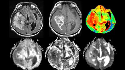
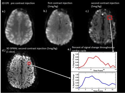
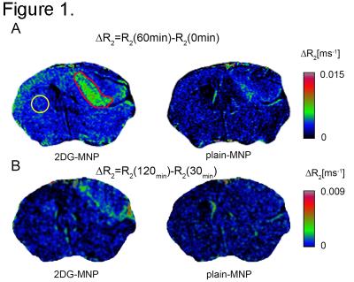
2-deoxy-D-glucose-conjugated magnetonanoparticles (2DG-MNP) uptake as a measure of metabolic activity in glioblastoma murine model 
Jelena Lazovic, Whitney Pope, Massoud Akhtari
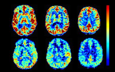
In the current study we tested if accumulation of non-radioactive 2-deoxy-D-glucose (2DG)-conjugated magnetonanoparticles (2DG-MNP) can be used as a measure of metabolic activity in glioblastoma. To quantify 2DG-MNP uptake across brain ΔR2 maps are proposed. The difference in R2 relaxation rates prior to and at different times following 2DG- or unlabeled (plain) MNP administration represents ΔR2. Significant changes in ΔR2 between brain and glioblastoma, reflecting increased metabolic rate of glioblastoma were found for glucose labeled MNP and not for plain-MNP. Our results suggest that 2DG-MNP have potential to be utilized in metabolic imaging as non-radioactive 2-18[F]-fluoro-deoxy-D-glucose analogue.
|
|
4634.
 |
2 |
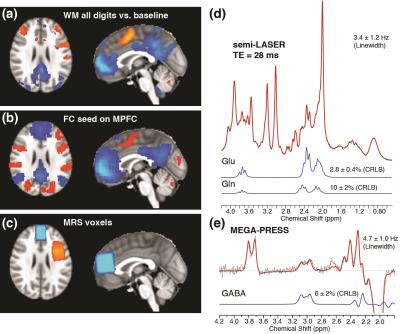
Regional GABA concentrations modulate inter-network resting-state functional connectivity
Xi Chen, Xiaoying Fan, Yuzheng Hu, Chun Zuo, Dost Ongur, Fei Du
The resting-state functional connectivity (rsFC) and task evoked fMRI, involving the default mode network (DMN) and control network (CN) was performed, with GABA, glutamate and glutamine measured at MPFC and DLPFC, in order to explore the underlying molecular mechanism of the DMN-CN interaction. We found that MPFC GABA concentrations significantly modulate DMN deactivation during a working memory task, and resting state anti-correlation between DMN and CN, while DLPFC GABA correlations modulate DMN-CN anti-correlation in the opposite direction. These findings suggest that MPFC and DLPFC GABA make a major but differential impact on task related activation and inter-network rsFC. The neurochemical characteristics of DMN and CN may provide novel insights into abnormal network activity in neuropsychiatric diseases and provide opportunities for novel interventions.
|
|
4635.
 |
3 |
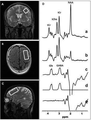
Sleep-Disordered Breathing Severity in Elderly is Associated with Decreased GABA in the Dorsolateral Prefrontal Cortex: A J-edited 1H MRS and Polysomnography Study
Ana Pereira, Xiangling Mao, Caroline Jiang, Guoxin Kang, SAra Milrad, Bruce McEwen, Ana Krieger, Dikoma Shungu
Sleep-disordered breathing (SDB) is a disorder characterized by repeated episodes of hypopnea and apnea during sleep that lead to sleep fragmentation and intermittent hypoxia. A leading model of model of SDB that posits GABAergic and glutamatergic dysregulations that lead to hyperexcitability and neuronal damage, which this study aimed to investigate using J-edited MRS and polysomnography. The main results were a robust DLPFC GABA decrease and associations between GABA and hypoxia as well as disease severity. The state of hyperexcitability observed in SDB is interpreted as likely the result of disinhibition (GABA) that might lead to excitotoxicity and neuronal damage.
|
|
4636.
 |
4 |
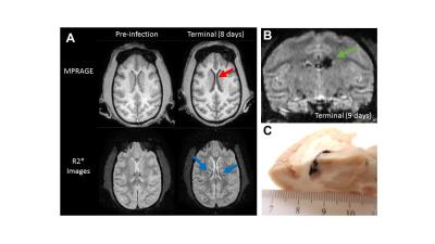
MR Imaging & Spectroscopy in a Non-Human Primate Model of Ebola Makona Aerosol Exposure - video not available
Margaret Lentz, Anna Honko, Jordan Bohannon, Matthew Lackemeyer, Jeffrey Solomon, Louis Huzella, Gene Olinger, Lisa Hensley, Peter Jahrling
The purpose of this study was to use MRI and magnetic resonance spectroscopy (MRS) to determine if structural or metabolic alterations occur in the brain of rhesus macaques exposed to Ebola virus via inhalation of aerosolized small particles. Unlike intramuscular inoculation with Ebola virus, small-particle aerosol exposure of macaques did not result in uniform changes in brain volume or vascular alterations 8-9 days after exposure. However, most animals had reductions in N-acetyl aspartate and increases in choline levels, indicating spectroscopy may be useful in identifying early alterations in brain metabolism due to Ebola virus disease.
|
|
4637.
 |
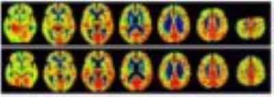
5 |
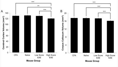
The impact of inflammation on grey matter metabolism, quantified using a novel NIRS/MRI system
Thomas Johnson, James Rogers, Jeff Dunn
Cerebral metabolic rate for oxygen (CMRO2) in gray matter (GM) is a sensitive marker for abnormalities in oxidative metabolism. We combined a near-infrared spectroscopy (NIRS) system with 9.4.T MRI to quantify regional CMRO2 in the experimental autoimmune encephalomyelitis (EAE) mouse model of multiple sclerosis. Increases in CMRO2 were seen in EAE mice and positive inflammation (CFA) controls at day 35 post-induction when compared to naïve controls. In addition, EAE and CFA mice showed increased CMRO2 from day 14 to 35. These data indicate that inflammation alone, not necessarily linked to a white matter autoimmune disease, could cause abnormal CMRO2 in GM.
|
|
4638.
 |
6 |
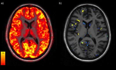
Individual mapping of neuronal damage in early relapsing-remitting MS using [11C]Flumazenil PET
Emilie Poirion, Benedetta Bodini, Marco Battaglini, Theodore Soulier, Léorah Freeman, Daniel Lorenzo-Garcia, Géraldine Bera, Michel Bottlaender, Bruno Stankoff
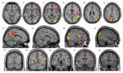
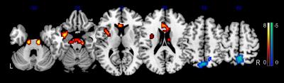
We explored the neuronal component of grey matter damage in the earliest phase of multiple sclerosis (MS) with [11C]Flumazenil positron emission tomography (PET). Using a novel post-processing approach based on the generation of individual maps of neuronal pathology, we found a significant neuronal damage in the cortical lesions of patients with MS which preceded the occurrence of cortical atrophy, and correlated with white matter lesion load. These results suggest that cortical demyelination, together with retrograde degeneration of transected axons within white matter lesions, are among the key pathogenic contributors to neuronal cortical damage at the earliest stage of the disease.
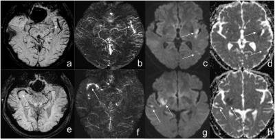
|
|
4639.
 |
7 |
Proton Spectroscopic Imaging of Secondary Progressive Multiple Sclerosis in the MS-SMART trial
Ian Marshall, Michael Thrippleton, Mark Bastin, Daisy Mollison, David Dickie, Francesca Chappell, Scott Semple, Annette Cooper, David Miller, Sue Pavitt, Gavin Giovannoni, Claudia Gandini Wheeler-Kingshott, Bhavana Solanky, Christopher Weir, Nigel Stallard, Clive Hawkins, Basil Sharrack, Moira Ross, Jeremy Chataway, Peter Connick, Siddharthan Chandran, the MS-SMART trialists
1H MR Spectroscopy yields metabolic information and has proved to be a useful addition to structural imaging in neurological diseases. We applied short-TE Spectroscopic Imaging combined with linear modelling with respect to brain tissue type in a homogeneous cohort of 42 patients with Secondary Progressive Multiple Sclerosis. Metabolite levels were significantly different in lesions compared with normal appearing tissues, suggesting axonal damage (reduced NAA) and increased glial activity (increased myo-inositol) yet relatively stable lesions (reduced Glx).
|
|
4640.
 |
8 |
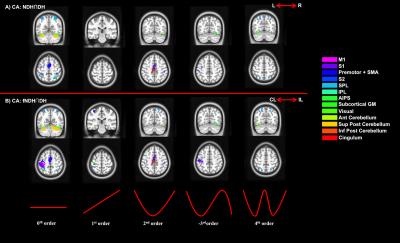
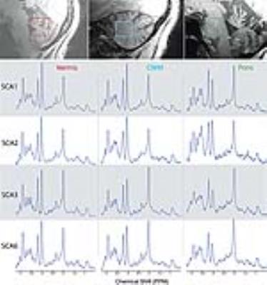
MRS-based classification of Spinocerebellar Ataxias 1, 2, 3, and 6 at 7T: A distance-weighted discrimination approach
James Joers, Dinesh Deelchand, Tianmeng Lyu, Uzay Emir, Diane Hutter, Christopher Gomez, Khalaf Bushara, Lynn Eberly, Gulin Oz
We investigated the sensitivity of ultra-high field MRS to distinguish 4 hereditary neurodegenerative diseases with substantial overlap in clinical presentation and conventional MRI. We carried out pairwise classifications of spinocerebellar ataxias (SCAs) 1,2,3 and 6 using distance weighted discrimination (DWD) on 7T MRS from 3 brain regions of 68 SCA patients. Each subject contributed to the DWD model a 50-dimensional vector of concentrations. SCA6 was classified from SCAs 1-3 with high reliability (90%) and SCA2 was classified from SCA3 with moderate success (84%), while SCA1 could not reliably be classified from SCA2 (62%) or SCA3 (48%) with the current model.
|
|
4642.
 |
10 |
The influence of mild hypercapnia on brain intracellular pH, phosphate metabolites and cerebral blood flow a multinuclear (1H/31P) MR study at 7T.
Devashish Das, Aneurin Kennerley, Samuel Harris, Jason Berwick
Permissive hypercapnia is commonly used as vasodilatory challenge in clinical applications and basic research. During fMRI experiments continuous exposure to mild (3-10%) CO2 can be applied to derive stimulus induced changes in the cerebral rate of oxygen consumption (CMRO2) by measuring cerebral blood flow and blood oxygenation dependent (BOLD) signal. Previous data from anesthetized primate during hypercapnia suggested increase in CBF are accompanied by decreases in neuronal activity. In this context, using multinuclear (31P/1H) and multi-parametric MR we show that mild exposure to hypercapnia elevates regional CBF, and can cause marginal but consistent drop in intracellular pH of rat brain, despite constant [ADP ~25-35µM] and [ATP ~3-2.4mM] to that of resting brain. Our findings support the view that unspecific drop in brain pH may likely elevate regional CBF, thereby sustain oxygen supply-to-demand ratio in rat brain.
|
|
4641.
 |
9 |
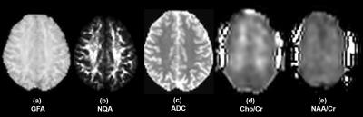
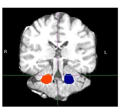
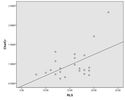
Abnormal Thalamic Metabolism in End-Stage Renal Disease Patients with Restless Legs Syndrome - video not available
Xueying Ma, Dun Ding, Peng Li, Fengli Liang, Zhuonan Wang, Yingxiang Sun, Haining Li, Ming Zhang
Restless legs syndrome (RLS) was very common among ESRD patients. However, the physiopathologic mechanisms of RLS in ESRD patients were largely unclear. Here we analyzed the thalamic metabolites using 1H MRS and the correlation between sleep/RLS score and metabolites, compared with corresponding normal controls. The results showed that the Cho/Cr ratio increased and NAA/Cr ratio decreased in thalamus of ESRD patients. The correlation between Cho/Cr ratio and RLS score was significant. It indicated that the sleep disturbance in ESRD patients with RLS might be associated with the abnormal thalamocortical activation of human sleep regulation system.
|
|
4643.
 |
11 |
The influence of mild hypercapnia on brain intracellular pH, phosphate metabolites and cerebral blood flow a multinuclear (1H/31P) MR study at 7T
Devashish Das, Aneurin Kennerley, Samuel Harris, Jason Berwick, Devashish Das
Permissive hypercapnia is commonly used as vasodilatory challenge in clinical applications and basic research. During fMRI experiments continuous exposure to mild (3-10%) CO2 can be applied to derive stimulus induced changes in the cerebral rate of oxygen consumption (CMRO2) by measuring cerebral blood flow and blood oxygenation dependent (BOLD) signal. Previous data from anesthetized primate during hypercapnia suggested increase in CBF are accompanied by decreases in neuronal activity. In this context, using multinuclear (31P/1H) and multi-parametric MR we show that mild exposure to hypercapnia elevates regional CBF, and can cause marginal but consistent drop in intracellular pH of rat brain, despite constant [ADP ~25-35µM] and [ATP ~3-2.4mM] to that of resting brain. Our findings support the view that unspecific drop in brain pH may likely elevate regional CBF, thereby sustain oxygen supply-to-demand ratio in rat brain.
|
|
4644.
 |
12 |
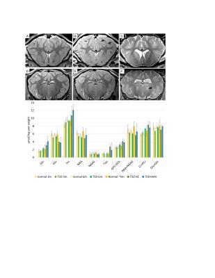
Longitudinal 7T MRI and MRS in a sheep model of Tay-Sachs disease and the effect of AAV gene therapy.
Heather Gray-Edwards, Elise Diffie, Ashley Randle, Amanda Gross, Nouha Salibi, Lauren Ellis , Ronald Beyers, Miguel Sena-Esteves, Thomas Denney, Douglas Martin
Tay-Sachs Disease (TSD) is a fatal neurodegenerative disorder of children and the sheep model of TSD is a powerful tool to study the disease and evaluate novel therapies. One such therapy, adeno associated viral (AAV) gene therapy has resulted in a 2-fold increase in lifespan and biomarkers are needed for clinical trials. 7T MRI shows white and gray matter alterations and MR spectroscopic abnormalities that worsen with TSD disease progression. At humane endpoint the AAV treated sheep has normalization of gray and white matter intensities, but cortical atrophy and MRS alterations persist. 7T MRI and MRS reflect TSD disease severity.
|
|
4645.
 |
13 |
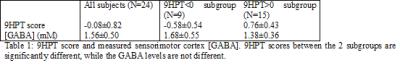
Dependence of degree of motor impairment on the association between motor performance and sensorimotor GABA level in relapsing remitting multiple sclerosis
Pallab Bhattacharyya, Micheal Phillips, Lael Stone, Mark Lowe
Gamma aminobutyric acid (GABA), a major inhibitory neurotransmitter, has been implicated as a metabolic marker of multiple sclerosis (MS). Previously it has been shown that sensorimotor cortex GABA level is higher in relapsing remitting MS (RRMS) patients with poorer motor performance. In this study, the association between cortical GABA level and motor performance, as measured by 9-hole peg test score, has been studied for groups of patients with RRMS with different degrees of motor impairment. The results suggest that cortical GABA has more involvement in motor performance in early stage of RRMS or in less impaired patients.
|
|
4646.
 |
14 |
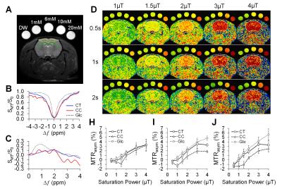
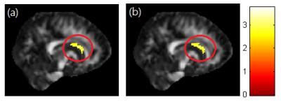
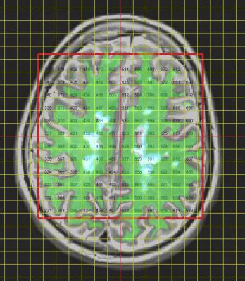
Baseline of Chemical Exchange Saturation Transfer Imaging for Brain
Yuki Kanazawa, Masafumi Harada, Mitsuharu Miyoshi, Yuki Matsumoto, Hiroaki Hayashi, Toshiaki Sasaki, Natsuki Ikemitsu
The purpose of this study is to assess the CEST effect and contrast of various brain regions in normal for phase cycle-CEST MR imaging. Subjects were five healthy volunteers. All slice positions were set on the nucleus basalis level. ROIs were set as thalamus, frontal, occipital, putamen, and gray matter on right and left brain hemispheres of each subject There was no significant difference in mean MTRasym values among each brain region (P > 0.01 for all). CEST effects for normal brain tissue had no dependency on region and/or left-right hemispheres.
|
|
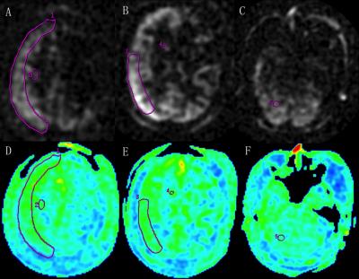
4647.
 |
15 |
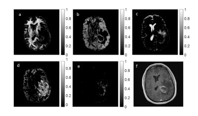
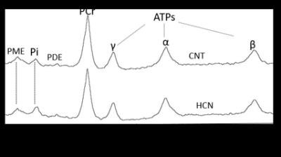
31P MR Spectroscopic Imaging of the Human Brain at 7T
Jimin Ren, Ty Shang, A. Dean Sherry, Craig Malloy
31P MRS is capable of probing cerebral metabolism in vivo, but clinical applications are limited by poor spatial resolution. This study demonstrates that 31P MRS imaging (MRSI) at 7T can offer high quality spectral data that enables cross-sectional mapping of various phosphorus-bearing metabolites in normal human brain. In a patient with prior ischemic stroke, high-energy phosphates were depleted and extracellular inorganic phosphate was increased in the stroke lesion.
|
|
4648.
 |
16 |
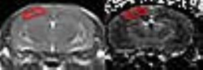
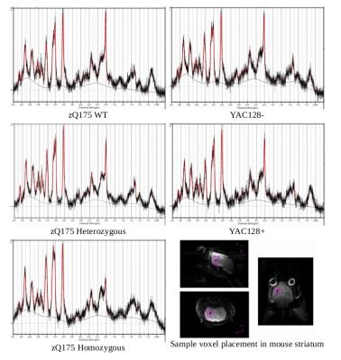
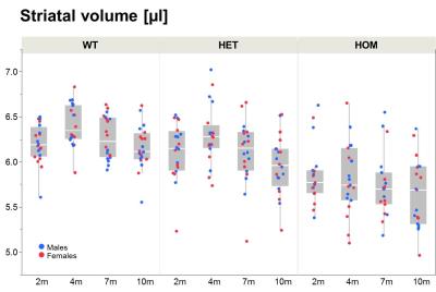
Striatal 7T 1H-MRS in two Huntington's Disease Mouse Models (zQ175 and YAC128)
Bretta Russell-Schulz, Andrew Yung, Austin Hill, Alex MacKay, Piotr Kozlowski, Blair Leavitt
Two Huntington’s disease (HD) mouse models with difference rates of disease progression, zQ175 and YAC128, were examined using 7T proton magnetic resonance spectroscopy of mice aged 6-7 months. zQ175 heterozygous and homozygous genotypes showed significantly decreased [tNAA] and increased [mI] compared to wild type. No differences in metabolite concentrations were found between YAC128- (wild type) and YAC128+ mice. A slow HD disease course in YAC128+ genotype may be the reason for lack of measured metabolite concentration differences.
|
|
4649.
 |
17 |
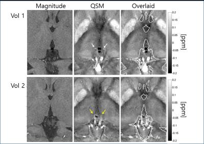
QSM meets MRS: The influence of subcortical iron on glutamatergic neurotransmission in a movement disorder population
Ahmad Seif Kanaan, Alfred Anwander, Andreas Schäfer, Berkin Bilgic, Torsten Schlumm, Jamie Near, Kirsten Müller-Vahl, Harald Möller
We use a combination of Quantitative Susceptibility Mapping and 1H-MRS to examine the role of iron and its association with glutamatergic signalling in Gilles de la Tourette syndrome (GTS). GTS is a neurodevelopmental movement disorder with abnormalities in the neurotransmission of dopamine and GABA and, as shown more recently, also in subcortical glutamate (Glu) and glutamine (Gln). In this work, we observed that GTS patients exhibit reductions in cerebral iron levels and report a general association between iron and the Gln:Glu ratio. This work provides a good example of utilizing multi-modal neuroimaging methods to interrogate pathophyiology at multiple scales.
|
|
4650.
 |
18 |
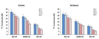
Effects of astrocytic Nrf2 activation on recently-identified 13C and 1H MRS flux-based biomarkers of mitochondrial energetics and neurotransmitter cycling in Huntington's Disease
Golam Chowdhury, Peter Dixon, Robin de Graaf, Xiaoxian Ma, Johnson Johnson, Jeffrey Johnson, Larry Park, Gerard Sanacora, Douglas Rothman, Kevin Behar
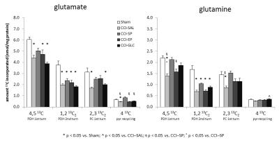
Alterations in brain glucose and energy metabolism is observed in Huntington’s Disease (HD) and HD animal models. 1H-[13C]-MRS can be readily adapted to measure metabolic pathway flux by use of 13C-labeled substrates. In this study we assessed whether activation of the astroglial Nrf2-ARE pathway in the R6/2 mouse model of HD, which has shown therapeutic potential in HD animal models, can reverse the reduction in 13C labeling seen previously in R6/2 mice. In cortex and striatum, astroglial Nrf2 activation led to increased amino acid 13C labeling, suggesting a degree of improvement in mitochondrial and neurotransmitter fluxes in the R6/2 mice.
|
|
4651.
 |
19 |
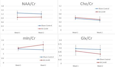
Longitudinal Changes of Metabolites Measured by Proton Magnetic Resonance Spectroscopy and Their Correlations with Behavioral Outcomes in a Rat Model of Kainic Acid Induced Spinal Cord Injury
Mingming Zhu, Alice Shum-Siu, Emily Martin, Abby Wade, Darlene Burke, David Magnuson, Chin Ng
We measured the profile of major metabolites longitudinally using MRS in spinal cord gray matter of kainic acid (KA) injured rats at both 7 and 14 days post KA administration and correlated their concentrations with functional outcomes assessed by the Basso, Bresnahan, and Beattie (BBB) Open Field Locomotor scores. Our preliminary findings indicate that metabolite concentrations in the lower lumbar spinal cord gray matter, caudal to the injury epicenter, as detected by in vivo 1H-MRS, could be used as injury and recovery biomarkers in SCI animal models.
|
|
4652.
 |
20 |
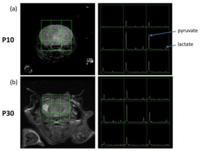
A metabolic study of hypoxic ischemia during mouse brain development using hyperpolarized 13C
Yiran Chen, Byongsop Lee, Robert Bok, Ilwoo Park, Subramaniam Sukumar, R Sheldon, A Barkovich, Donna Ferriero, Duan Xu
In this study, we applied dynamic nuclear polarization (DNP) technique to investigate C1 labeled 13C pyruvate to lactate conversion on hypoxic ischemia (HI) injured neonatal mouse brains during development. Our results showed that lower pyruvate level and higher lactate to pyruvate ratio on the injured hemisphere in comparison to the non-injured hemisphere at the day of injury (P10). This difference narrows as the brain matures. With this technique, we are able to examine individuals’ response to HI in vivo during brain development.
|
|
4653.
 |
21 |
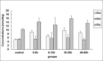
Correlation between dynamic changes of glutamate metabolism and microcirculatory perfusion in basal ganglia after hypoxic–ischemic brain damage - video not available
Yuxue Dang, Xiaoming Wang, Kaining Shi
The excitotoxicity of glutamate metabolism as well as hemodynamic disorders of the brain are both risk factors for neonatal hypoxic–ischemic brain damage (HIBD). To investigate the combined application of intravoxel incoherent motion (IVIM) and 1H-magnetic resonance spectroscopy (MRS) in exploring the possible mechanisms. This study was undertaken to examine basal ganglia metabolites (by means of 1H MRS) and microcirculation (by means of IVIM) in a piglet model of hypoxic-ischemia (HI). It is concluded that elevation of glutamate occurs in parallel to perfusion disruption reflected by changes in perfusion fraction f after HI.
|
|
4654.
 |
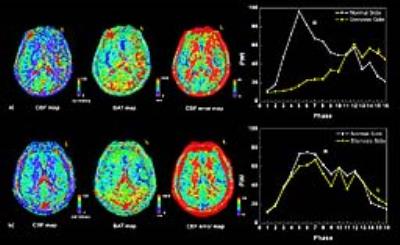
22 |
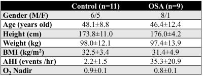
Implications of Obstructive Sleep Apnea on Global Cerebral Metabolic Rate of Oxygen
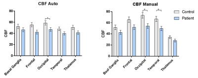
Ana Rodríguez-Soto, Yulin Chang, Wen Cao, Zachary Rodgers, John Detre, Erin Englund, Sarah Leinwand, Richard Schwab, Michael Langham, Felix Wehrli
Obstructive sleep apnea (OSA) is a chronic disorder caused by intermittent obstruction of the upper airways during sleep. Neurocognitive deficits in this population have previously been associated with altered brain metabolism. In fact, a recent pilot study suggests that global cerebral metabolic rate of oxygen (CMRO2) may be a potential marker of oxygen metabolic dysfunction in this population. Here, we present preliminary results from an ongoing study designed to quantify CMRO2 in subjects with OSA and matched controls. Initial results suggest that apneics have overall lower baseline CMRO2 and increased response to volitional apnea, a paradigm to mimic spontaneous apneas.
|
|
4655.
 |
23 |
Mutant huntingtin dosage effect on incidence of high striatal Glutamine in a mouse model of Huntington’s disease – a symptom of liver pathophysiology?
Thomas Mueggler, Andreas Bruns, Laurence Ozmen, Herve Schaffhauser, Basil Künnecke, Markus von Kienlin
In contrast to transgenic models of Huntington’s disease (HD) newly available knock-in models, such as the HttQ175, recapitulate the heterozygosity present in the clinically observed pathophysiology. Here we report a longitudinal assessment of striatal metabolites using 1H-MRS and volumetry in 30+ brain regions in both male and female HttQ175 mice. Apart from a genotype-specific, progressive neurodegenerative phenotype present in both genders, we discovered an entirely novel feature in mouse models of HD, namely individuals with abnormally high glutamine (Gln) levels, whose incidence increased with mutant Htt gene dosage. In line with emerging data on a widespread peripheral metabolic perturbation in HD, we hypothesize that this excessive Gln is a consequence of disordered hepatic metabolism.
|
|
4656.
 |
24 |
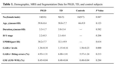
Inhibitory Motor Dysfunction in Parkinson’s Disease Subtypes - permission withheld
Tao Gong, Guangbin Wang, Richard Edden, Fei Gao, Yuanyuan Xiang, Weibo Chen
This study was aimed to evaluate the differences between PD motor subtypes of GABA+ levels using MEGA-PRESS. PD patients were classified into PIGD and TD groups; sixteen healthy controls were recruited. All subjects underwent 3T MR examination including MEGA-PRESS. We found that GABA+ concentration was lower in PD compared with controls; furthermore, the TD group was lower than PIGD. In PD patients, the GABA+ levels were correlated with UPDRS scores. The results suggest that GABAergic dysfunction may play an important role in the pathogenesis of Parkinson’s disease. MEGA-PRESS provides a valuable examination method to discriminate between PD motor subtypes.
|
|