ISMRM 25th Annual Meeting & Exhibition • 22-27 April 2017 • Honolulu, HI, USA

Electronic Poster Session: Body: Breast, Chest, Abdomen, Pelvis
4776 -4799 Prostate Cancer
4800 -4823 Female Pelvis, Fetal & Placenta
4824 -4847 Body: Emerging Techniques
4848 -4871 Body: Animal Studies
4894 -4917 Thoracic MRI
4918 -4941 Breast Imaging
4942 -4965 Body: Cancer
4966 -4989 Gastrointestinal MRI
 
Prostate Cancer
Electronic Poster
Body: Breast, Chest, Abdomen, Pelvis

 
Wednesday, 26 April 2017
Exhibition Hall  13:45 - 14:45


    Computer #

 
4776.   
25 FOCUS diffusion-weighted imaging for prostate cancer at high b-values: An analysis of image quality, diagnostic accuracy and observer agreement.
Tom Syer, Keith Godley, Donnie Cameron, Paul Malcolm
To assess FOCUS diffusion-weighted imaging (DWI) for prostate cancer assessment, 30 consecutive biopsy-proven patients underwent both FOCUS and conventional DWI. Sensitivity and specificity was not significantly different between sequences but inter-observer agreement improved from moderate to substantial when using FOCUS. There was significantly lower SNR and CNR for FOCUS on b-value images, but similar CNR on ADC maps. Mean ADC values were significantly lower using FOCUS and both sequences showed excellent discrimination between malignant and benign prostate with no statistical difference. FOCUS DWI improves agreement between observers of varying experience while maintaining diagnostic accuracy despite lower SNR and CNR. 


4777.   
26 Target performance of MRI-ultrasound fusion guided prostate biopsy in a cohort of patients suspicious for prostate cancer
Matthias Zadory, Jean-Luc Fehr, Claudius Moeckel, Seife Hailemariam, Johannes Froehlich, Michael Patak
Conventional systematic core biopsies might fail to detect clinical significant prostate cancer. ARTEMIS MRI-ultrasound fusion guided prostate biopsy (ART-PBx) might overcome this issue by improving the targeting of suspicious lesions. In a retrospective clinical study including 194 patients (243 lesions) we determined a target performance of 56.3 % positive biopsies related to histopathology. The detection rate rises up to 71.4% for high score lesions (PIRADS 5) but did not show any correlation with lesion’s size. This target methodology based on MRI achieves greater detection rate of clinical significant prostate cancer, improving the ability to appropriately counsel patients regarding therapy.


4778.   
27 Role of DWI in guiding MRI-TRUS fusion biopsy in patients with prostate cancer: a prospective cohort study
Chandan Das, Rohit Kaushal, Sanjay Sharma, Rajeev Kumar, P Dogra, S DattaGupta
Transrectal ultrasound (TRUS) guided 12 core biopsy of prostate has a sensitivity of 39-52%. We prospectively evaluated the role of DWI in guiding MRI-TRUS fusion biopsy. MRI was performed on a 3 Tesla system.  PIRAD score was assigned and PIRAD 3-5 score were subjected to targeted fusion biopsy using the Artemis device along with standard 12 core biopsies. Targeted biopsy detected a higher number (93%) of clinically significant cancers and 71% cancers were upgraded to significant cancer on targeted biopsy. Fusion biopsies guided by DWI thus provide incremental information over standard TRUS biopsies in the diagnosis of significant prostate cancer.


4779.   
28 MR-based machine learning analysis can help to predict pathological outcome of biopsy-proven Gleason score 3+3 prostate cancer - video not available
Chen-Jiang Wu, Yu-Dong Zhang, Hai-Bin Shi
Machine learning-based analysis of multi-parametric was used to predict pathological outcome of biopsy-proven Gleason score 3+3 prostate cancer and proven to be better compared with single any MR or clinical paremeter  in predicting pathological outcome.


4781.   
30 DWI of prostate cancer beyond ADC: correlation with histopathology
Stefanie Hectors, Sahar Semaan, Christopher Song, Ashutosh Tewari, George Haines, Bachir Taouli
In this study, we correlated advanced DWI [diffusion kurtosis imaging (DKI), stretched-exponential (SE) DWI and diffusion tensor imaging DTI)] parameters with fractional tissue fractions of nuclei, cytoplasm, cells, stroma and lumen and the nuclear-cytoplasmic ratio in prostate cancer (PCa) lesions. We found that all assessed diffusion methods showed significant correlations with cytoplasmic, cellular and/or stromal tissue fractions. Specifically, DKI seems promising for characterization of PCa tissue composition, since multiple significant correlations between histology parameters and both ADCDKI and kurtosis parameter K were observed.


4782.   
31 Mixed performance of PIRADS v2: a validation study - permission withheld
James Walton, Sajal Pokharel, E. Crawford, Kavita Garg, Kimberly Lind, Emma Murugaverl, Nayana Patel

The second version of the Prostate Imaging Reporting and Data System (PIRADSv2) is the new standard for interpreting prostate MRI.  Validation studies have been performed of this scheme, but many have used targeted biopsies as the reference standard.  Our validation study is unique in utilizing 3D trans-perineal mapping biopsy (3DTMB) as the reference standard.  With a total of 41 MRI lesions, PI-RADSv2 score = 5 lesions had a PPV for cancer of 1.0.  However, when all significant lesions (PI-RADSv2 score ≥3) were included, the PPV is 0.29, with equal PPV of PI-RADS 3 and 4 lesions.


4780.   
29 Diffusion-Kurtosis imaging predicts tumor upgrading in biopsy-proven Gleason score = 6 prostate cancers - video not available
Chen-Jiang Wu, Yu-Dong Zhang, Hai-Bin Shi
The study investigated the feasibility of Diffusion Kurtosis Imaging (DKI) in predicting surgical result upgrading of biopsy-proven Gleason Score (GS) = 6 prostate cancer.  The predicting efficiency of clinical variables (tumor volume, PSA level) and MRI variables (ADC, Dapp, Kapp) were compared by ROC analysis. DKI was found feasible to predict surgical result upgrading of biopsy-proven GS = 6 prostate cancer and Dapp-minimun had the highest Az value.


4783.   
32 Single Slice vs. Volumetric Analysis of Multiparametric Prostate MRI Metrics
Edward Johnston, Clare Allen, Michela Antonelli, Nikolaos Dikaios, Sebastien Ourselin, Shonit Punwani
Despite consensus guidelines advising volumetric analysis of tumours over single slice region-of-interest analysis, there is little data in the literature to support this recommendation. In this study we compare the reproducibility between each of these two methods in 20 patients with prostate cancer and also determine the intraobserver repeatability of each method. We show high levels of agreement and intraobserver repeatability in both of these methods. This study suggests that region-of-interest analysis is a perfectly acceptable analytical method and may have higher intraoberver repeatability than volumetric analysis, and could expedite the analysis of multiparametric prostate MRI datasets in clinical trials. 


4784.   
33 Improving the Reproducibility of Quantitative Imaging Metrics for Multicentre Multiparametric Prostate MRI Trials
Edward Johnston, Michela Antonelli, Nikolaos Dikaios, Sebastien Ourselin, David Atkinson, Shonit Punwani
Whilst multi-scanner studies provide the most robust evidence for quantitative imaging trials, they tend to be limited by poor reproducibility of scans performed on different scanners. In this study, 14 patients underwent paired multiparametric prostate MRI within 3 months of each other. We found that normalisation of T2 signal to the bladder improved the reproducibility of both peripheral zone and transition zone metrics considerably when compared with the current convention of using obturator internus. Whilst ADC also met sufficient levels of reproducibility, semiquantiative and quantitative DCE analysis and histographic features failed to do so.


4786.   
35 Preliminary application of magnetization transfer imaging and amide proton transfer imaging of prostate cancer at 3.0 tesla - video not available
Xiangde Min, Zhaoyan Feng, Liang Wang, Zhongping Zhang
Magnetization transfer (MT) imaging and amide proton transfer (APT) imaging have reported many promising results. However, little is known about their usefulness for prostate cancer (PCa). In this study, MT-APT imaging were performed for 39 patients with pathological proven PCa. The feasibility of MT imaging and APT imaging for PCa detection was assessed, and their differential diagnostic values for PCa were compared. The results revealed that MTR(16.5 ppm) increased in cancerous tissues compared with normal PZs, and MT imaging outperformed APT imaging for PCa diagnosis. MT imaging showed promising role in the diagnosis of PCa.


4787.   
36 Evaluation of prostate cancer on diffusion weighted imaging; Can FOCUS and synthetic diffusion weighted imaging with FOCUS contribute to the Prostate Imaging Reporting and Data System (PI-RADS) version 2.0?
Motoyuki Katayama, Takayuki Masui, Kei Tsukamoto, Mitsuteru Tsuchiya, Masako Sasaki, Yuki Hayashi, Takahiro Yamada, Mitsuharu Miyoshi, Tetsuya Wakayama, Harumi Sakahara
We evaluated 47 patients suspected of having prostate cancers on synthetic DWI calculated from FOCUS DWI with PI-RADS version 2.0. Compared with conventional FOV DWI, FOCUS DWI is more useful for evaluation of prostate cancer with high spatial resolution and less distortion. S-DWI is able to enhance diagnostic ability of FOCUS without image degradation, which might be one of the best combinations, and contribute to the Prostate Imaging Reporting and Data System (PI-RADS) version 2.0. 


4788.   
37 Prostate Cancer Diffusion Signal Analysis: Combination of Multiple Fit Parameters Improves Tissue Discrimination
Stephan Maier, Thiele Kobus, Andriy Fedorov, Fredrik Langkilde, Ruth Dunne, Robert Mulkern, Clare Tempany
Diffusion signals over an extended b-factor range 0-3500 s/mm2 were measured with an endorectal coil at 3 Tesla in 56 prostate cancer patients. For each pixel, signal decay fits were computed assuming biexponential, kurtosis, stretched exponential and gamma distribution diffusion signal models. The potential of individual parameters and linear parameter combinations to differentiate normal from cancerous tissue was evaluated with ROC analysis. For the kurtosis and stretched exponential models, single parameters yield the highest AUCs, whereas for the biexponential and gamma distribution models, only combinations of parameters produce the comparably high AUCs.


4789.   
38 3T DCE-MRI Performance in Prostate Cancer Detection: Correlation of Different Kinetic Parameters in the Transition and Peripheral Zone Stratified by Gleason Score and Scanner B1+ field Heterogeneity
Nazanin Asvadi, Kyung Sung, Pooria Khoshnoodi, Pornphan Wibulpolprasert, Tristan Grogan, Anthony Sisk, Robert Reiter, Steven Raman
To correlate 3T dynamic contrast-enhanced MRI (DCE-MRI) kinetic parameters in prostate cancer (PCa) in the transition zone (TZ) and peripheral zone (PZ) confirmed by whole mount histopathology (WMHP) stratified by Gleason Scores (GS) and inter-scanner B1+ field variation. This study demonstrates that clinically relevant heterogeneity between kinetic parameters in low and high grade PCa in the TZ & PZ exists. 


4790.   
39 Using Natural Language Processing to explore the correlation of prostate MR findings and prostate biopsy
Yi Liu, Shuai Ma, Rui Wang, Ge Gao, En Ouyang, Zuofeng Li, Juan Wei, Xiaoying Wang
Natural language processing (NLP) provides techniques that aid the conversion of text into a structured representation, which is potentially a valuable source of information for improving clinical care and supporting research in medical domain.1,2 Used on radiology reports and biopsy results, NLP techniques enable automatic identification and extraction of information. For most patients suffer from prostate diseases, they always conduct tests including radiology and biopsy. And it is an interesting task to explore the prostate MR findings and prostate biopsy, which have not been studied in other research center.


4791.   
40 Ex vivo MRI evaluation of prostate cancer: localization and margin status prediction of prostate cancer in fresh radical prostatectomy specimens
Jan Heidkamp, Martijn Hoogenboom, Iringo Kovacs, Andor Veltien, Arie Maat, Michiel Sedelaar, Christina Hulsbergen-van de Kaa, Jurgen Fütterer
This study investigated the ability of high field ex vivo MRI to localize prostate cancer (PCa) and to predict the margin status in fresh radical prostatectomy (RP) specimens using histology as gold standard. In twelve specimens, ex vivo MRI localized 17 (47%) of 36 PCa lesions confirmed by histological examination. Ex vivo MRI identified none of the 4 histological positive surgical margins (sensitivity 0%) and 9 of the 13 negative margins (specificity 69%). Our results indicate accurate localization of PCa in fresh RP specimens by ex vivo MRI, yet the technique did not perform well in predicting the margin status.


4792.   
41 Clinical Evaluation of a Simple Approach for Improving Shear Wave Illumination in Magnetic Resonance Elastography of the Prostate
Jin Wang, Tianhui Zhang, Kevin Glaser, Jingbiao Chen, Jun Chen, Phillip Rossman, Bingjun He, Arvin Arani, Ziying Yin, Zhuang Kang, Qungang Shan, Jun Pang, Richard Ehman
Conventional prostate magnetic resonance elastography (MRE) is performed using an external driver to transmit shear waves into the prostate. It is a challenge to produce shear waves in the prostate with adequate amplitude at a suitably high frequency because the prostate is a deep-seated organ. We evaluated the hypothesis that the placement of a urinary catheter would improve the shear wave illumination of the prostate when waves are emitted from an external driver. Our results in 30 BPH patients show that the commercially available liver MRE driver used in combination with a catheter can improve MRE image quality at higher frequencies.


4793.   
42 Linking a multi-compartment T2 model to diffusion microstructure in prostate cancer
William Devine, Edward Johnston, Elisenda Bonet-Carne, Shonit Punwani, Daniel Alexander, David Atkinson
This work develops a multi-compartment T2 model for prostate imaging.  We investigate whether this model can provide information about differences in tissue microstructure, such as those between normal prostate tissue and tumour, by comparing it to the VERDICT diffusion model6. The high correlations found between a number of the parameters suggest that the proposed model is capable of detecting some microstructural differences. In the future this method may be able to provide different and complementary microstructural information to current diffusion models.


4794.   
43 Magnetic Resonance Elastography of the Prostate: Impact of Driver Size on Image Quality
Tianhui Zhang, Jin Wang, Kevin Glaser, Phillip Rossman, Jun Chen, Bingjun He, Jun Pang, Ziying Yin, Zhuang Kang, Qungang Shan, Jingbiao Chen, Arvin Arani, Richard Ehman
Conventional prostate MRE uses an external driver located on the surface of the body to transmit shear waves into the prostate. However, the conventional large-diameter driver produces poorer image quality at higher frequencies. In this study we compared the performance of a large- and small-diameter Pelvic Wall driver at multiple frequencies using 2D and 3D MRE. Our results show that the smaller Pelvic Wall driver performs better than the larger driver in terms of image quality and success rate.


4795.   
44 TRUS Biopsy Has a Lower Cancer Detection Rate in Large Prostate Volumes: Can In-Bore MRI-Guided Biopsy Do Better?
Kareem Elfatairy, Christopher Filson, Adeboye Osunkoya, Rachel Geller, Sherif Nour
TRUS biopsy known to have low cancer detection rates in patients with large prostate volumes. With the proved advantages of MRI guided biopsy (MRGB), it may offer a better alternative to those patients. We compared between cancer detection rates of TRUS biopsy and MRGB in 49 patients as related to their prostate volumes. MRGB showed better detection of clinically significant cancers in prostate volumes between 30-59 mL . Adopting MRGB in patients with gland volumes ≥ 30 mL may save patients the need for repeated TRUS biopsies, provide better disease risk stratification, and reduce the healthcare costs associated with unnecessary biopsies.


4796.   
45 Quantitatively Differentiating Prostate Cancer, Prostatitis and Benign Prostatic Hyperplasia (BPH) by Diffusion MRI Histology (D-Histo)
Ze-Zhong Ye, Qingsong Yang , Peng Sun , Yasheng Zhu, Chunyu Song, Joshua Lin, Jianping Lu, Yinghao Sun, Sheng-Kwei Song
A recent consensus established mpMRI for PCa detection has not had the needed sensitivity or specificity to distinguish prostatitis from PCa. Thus, PCa diagnosis by MRI remains uncertain, resulting in over-diagnosis and over-treatment.  For the first time, we demonstrate that the new method, diffusion MRI histology (D-Histo), accurately localizes and quantifies PCa, prostatitis, and BPH. With improved diagnosis accuracy, D-Histo affords effective guidance of treatment planning, and assessment of treatment efficacy. 


4797.   
46 Modified Dispersion Model in Prostate Dynamic Contrast-Enhanced MRI
Kyunghyun Sung, Xinran Zhong, Holden Wu
The dispersion imaging has shown great promise in prostate DCE-MRI, but there still exist practical limitations due to the complex model fitting. We describe a modified dispersion model to overcome these limitations by adapting a simple dispersion factor into the existing population-averaged arterial input function. We use the regions of interest, derived from the histological analysis, to evaluate both the quality of the model fitting and the ability of DCE-MRI parameters to delineate between cancerous and normal prostate tissues using 25 prostate patient cases available with the whole-mount histopathology. 


4798.   
47 A Transverse External RF Surface Coil for Prostate MR Imaging at 3T
Keith Hulsey, Alexander Ivanishev, Ivan Dimitrov, Robert Lenkinski
MRI is playing an increasingly important role in the work up of prostate cancer. This increased demand for prostate imaging has caused a debate in the field regarding the necessity of an endo-rectal coil for these studies. While the endo-rectal coil has distinct advantages it also has some drawbacks in both work-flow and cost. Current external coils do not provide images at comparable SNR to endo-rectal coil images (at the same field strength). Here we describe our initial experience with a transverse external RF coil designed to be placed on the perineum for high quality images.


4799.   
48 3T Restriction Spectrum Imaging Association with Prostate Cancer Gleason Score, PI-RADS v2 Score and Tumor Diameter on Whole Mount 3D-Mold-Sectioned Histopathology
Pooria Khoshnoodi, Sepideh Shakeri, Ashkan Shademan, Naznin Asvadi, Leila Mostafavi, Nathan White, David Karow, Daniel Margolis, Anthony Sisk, Robert Reiter, Steven Raman
Multiparametric MRI is becoming a crucial imaging for prostate cancers. A novel advanced, diffusion-based technique, restriction spectrum imaging (RSI) has been applied for prostate cancer imaging recently. In this work we will investigate the RSI performance in prostate cancer evaluation verified by post-surgery whole mount histopathology slides. 


4785.   
34 Lower Normalised T2 Signal Intensity is Associated with Higher Intratumoural Heterogeneity: A Radiogenomic Study in High-Risk Prostate Cancer
Edward Johnston, Mark Linch, Gerald Goh, Crispin Hiley, Yaalini Shanmugabavan, Michela Antonelli, Marco Gerlinger, Andrew Rowan, Yien Ning Wong, Helen King , Andrew Furness, Alexander Freeman, Linares Linares, Ayse Akarca, Javier Herrero, Stephan Dentro, Nathalie Harder, Guenter Schmidt, Gareth Gareth, Nicholas McGranahan, Nicolai Birkbak, Richard Mitter, Paul Cathcart, Rebecca Scott, Michelle Hung, Mark Emberton, Gert Attard, Zoltan Szallasi, Sergio Quezada, Teresa Marafioti, Sebastien Ourselin, Hashim Ahmed, Charles Swanton, Shonit Punwani
Intratumoural heterogeneity (ITH) has been shown to predict overall survival in prostate cancer, and non-invasive biomarkers that can measure this genetic diversity would be welcome. In this study, we correlated imaging metrics derived from multiparametric prostate MRI with genomic heterogeneity indices and showed that low values of normalised T2 signal intensity within tumours are associated with a higher degree of mutational ITH. Our study shows the potential to use diagnostic imaging as a surrogate for genomic ITH in order to risk stratify patient for guiding management decisions.
 
Female Pelvis, Fetal & Placenta
Electronic Poster
Body: Breast, Chest, Abdomen, Pelvis

 
Wednesday, 26 April 2017
Exhibition Hall  13:45 - 14:45


    Computer #

 
4800.   
49 DECIDE: Diffusion-rElaxation Combined Imaging for Detailed Placental Evaluation
Andrew Melbourne, Rosalind Pratt, David Owen, Magdalena Sokolska, Alan Bainbridge, David Atkinson, Giles Kendall, Jan Deprest, Tom Vercauteren, Anna David, Sebastien Ourselin
We propose a new multi-compartment model for the tissue signal in MRI and apply this to images of liver and placenta. Motivated by different flow characteristics in these organs, a three compartment model comprising fast and slowly circulating fluid pools and a tissue pool is fitted to overlapping multi-echo T2 relaxometry and an intra-voxel incoherent motion diffusion acquisition with low b-values. We compare and contrast parametric maps for regions of interest in liver and placenta.


4801.   
50 Perfusion MRI of the Placenta: Preliminary Results using ASL FAIR and Ferumoxytol DCE MRI in the Rhesus Macaque
Kai Ludwig, Sean Fain, Sydney Nguyen, Thaddeus Golos, Scott Reeder, Ian Bird, Oliver Wieben, Dinesh Shah, Kevin Johnson
Non-contrast enhanced methods are needed to quantify placental perfusion to detect and monitor pathophysiological changes during pregnancy. We evaluate the potential of an endogenous perfusion labeling perfusion technique, Arterial Spin Labeled Flow-sensitive Alternating Inversion Recovery (ASL FAIR), and compare with ferumoxytol dynamic contrast enhanced (DCE) MRI. Three pregnant rhesus macaque were imaged with both FAIR and ferumoxytol DCE. Localized regions of ASL perfusion were observed that coincided with regions of early contrast enhancement seen in DCE. Ferumoxytol DCE measured perfusion with extended transit times across the placenta beyond those typically used for ASL labeling.


4802.   
51 Evaluation of Placenta Motion throughout Gestation
Thomas Martin, Dapeng Liu, Teresa Chanlaw, Sherin Devaskar, Carla Janzen, Tess Armstrong, Yutaka Natsuaki, Daniel Margolis, Rinat Masamed, Holden Wu, Kyunghyun Sung
Proper placental function is essential for normal fetal development.  MRI can easily assess the growth and development of the placenta throughout gestation due to its capabilities in functional imaging.  However, uterine contractions, and fetal and maternal motion can lead to inaccuracies in the quantitative assessments. In this we assessed the impact of extraneous motion on placental imaging by using a 3D multi-echo golden angle radial sequence to generated dynamic images of the placenta.


4803.   
52 Differentiating metastatic from nonmetastatic lymph nodes by using monoexponential, biexponential, and stretched exponential diffusion-weighted imaging in cervical cancer patients
Qingxia Wu, Dandan Zheng, Ligang Shi, Mingbo Liu, Meiyun Wang, Dapeng Shi
The aim of our study was to judge whether diffusion parameters derived from different models of multi-b value DWI could be used to discriminate metastatic from nonmetastatic pelvic lymph node (LN) status in patients with cervical cancer. A statistical significant difference in the mean D, f and α values between metastatic and nonmetastatic LNs, with metastatic LNs presenting with higher D values, lower f values and higher α values than nonmetastatic ones. This study revealed that metastatic LNs had their respective diffusion parameters compared with nonmetastatic LNs.


4818.   
67 Radial segmented echo-planar readout for fast fetal angiography – feasibility test
Brijesh Yadav, Uday Krishnamurthy, Pavan Jella, Edgar Hernandez-Andrade, Swati Mody, Feifei Qu, Anabela Trifan, Sonia Hassan, Roberto Romero, Ewart Haacke, Jaladhar Neelavalli
A simulation study showing the potential benefit of a radial-trajectory based data acquisition technique named as "radial segmented echo-planar readout (radialSEPI)" for faster fetal angiography is presented. The results indicate that at echo train length of 2 and echo-spacing of 6.1 ms can provide good quality fetal/adult MRA reconstructions providing a potential factor 2 improvement in time. 


4804.   
53 Comparison of 18F-FDG PET/MRI and MRI alone for pretherapeutic tumor staging of patients with primary cancer of the uterine cervix. - video not available
Johannes Grueneisen, Lino Sawicki, Axel Wetter, Julian Kirchner, Michael Forsting, Verena Ruhlmann, Lale Umutlu
This study demonstrates a successful attempt to utilize and evaluate the diagnostic potential of integrated PET/MRI for staging patients with primary cervical cancer. According to the results, 18F-FDG PET data do not seem to provide useful additional information to MRI for the determination of the local extent of the primary tumors. However, the present results show a better performance of simultaneously acquired 18F-FDG PET and MR datasets for the detection of nodal and distant metastases if compared to MRI alone. Therefore, integrated PET/MR imaging may provide valuable information for treatment planning and to predict prognosis.


4805.   
54 Full 3D high-resolution BOLD imaging of the human placenta with prospective navigation and 2D spatially selective excitation
Glen Morrell, Matthias Schabel, Robert Silver, Christopher Kroenke, Antonio Frias
Full 3D high-resolution human placenta BOLD imaging at 3T was performed with a free-breathing prospectively navigated sequence using 2D spatially selective excitation.  Advantages of this sequence over conventional multi-slice breath hold BOLD include increased SNR, no breath hold recovery periods leading to better time efficiency, and elimination of artifacts from respiratory and fetal motion.  T2* maps clearly show the cotyledon architecture of the human placenta.


4806.   
55 Non-invasive placental perfusion imaging in pregnancies complicated by fetal heart disease using velocity-selective arterial spin labeling
Zungho Zun, Greg Zaharchuk, Nickie Niforatos-Andescavage, Samantha Bauer, Mary Donofrio, Catherine Limperopoulos
Recent data have reported that placental dysfunction may be present in the setting of complex fetal congenital heart disease (CHD). We performed placental perfusion imaging using velocity-selective arterial spin labeling in pregnancies complicated by fetal CHD and healthy pregnancies. We demonstrated that global placental perfusion and regional variation of perfusion were significantly correlated with GA in pregnancies complicated by fetal CHD, but not in healthy controls. Our findings suggest that placental ASL may have the potential to serve as an early biomarker of placental dysfunction in fetal CHD.


4807.   
56 Measuring human placental blood flow with multi-delay 3D GRASE pseudo-continuous arterial spin labeling at 3 Tesla
Xingfeng Shao, Dapeng Liu, Thomas Martin, Teresa Chanlaw, Sherin U. Devaskar, Carla Janzen, Daniel Margolis, Kyunghyun Sung, Danny JJ Wang
We presented a multi-delay pCASL combined with inner-volume GRASE imaging technique to measure placental blood flow (PBF) and arterial transit time (ATT) simultaneously, and report PBF and ATT evolution along different gestational ages during the second trimester. More blood flew through placenta with a slightly shorter transit time was seen with fetal development. Overall, PBF and ATT for the second trimester were 129.8±44.7 ml/min/100g and 786.8±174.1 ms respectively. 


4808.   
57 Pelvic Floor Structural Alterations of Primipara with Stress Urinary Incontinence After Vaginal Delivery:A MRI Study
yujiao zhao, zhizheng zhuo, wen shen
Vaginal childbirth women have an increasing incidence of stress urinary incontinence(SUI). There are few studies on pathogenesis of SUI and the relationship between the SUI and pelvic floor structure changes. In this study, static and dynamic MRI imaging are performed to describe and assess pelvic floor structure changes in patients with stress urinary incontinence. The results showed that pelvic floor structures changed significantly in the primipara suffering from SUI after vaginal delivery, which suggest a series of pathological state.


4809.   
58 Characterization of the placental vascular tree using MRI: an ex-vivo study
Daphna Link, Ariel Many, Liat Ben Sira, Shaul Harel, Dafna Ben Bashat
Placental vascular dysfunction is a major cause of pregnancy complications, such as intrauterine growth restriction (IUGR). However, current knowledge on human placental vascular architecture is limited. In this study, for the first time, we characterized the structure of the placental vascular system ex-vivo, using MRI. Fifteen normal placentas and one with IUGR were studied using a novel method, and the vascular structure was analyzed with an automatic algorithm. Results provided information regarding: cord insertion location; branching pattern; branching generation; and daughter to mother diameters of normal placentas. Preliminary results from one IUGR placenta suggest significant differences from normal placentas.


4810.   
59 Safety of 3T MRI Scan for pregnant women: Effect of Maternal Size, Maternal Position and Twin Pregnancy
Esra Abaci Turk, Filiz Yetisir, Borjan Gagoski, Bastien Guerin, Natalie Copeland, Lawrence Wald, Elfar Adalsteinsson, P. Ellen Grant
Possible temperature increase due to RF exposure during MRI scan of pregnant women can be critical for the fetus. In this study, we perform electromagnetic and temperature simulations using different pregnant women models with different postures. We assess the variability of in-utero RF induced heating in a 3T birdcage coil for different models generated by segmenting structural MR images. 


4811.   
60 4D Flow MRI in the Rhesus Macaque Fetus
Jacob Macdonald, Philip Corrado, Sydney Nguyen, Kevin Johnson, Christopher Francois, Ian Bird, Dinesh Shah, Thaddeus Golos, Oliver Wieben
4D flow measurements were performed with PC-VIPR in rhesus macaque monkeys undergoing healthy pregnancies to determine the feasibility of flow measurements in the fetal vasculature and umbilical cord. Flow measures appeared to be viable for the larger fetal vessels (aorta and IVC), but more variable in smaller vessels with slower flow (umbilical vessels). Image quality improved for later gestational ages as a result of increased vessel area. 


4812.   
61 Ferumoxytol MRA in the Pregnant Rhesus Macaque
Jacob Macdonald, Philip Corrado, Sydney Nguyen, Christopher Francois, Scott Reeder, Ian Bird, Dinesh Shah, Thaddeus Golos, Oliver Wieben, Kevin Johnson
Both maternal and fetal complications arise from poor vascular adaptation to pregnancy. Assessing utero-placental vessels with contrast-enhanced MR Angiography may be valuable, but Gadolinium based contrast agents commonly used for MR angiography are contraindicated during pregnancy. In this work, we tested the feasibility of ultrashort echo time contrast-enhanced angiography with Ferumoxytol in a small cohort of pregnant rhesus macaques. Ferumoxytol allowed for detailed visualization of utero-placental vessels without detectable uptake in fetal tissues. 


4813.   
62 Myometrial invasion in endometrial cancer: Comparison of reduced field-of-view diffusion-weighted imaging and dynamic contrast-enhanced magnetic resonance imaging
Mayumi Takeuchi, Kenji Matsuzaki, Masafumi Harada
The depth of myometrial invasion was evaluated in 25 patients with surgically proven endometrial cancer by T2WI, reduced field-of-view DWI (rFOV-DWI) and 3D dynamic contrast-enhanced MRI (DCE-MRI). The depth of myometrial invasion (stage S: <50% vs stage D: ≥50%) on MRI was correlated with surgical pathology results. The staging accuracy was 68% for T2WI, 92% for DCE-MRI, and 96% for rFOV-DWI. Combination of rFOV-DWI reading together with T2WI improved the assessment of myometrial invasion with a diagnostic accuracy of up to 100%. Especially, rFOV-DWI has an advantage in assessing the depth of myometrial invasion in cases with coexisting adenomyosis.


4815.   
64 Preliminary Experience Using Motion-Robust Dynamic MRI to Visualize Fetal Congenital Heart Disease: Comparison to Static MRI
Christopher Roy, Mike Seed, Christopher Macgowan
Recent advances in cardiac MRI have enabled powerful new methods for assessing the fetal heart in utero. Using a novel reconstruction framework, combining methods for motion correction, retrospective gating, and accelerated imaging, motion-robust CINE images are reconstructed and compared to conventional static MRI of the fetal heart. Preliminary evaluation of fetal congenital heart disease with this technique is demonstrated in multi-slice axial acquisitions of four subjects.


4814.   
63 Red degeneration of uterine leiomyoma: Clinical utility of susceptibility-weighted MR imaging
Mayumi Takeuchi, Kenji Matsuzaki, Masafumi Harada
Red degeneration of uterine leiomyoma (RDL) is hemorrhagic infarction caused by peripheral venous thrombosis. Peripheral high intensity rim on T1WI due to methemoglobin of blood products confined to thrombosed vessels is characteristic, however, it may not be observed at acute phase. We evaluated MR images including SWI of 17 RDL and 12 usual leiomyomas (UL). High intensity rim on T1WI, low intensity rim on T2WI and on SWI were observed in 47%, 47%, and 100% of RDL, whereas 0%, 8%, and 0% of UL, respectively. SWI may be helpful for the diagnosis of RDL in distinguishing from UL or sarcomas.


4816.   
65 MR non-Gaussian diffusion model of female cervix: a diffusion kurtosis imaging study
Wenhui Guo, Kuang Fu, Lizhi Xie
To assess the fitted parameters of DKI in female cervix and to investigate their potential in distinguishing tumors from diverse healthy tissues. The presence of rich collagen and fibers in healthy cervix helps to differentiate itself from the heterogeneious and cell-rich malignancies. It is concluded that DKI can be performed as a feasible technique to depict the real phenomenon of non-Gaussian water diffusion behavior in female cervix.


4817.   
66 Histogram analysis of intravoxel incoherent motion parameters in assessing tumour diffusion and perfusion heterogeneity in cervical cancer before and after chemoradiotherapy
Jose Perucho, Elaine Lee, Wing Chi Chan, Nanjie Gong, Queenie Chan
Histogram analysis of intravoxel incoherent motion (IVIM) diffusion-weighted MRI (DWI) could be a promising quantitative approach in assessing tumour heterogeneity. We retrospectively studied twenty-five patients with cervical cancer who had paired IVIM MRI examinations before and at week-4 of chemoradiotherapy treatment (CRT). We observed histogram skewness and kurtosis significantly decreased while mean and all percentiles significantly increased in apparent diffusion coefficient (ADC), true diffusion coefficient (D) and perfusion fraction (f) following treatment. Furthermore, these significant differences were not correlated with a change in primary tumour volume (PTV) following treatment.


4819.   
68 Radial-SWI in Human Fetal Imaging
Brijesh Yadav, Uday Krishnamurthy, Pavan Jella, Edgar Hernandez-Andrade, Swati Mody, Feifei Qu, Anabela Trifan, Sonia Hassan, Roberto Romero, Ewart Haacke, Jaladhar Neelavalli
Radial-SWI in human fetal imaging is presented which includes: (a) determining the minimum number of radial projections necessary for fast measurement of intravascular phase in the blood vessels in human adult and fetus, without loss of accuracy; and (b) exploring the feasibility of fetal venography using radial-SWI. Results of this study illustrate that (a) in both fetal and adult imaging, accurate quantification of intravascular phase from the superior-sagittal-sinus is possible from radial SWI with just 161 projections, and (b) venograms in fetal brain were presented using radial-SWI.


4820.   
69 Accelerated HASTE-Based Fetal MRI with Low-Rank Modeling
Bo Zhao, Borjan Gagoski, Elfar Adalsteinsson , P. Ellen Grant, Lawrence Wald
HAlf-fourier Single-shot Turbo spin Echo (HASTE) sequence is one of the most common acquisitions in fetal MRI due to its T2 contrast and relative robustness to fetal motion. In this work, we present a low-rank model-based imaging method to accelerate HASTE acquisitions. The proposed method is fully compatible with the k-space sampling strategy implemented by vendor-provided pulse sequences, and provides improved image quality and/or noise robustness compared to the conventional half-Fourier and GRAPPA reconstruction, and compressive sensing reconstruction.    


4821.   
70 Pseudo-fat in the fetal liver with two-point Dixon water-fat separation
Stephanie Giza, Barbra de Vrijer, Charles McKenzie
2-point Dixon water-fat imaging is a widely available sequence that can be used to generate fat images.  Increased fat was seen in the livers of a 35+4 week anencephalic fetus and a 37 week normal fetus on 2-point Dixon images, but not with IDEAL water-fat imaging. When applied to the fetus late in gestation, caution should be taken as a shortened fetal liver T2* may appear as false liver fat. IDEAL water-fat imaging corrects for changes in T2* and is a more appropriate sequence for fetal water-fat separated imaging in the third trimester.


4822.   
71 Application of Texture Analysis to Apparent Diffusion Coefficient Images of the Normal Human Placenta
Quyen Do, Matthew Lewis, Ananth Madhuranthakam, Yin Xi, April Bailey, Diane Twickler
The human placenta is a complex structure with unique capabilities. It has a 40-week average life span, during which it facilitates the exchange between the maternal and fetal cardiovascular systems. There is little known about the development and maturation of the placenta throughout the gestational period during normal pregnancy.   It is however recognized that the placenta appears more heterogeneous on various imaging modalities as the pregnancy progresses. In this work, we propose to characterize the placenta heterogeneity as a function of gestational age through the application of grey-scale texture analysis to ADC maps using a large retrospective MRI database.


4823.   
72 Potential of parallel transmission for fetal imaging in reducing SAR and mitigating flip angle inhomogeneities: a simulation study at 3T
Filiz Yetisir, Esra Turk, Bastien Guerin, Ellen Grant, Lawrence Wald, Elfar Adalsteinsson
In this work we evaluate the potential benefits of parallel transmission for fetal imaging in reducing local SAR and mitigating flip angle inhomogeneities. Our results show that compared to single channel transmission, using 2 channel parallel transmission with a 2 port birdcage coil, local SAR can be reduced by a factor of up to 5 and flip angle inhomogeneity can be mitigated by up to 66% for realistically long RF pulses. 
 
Body: Emerging Techniques
Electronic Poster
Body: Breast, Chest, Abdomen, Pelvis

 
Wednesday, 26 April 2017
Exhibition Hall  13:45 - 14:45


    Computer #

 
4824.   
73 Comparing the invasive depth of esophageal carcinoma in 3.0 T ex vivo T2-mapping MR imaging with histopathological findings
Dandan Zheng, Yi Wei, Shaocheng Zhu
Esophageal carcinoma is the eighth most common cancer worldwide with a rising incidence.  It has been demonstrated that the normal esophageal wall can be depicted as eight layers in T2 weighted MRI , however only qualitative assessment has been made so far. In this work, an ex vivo experiment was conducted on 3.0T clinical scanner to prospectively establish the quantitative T2 value as a means of depicting the normal esophageal wall by T2-mapping and evaluate the depth of the carcinoma invasion using histopathological as a reference. 


4845.   
94 A novel portable perfusion phantom for quantitative DCE-MRI of the abdomen
Harrison Kim, Mina Mousa, Patrick Schexnailder, Mark Bolding, Robert Hergenrother, Vinoy Thomas, Desiree Morgan
We developed a perfusion phantom that reduced variability in quantifying perfusion parameters of human abdominal tissues across different MR units.  The phantom is compact enough to be imaged with the human subject and large enough to not suffer from partial volume effect, thus MR system calibration can be implemented simultaneously with patient imaging.  Since it is composed of inexpensive materials, the phantom can be constructed as a disposable device.  It is simple to use, so clinical MRI technologists should be able to operate it routinely.  This phantom has the potential to facilitate multi-institutional clinical trials employing quantitative DCE-MRI to evaluate various abdominal malignancies. 


4825.   
74 Whole body functional and anatomical MRI: Accuracy in staging of Childhood and Adolescent Hodgkin’s Lymphoma compared to conventional multimodality imaging
Arash Latifoltojar, Shonit Punwani, Paul Humphries, Leon Menezes, Deena Neriman, Stephen Daw, Ananth Shankar, Bilyana Popova, Paul Smith, Ka Man Condne, Andre Lopes, Stuart Taylor
Current gold standard imaging for assessment of paediatric Hodgkin's lymphoma is PET-CT. Anatomical and functional whole-body MRI (WB-MRI) provides an alternative/adjunct radiation free imaging technique.

The aim of this prospective single centre study is to investigate the diagnostic accuracy of WB-MRI against reference standard imaging for staging of paediatric Hodgkin's lymphoma.


4826.   
75 A Novel Retrospective Sorting Strategy Utilizing both Respiratory Profile and Internal Surrogate Position for Generating Free-Breathing Abdominal 4D-MRI
Yihang Zhou, Oi Lei Wong, Jing Yuan, George Chiu, Kin Yin Cheung, Siu Ki Yu
Four-dimensional (4D) image are widely used to capture the respiration-induced motion of the abdominal organs in free-breathing radiotherapy. However, due to the breathing variability, appropriate assignment of images to each phase is critical in revealing the motion of anatomy structures. In this study, we proposed a novel approach to develop high-quality retrospective 4D-MRI. Instead of sorting images only based on their respiratory phases, the proposed strategy sorted the images using both the respiratory phases and the internal surrogate positions. The proposed strategy was tested using in-vivo human abdominal images.


4827.   
76 Complex Chemical Shift-Encoded MRI to Estimate Fat in Livers with Elevated Iron Content
Sarah Eskreis-Winkler, Simone Krebs, Alessandra Borgheresi, Davinia Ryan, Scott Reeder, Lorenzo Mannelli
In patients with elevated liver iron, in-and-out-of-phase and IDEAL-IQ methods yield very different proton density fat fractions. In these patients, IDEAL-IQ could potentially serve as a more sensitive marker of hepatic steatosis than in-and-out-of-phase imaging, although larger studies are needed to confirm these findings. Irrespective of liver fat and liver iron content, conventional and IDEAL-IQ R2* values are highly correlated.  


4828.   
77 Amplitude-probability sorting for single-pass, retrospective 4D-MRI using 2D bSSFP MRI with interleaved cylindrical navigators
Erik Tryggestad, Ersin Bayram, Yanle Hu, Daniel Litwiller, Dan Rettmann, Matthew Walb, Darin White, Kiaran McGee
With MRI being increasingly incorporated in the radiotherapy workflow, the multidisciplinary community has a strong interest in developing “4D-MRI” techniques for both offline (tumor motion characterization for treatment planning) and online (tumor motion tracking) applications. In a cohort of 10 volunteers, the present study applied 2D (coronal) bSSFP with interleaved cylindrical navigators monitoring the liver dome to retrospectively derive 4D-MRI comparing two sorting methods, namely phase and amplitude-probability. Amplitude-probability binning was shown, both qualitatively and quantitatively, to reduce “volume inconsistencies” caused by variable breathing.


4829.   
78 3D MSVAT-SPACE-STIR or 2D SEMAC-STIR for high resolution dental MRI?
Tim Hilgenfeld, Marcel Prager, Alexander Heil, Daniel Gareis, Mathias Nittka, David Grodzki, Martin Bendszus, Sabine Heiland
Dental MRI is a new and promising diagnostic tool. Unfortunately, in presence of implants image quality is impaired by failure of fat suppression and susceptibility artifacts. Here, we for the first time systematically evaluated fat saturated MR sequences for artifact reduction for dental MRI. Smallest artifact volume was noted for SEMAC-STIR and TSE-STIR sequences. But, higher and isotropic resolution was only achieved with MSVAT-SPACE-STIR sequence. No artifact reduction was measured for SEMAC-STIR compared to standard TSE-STIR. In contrast, MSVAT-SPACE-STIR reduced artifacts up to 70% compared to standard SPACE-STIR. Since imaging of dental structures benefit from isotropic high resolution MSVAT-SPACE-STIR is advantageous.


4830.   
79 SAR-Constrained kT-Points Pulse Design Applied to B1 Inhomogeneity Mitigation in the Human Abdomen at 3T
Raphaël Tomi-Tricot, Vincent Gras, Franck Mauconduit, Nicolas Boulant, Pierre Zerbib, Alain Rahmouni, Alexandre Vignaud, Alain Luciani, Alexis Amadon
High field MRI systems offer better performance in terms of signal-to-noise ratio but are burdened with dielectric resonance artefacts inducing zones of weak excitation with major consequences on Signal and Contrast to Noise Ratios. In this work, the interest of subject-tailored kT-points pulse design with joint SAR control over current patient-specific RF-shimming technique is investigated, in the context of human liver imaging at 3T. T1w acquisitions are performed in-vivo to compare quadrature, tailored RF-shimming, and kT-points pulses. The interest of kT-points is clearly demonstrated in terms of signal, contrast and diagnostic power. 


4831.   
80 Compressed Sensing Black Blood SPACE for Abdominal Aortic Aneurysmal Vessel Wall Imaging
Sinyeob Ahn, Chengcheng Zhu, Esther Raithel, Christoph Forman, Gerhard Laub, David Saloner
Abdominal aortic vessel wall imaging has been interested and used for studying pathological vasculature. In this study, compressed sensing (CS) 3D SPACE DANTE technique is proposed for imaging abdominal aortic aneurysm (AAA) vessel wall to accelerate imaging acquisition. Its scan/reconstruction parameters were optimized on normal volunteers then, used for patient scan. DANTE provided sufficiently dark blood signal at a large vessel like the aorta, which is essential to delineate the vessel wall. CS SPACE DANTE provided comparable image quality and vessel wall-lumen signal contrast as compared to non-accelerated SPACE DANTE technique.


4832.   
81 Quantitative comparison of time-SLIP and Triple Inversion Recovery (TIR) non-contrast enhanced MRI for renal angiography
Suzanne Franklin, Torben Schneider, Marcelo Andia, Markus Henningsson, Rene Botnar
Renal artery stenosis (RAS) has been associated  with hypertension, chronic kidney disease and an increased  risk of vascular events. This study quantitatively  compares two non-contrast enhanced magnetic resonance angiography (NC-MRA) techniques for renal angiography, based on SNR, CNR, vessel sharpness and number of renal branches. TIR has shown promising results in suppressing background signal and provided overall better image quality than outflow time-SLIP. TIR has the advantage over time-SLIP that tissues can be nulled over a wide range of T1 -values. To further shorten scan time the TIR technique could be combined with more advanced image based motion correction techniques.


4833.   
82 Comparison of diffusion imaging microstructure models of the human placenta at 3T
Paddy Slator, Jana Hutter, Laura McCabe, Ana Dos Santos Gomes, Anthony Price, Mary Rutherford, Joseph Hajnal, Daniel Alexander
Developmental abnormalities in placental vascular formation are associated with major complications of pregnancy, such as fetal growth restriction and early onset pre-eclampsia. Multi-shell diffusion MRI at 3T has been shown to be capable of capturing fine microstructural information in a wide variety of organs and disease states, and could prove valuable for studying the human placenta in-vivo. We develop a rich scanning protocol, and use the data to investigate the complexity of models supported­­ by the placental diffusion MRI signal. We demonstrate the feasibility of using this non-invasive approach to quantify microstructure in the human placenta and surrounding tissue.


4834.   
83 Quantitative Assessment of Markers of Oxidative Stress in Mice Model of Renal Artery Stenosis
Behzad Ebrahimi, Alvin Ihsani, Arkadiusz Sitek, Slobodan Macura, Lilach Lerman
Inability of cells to detoxify reactive oxidative species (ROS) is responsible for numerous degenerative pathological conditions. Currently there is no clinical method to assess reduction-oxidation (redox) state in vivo. In this study, using a cyclic nitroxide T1 MR probe with unique characteristics, we propose a two-tissue compartment model which provides quantitative information of markers of oxidative stress. Results demonstrated the feasibility of redox status assessment in vitro and in vivo in the stenotic mouse kidney. In the stenotic kidney, our method indicated increased renal ROS production, accompanied by preserved ability to detoxify ROS compared to the contralateral kidney.


4835.   
84 Quantitative BOLD MRI and T1 Mapping in Acute Kidney Injury Patients: A preliminary study - video not available
Yingjie Mei, Xiang Xiao, Zihan Lei, Jie Feng, Yuankui Wu, Ruiying Chen, Qiangqiang Gang, Min Liang, Yikai Xu, Yanqiu Feng
Hypoperfusion and hypoxia are thought to be important factors in the pathogenesis of AKI and in the progression from AKI to chronic kidney disease. Previous studies have demonstrated the correlation between MR relaxation time (T1 and T2*) and tissue oxygenation. The result of this preliminary study suggests BOLD and T1 mapping are potential to diagnose AKI at early stage and predict the progression of AKI non-invasively.


4836.   
85 Fast, Motion-Robust T1-weighted Body Imaging with Single-Shot Fast Spin Echo via Centric Partial Fourier Encoding and Variable Refocusing Flip Angle
Daniel Litwiller, Valentina Taviani, Lloyd Estkowski, Ersin Bayram
Here we present a single-shot fast spin echo sequence optimized for fast, motion-robust T1-weighted body imaging through the use of inversion recovery preparation, and the introduction of variable refocusing flip angle configured for centric, partial Fourier encoding.


4837.   
86 Automated scan prescription for an oblique plane through the aortic arch for bolus tracking in DCE-MRI
Takao Goto, Miki Araki, Kenji Asano
Accurate placement of a 2D plane across the aorta arch while examining scout images is a complex task that makes the operator’s workflow difficult when bolus tracking in DCE-MRI. We present a novel method for automated scan prescription for an oblique plane delineating the entire aortic arch used to monitor bolus arrival. The oblique plane was prescribed automatically by selecting the optimal oblique angle using regression forests. A dataset with 31 volunteers was tested, and all cases depicted the cross section of the aortic arch clearly. This automation will assist the operator and decrease the total examination time.


4838.   
87 The direct and indirect findings of pulmonary embolism found on contrast enhanced pulmonary Magnetic Resonance Angiography exams: A   pictorial essay approach for the imager based on the real world findings found in over 600 patients - permission withheld
Mark Schiebler, Donald Benson, Christopher Francois, Scott Nagle
           The use of contrast enhanced pulmonary magnetic resonance angiography (CE-MRA) is being used as a primary modality for the diagnosis of pulmonary embolism (PE) at our institution. We have found both direct and indirect findings of PE on CE-MRA images. It is of critical importance to have a thorough understanding of the pitfalls in the diagnosis of PE using this modality. This pictorial essay will help to educate imaging physicians about these pitfalls and the associated indirect findings that can point to the presence of this important disease.


4839.   
88 The role of Multiparametric prostate MRI in the detection, biopsy, and staging of prostate cancer.
Mathew Cherny, Robert Villani
Prostate cancer is the second cause of cancer related death in men. Historically, screening has consisted of serum PSA, digital rectal exams, and random US guided transrectal biopsy. Compared to older methods, mp-MRI is superior for the detection and staging of prostate cancer due to its improved visualization and lesion characterization. The PI-RADS classification system is a schema developed concordantly with mp-MRI in order to better characterize the clinical significance of imaging findings. Increasingly, mp-MRI is taking a central role in detection, staging and biopsy of prostate neoplasm. In the future it may emerge as a primary screening tool.


4840.   
89 Can Thoracic MRI Add Value to Your Practice?
Chi Wan Koo, Darin White, Geoffrey Johnson
After reviewing state-of-the art clinical applications of thoracic MRI, reader will become cognizant of potentials and limitations of thoracic MRI and be the judge of whether thoracic MRI can add value to clinical practice. 


4841.   
90 Magnetic Resonance Imaging Features of Immunoglobulin G4 Related Kidney Disease
Qiang Huang, Jinpeng Liu, Feng Chen, Wenjie Liang, Wenbo Xiao
Immunoglobulin G4 related disease (IgG4-RD) is a recently recognized distinct disease entity that can affect many organs/tissues. The kidney is a frequently involved organ with tubulointerstitial nephritis (TIN), and the renal lesions are collectively referred to as IgG4-related kidney disease (IgG4-RKD). Although definitive diagnosis requires histopathologic analysis, imaging plays a crucial role in demonstrating the involved organs/tissues. We retrospectively reviewed the magnetic resonance (MR) imaging findings of 13 patients diagnosed as IgG4-RKD. Various features were assessed, including renal size, margin between cortex and medulla, signal intensity on T1, T2 and diffusion weighted MR images, contrast enhancement, collecting system and/or renal fascia changes. MR imaging features, such as morphology changes, signal intensity abnormality especially on DWI, collecting system and/or renal fascia involvement enables the diagnosis of IgG4-RKD.


4842.   
 
91 A Review of the Magnetic Resonance Findings in Abnormal Placental Implantation.
Caron Parsons, Charles Hutchinson
This presentation will cover the normal anatomy, variants and physiology of the human placenta, as well as magnetic resonance imaging techniques for the evaluation of the placenta, and the spectrum of magnetic resonance findings in abnormal placental implantation. The implications for the mother and foetus will be discussed.


4843.   
92 MRI of Non-obstetric Acute Pelvic Pain in Pregnant Patients
Francisco Lazaga, Laura Miller, Yogesh kumar
Acute abdominopelvic pain is a common concern during pregnancy. The differential diagnosis for pelvic pain is extensive and includes multiple non-obstetric etiologies. MRI is effective in evaluating pregnant patients with acute abdominopelvic pain.  

MRI provides a complete cross-sectional evaluation of the abdomen and pelvis without radiation exposure or the need for intravenous contrast.  MRI can help with the diagnosis in pregnant women with acute pelvic pain expediting treatment, especially when ultrasound results are indeterminate.  MRI can also prevent unnecessary surgical exploration which carries a high risk to the pregnancy secondary to preterm uterine contractions.       

We provide a list and MR imaging examples of differential diagnoses of acute non-obstetric pelvic pain in pregnancy.


4844.   
93 Quantitative Susceptibility Mapping to Characterise Hepatic Hyperoxia in Mice
Eoin Finnerty, Rajiv Ramasawmy, James O'Callaghan, John Connell, Karin Shmueli, David Thomas, Simon Walker-Samuel
Information gleaned from Hepatic Venous Oxygen Saturation (ShvO2) can be beneficial in the post-operative care of those who have undergone a partial hepatectomy. Previously this has been performed invasively. We hypothesise that Quantitative Susceptibility Mapping (QSM) can do so non-invasively. The ShvO2 of a healthy cohort of mice was manipulated with a hyperoxic gas challenge. Susceptibility was measured under normoxic and hyperoxic conditions, and ShvO2 was calculated from the measurements. Significant differences were measured in susceptibility and ShvO2 in response to the gas challenge. We conclude that QSM can non-invasively measure changes in ShvO2 in the pre-clinical liver in-vivo.


4846.   
95 Semiautomatic determination of arterial input function in breath-hold DCE-MRI of the abdomen
Harrison Kim, Desiree Morgan
We developed a semiautomatic technique to determine the arterial input function (AIF) in breath-hold DCE-MRI of the abdomen.  The error in AIF was significantly reduced by tracking the motion of aorta.  Also, we confirmed that the semiautomatic segmentation of the aortic region can reduce error in AIF induced by manual segmentation up to 15%. 


4847.   
96 Hepatocellular carcinoma (HCC) screening with contrast-enhanced liver MRI: View-sharing artifact reduction with retrospective compressed sensing reconstruction
Paul Stoddard, Evan Levine, Stephanie Chang, Qiong Song, Michael Muelly, Brian Hargreaves, Shreyas Vasanawala, Andreas Loening
Hepatocellular carcinoma (HCC) screening is a common indication for contrast-enhanced liver MRI. Dynamic contrast-enhanced (DCE) acquisitions often utilize view-sharing (VS) to optimize spatiotemporal resolution, but MRI can often be degraded by respiratory motion. VS introduces temporal blurring of high spatial frequencies that propagate coherent motion artifacts across phases. Compressed sensing (CS) can reduce the need for VS by recovering missing k-space data from pseudo-random undersampling, thus potentially reducing temporal blurring while maintaining spatial resolution. CS results in greatly reduced ghosting artifacts despite a more synthetic appearance.
 
Body: Animal Studies
Electronic Poster
Body: Breast, Chest, Abdomen, Pelvis

 
Wednesday, 26 April 2017
Exhibition Hall  13:45 - 14:45


    Computer #

 
4848.   
97 Assessing placenta injury with anatomical and IVIM-diffusion MRI in a mouse model of intrauterine inflammation
Dan Wu, Jun Lei, Solange Eloundou, Irina Burd
In this study, we investigated the placenta anatomy and function in a mouse model of intrauterine inflammation, using T2-weighted MRI and introvoxel incoherent motion (IVIM) MRI to measure placental perfusion. The high-resolution T2-weighted images demonstrated altered placenta anatomy in response to the acute inflammatory injury, which agreed with the histological measurements. IVIM of the mouse placenta was acquired with diffusion-weighted echo-planar imaging in a reduced field-of-view. The pseudo-diffusion fraction (f) and coefficient (D*) fitted from IVIM model indicated reduced perfusion volume (f) and velocity (f·D*) in the injured placentas compared to the shams.


4849.   
98 Water diffusion MRI as a biomarker of fetal lung development
Xuefeng Cao, Xiaojie Wang, Jinbang Guo, Nara Higano, Susan Wert, Christopher Kroenke, Jason Woods
In the developing fetal lung of both humans and rhesus macaques, the amount of interstitial tissue decreases during the transition from the canalicular to saccular stage. We hypothesize that this change corresponds to a decrease in restricted 1H diffusion in fetal lungs. 17 rhesus fetal lungs (in-vivo and ex-vivo) were imaged at gestation days 83-85, 110, and 133-135 with diffusion-weighted MRI. The apparent diffusion coefficients (ADCs, normalized by free-diffusion) significantly increased with gestational age for both in-vivo and ex-vivo experiments. These results demonstrate that ADC in the fetal lung can be used as a biomarker for the degree of alveolarization.


4870.   
119 Multi-parametric MRI of Murine Unilateral Ureter Obstruction
Feng Wang, Keiko Takahashi, Hua Li, Zhongliang Zu, Junzhong Xu, Raymond Harris, Takamune Takahashi, John Gore
Multi-parametric MRI techniques may allow the assessment of renal injury and function in a sensitive and objective manner. This study aimed to evaluate an array of MRI methods that exploit endogenous contrasts for their sensitivity in detecting abnormal features associated with kidney disease in a murine model of unilateral ureter obstruction (UUO). Diffusion weighted imaging (DWI), quantitative magnetization transfer (qMT), chemical exchange saturation transfer (CEST), and nuclear Overhauser enhancement (NOE) provide specific information about the cellular and molecular changes produced by UUO.


4850.   
99 Magnetic Resonance Spectroscopy of the Testis under Ischemic Condition - permission withheld
Masayuki Yamaguchi, Hidehiro Watanabe, Nobuhiro Takaya, Fumiyuki Mitsumori, Hirofumi Fujii
Testicular ischemia is an acute disorder, which requires an accurate diagnosis whether the testis function is reversible or irreversible at the time of presentation; however, diagnostic test that predicts the functional reversibility of the ischemic testis has yet been established. This paper reports temporal metabolite changes for up to 24 hours after the onset of experimental testicular ischemia by using a 9.4-tesla magnetic resonance spectroscopy. We found the reduction in creatine levels in the testis under prolonged ischemic condition; hence, the alteration in creatine levels could be a possible metabolite marker that indicates the functional reversibility of the ischemic testis.


4851.   
100 Imaging Seminiferous Tubules in a Mouse Model at 9.4 T – Feasibility for In Vivo Fertility Research
Mari Herigstad, Sofia Granados Aparici, Rachel Rodham, Allan Pacey, Martyn Paley, Steven Reynolds
Damage to gonads, including the seminiferous tubules or epididymis, can diminish male fertility. Fertility research often employs mouse models, yet biopsy procedures may sometimes be incompatible with longitudinal studies. A viable non-invasive alternative may be MR. We scanned 8 mice at 9.4T, showing that internal testicular structure can be clearly observed, accurate measurements of seminiferous tubules (volume, diameter) obtained, extra-testicular tissues (e.g. epididymis) identified and spectroscopy peaks spatially localized across different tissues. This indicates that MRI/MRS could be useful in mouse models of fertility and possibly extended to human fertility studies in the future. 


4853.   
102 Arterial Spin Labeling imaging of kidney after administration of 2 types of iodinated contrast medium: a time course study in CIN animal models
Kai Zhao, Xueqing Sui, Rui Wang, Zhiyong Lin, Xiaodong Zhang, Jian Luo, Xiaoying Wang
ASL imaging is a preeminent noninvasive method to quantify renal blood flow, which may be helpful to understand the pathogenesis of CIN. Our time course study indicates that the iodinated contrast medium can reduce the blood flow in the different zones of kidney. And some differences do exist on the renal blood flow after the two kinds of iodinated CM administration.


4852.   
101 MR- and optical-based multimodal and multiscale protocol for mice colorectal diseases diagnosis
Hugo Dorez, Raphaël Sablong, Hélène Ratiney, Laurence Canaple, Hervé Saint-Jalmes, Sophie Gaillard, Driffa Moussata, Olivier Beuf
A multimodal and multiscale protocol was defined for the diagnosis of mice colorectal diseases. Based on endoluminal MRI, using dedicated endorectal coils, and optical modalities the protocol was assessed on a mouse model of colorectal cancer for a six-month period. Optical modalities were used for microscopic characterization of the surface of the colon wall where endoluminal MRI was used for in-depth macroscopic characterization and staging of lesions from inflammation to cancer. The protocol was then used to support in vivo MRS signal analysis that was used to assess the biochemical content of various anatomical structures (colon wall, visceral fat…).


4854.   
103 BOLD imaging of kidney after administration of 2 types of iodinated contrast medium: a time course study in CIN animal models
Kai Zhao, Xueqing Sui, Rui Wang, Zhiyong Lin, Xiaodong Zhang, Jian Luo, Xiaoying Wang
BOLD is a preeminent noninvasive method to quantify renal function, which may be helpful to understand the pathogenesis of CIN. Our time course study indicates that the iodinated contrast medium can induce some affect to the different zone of kidneys. And some differences do exist on the renal oxygen consumption after the two kinds of iodinated CM administration.


4856.   
105 Comparison of Renal Blood Flow Measurements obtained using ASL-MRI and CT Perfusion
Vanessa Landes, Christopher Ferguson, Hung Do, John Woollard, James Krier, Lilach Lerman, Krishna Nayak
Computed tomography perfusion (CTP) is a validated method for assessment of single-kidney renal blood flow (RBF), but is limited by the use of exogenous contrast (i.e. iodine) injection and ionizing radiation. Arterial Spin Labeling (ASL)-MRI is an emerging technique that quantitatively measures RBF without any contrast agents or ionizing radiation. We compared renal perfusion (RBF/gram tissue) obtained using CTP and ASL-MRI in the left and right cortex and medulla of the same pigs (n=6), and observed a linear fit of ASL-MRI = 0.24*CTP+0.42 ml/ml/min with correlation r2=0.71. 


4857.   
106 Intravoxel Incoherent Motion MR Study of Rabbit Liver Fibrosis Model - video not available
Lisui Zhou, Guangnan Quan, Xiaocheng Wei
Staging of liver fibrosis is of great clinical value, because early stage fibrosis is reversible under proper treatment. Liver biopsy, which is currently the golden standard for fibrosis staging, has many limitations. In this study, we evaluated non-invasive IVIM diffusion imaging technique on rabbit liver fibrosis models. As a result, IVIM parameters show significant difference between normal, early and advanced fibrosis liver models. Our study suggests that IVIM parameters have potential to become the biomarker for liver fibrosis staging.


4858.   
107 Assessment of unilateral renal infarction with VTE-ASL in comparison with DCE-MRI
Hanjing Kong, Chengyan Wang, Fei Gao, Bihui Zhang, Haochen Wang, Xiaodong Zhang, Min Yang, Jue Zhang, Xiaoying Wang, Jing Fang
DCE-MRI and ASL are promising method in evaluating renal disease. However, their application on renal infarction is lagging behind. In this study, we aim to investigate the value of VTE-ASL and DCE-MRI in renal infarction assessment and further compare the results with  histological findings. 


4859.   
108 Whole body, high throughput mouse embryo 3D phenotyping using multi gradient echo and ultra short echo time with computed tomography validation.
Orlando Aristizabal, Dung Hoang, Sebastian Mendoza, Daniel Turnbull, Youssef Zaim Wadghiri
In this study a combination of 3D  multi gradient echo (MGE) and ultrashort echo time (UTE)  100 micron isotropic resolution where acquired from 6 fixed mouse embryos at embryonic day 16.  The embryos where littermates from an engrailed knockout mouse whose mutant embryos die at birth.  The data from the UTE was validated with a high resolution CT scan.  Results verify the utility of this approach to image both soft tissue and the skeletal system in a high throughput manner.   The expected phenotype was easily identifiable and the 3D reconstruction of the skeletal system was equivalent to CT.


4860.   
109 Evaluation of unilateral obstructive uropathy using co-polarized 13C-pyruvate and 13C-urea - video not available
Per Nielsen, Rikke Nørregaard, Christoffer Laustsen
Unilateral obstructive uropathy (UUO) is a cause of acute kidney injury and can also lead to chronic kidney diseases. A common cause for UUO is kidney stones and is also often the cause of end-stage renal diseases in children because of congenital development defects. Here we used a unilateral obstruction model with obstruction release after 5 days, and injection of co-polarized 13C-pyruvate and 13C-urea 2 days after release. We saw a marked elevation in lactate/pyruvate ration in the UUO kidney of the animals and also a reduction of approx. 50% in kidney blodflow measured by 13C-urea. The mechanisms involved must be investigated further. We believe that injection of co-polarized 13C-pyruvate and 13C-urea can be used as a clinical tool to follow kidney metabolic status and blodflow after surgical release of ureter obstruction.


4861.   
110 Evaluation of renal oxygenation change by functional MRI with administration of furosemide in diabetic nephropathy model
Rui Wang, Zhiyong Lin, Xueqing Sui
Our study was to assess renal hemodynamics and oxygenation using blood oxygen level dependent (BOLD) and oxygen extraction fraction (OEF) imaging in diabetic nephropathy (DN) rabbits following administration of furosemide.


4862.   
111 A multicenter in vivo study to evaluate gadoxetate DCE-MRI as a preclinical biomarker of liver function
Paul Hockings, Anastassia Karageorgis, Stephen Lenhard, Brittany Yerby, Mikael Forsgren, Serguei Liachenko, Edvin Johansson, Richard Peterson, Xi Yang, Dominic Williams, Sharon Ungersma, Ryan Morgan, Kim Brouwer, Beat Jucker

Gadoxetate dynamic contrast-enhanced MRI (DCE-MRI) seems promising for non-invasive quantification of liver function. Here we tested the reproducibility of gadoxetate DCE-MRI at four MRI sites using an acute clinical dose of the antibiotic rifampicin. We found significant differences between sites in maximum relative enhancement (RE) in vehicle treated rats. However, highly significant differences in maximum RE between vehicle and rifampicin treated rats was detected at all sites. To our knowledge this is the first multicenter preclinical reproducibility study of an imaging biomarker.


4855.   
104 Oxygen Extraction Fraction imaging of kidney after administration of 2 types of iodinated contrast medium: a time course study in CIN animal models
Kai Zhao, Xueqing Sui, Rui Wang, Zhiyong Lin, Xiaodong Zhang, Jian Luo, Xiaoying Wang
MEGSE imaging is a preeminent noninvasive method to quantify renal oxygen extraction fraction, which may be helpful to understand the pathogenesis of CIN. Our time course study indicates that the iodinated contrast medium can inrease the OEF in different zones of kidney. And some differences do exist on the renal Oxygen Extraction Fraction (OEF) after the two kinds of iodinated CM administration.


4863.   
112 Characterization of renal parenchyma impairment in partial unilateral ureteral obstruction in mice with Intravoxel Incoherent Motion MR imaging
Maguelonne Pons, Benjamin Leporq, Liza Ali, Marianne Alison, Miguel Albuquerque, Michel Peuchmaur, Marie-Laurence Poli Mérol, Ulrich Blank, Simon Lambert, Alaa El Ghoneimi
Ureteropelvic junction obstruction constitutes a major cause of progressive pediatric renal disease. To date the follow-up of patients is difficult because there is a lack of non-invasive biomarkers. Here we propose to quantitatively characterize impairment of the kidney parenchyma after partial unilateral ureteral obstruction (pUUO) on mice using an intravoxel incoherent motion diffusion sequence. The results suggest that an f reduction is associated with a decrease in the volume of the renal parenchyma, which could be related to decreased renal vascularization. The later may occur before impairment by fibrosis and the findings are in accordance with the literature on pUUO.


4864.   
113 Evaluation of compressed sensing for 3D T1-weighted fat-suppressed breast MRI
Courtney Morrison, Jacob Johnson, Yuji Iwadate, Kevin King, James Holmes, Frank Korosec, Roberta Strigel, Kang Wang
Spatial resolution has typically been prioritized at the expense of temporal resolution in the setting of dynamic contrast-enhanced breast MRI. In this work, we evaluated the use of compressed sensing (CS) with intermittent fat suppression for improved temporal resolution by comparing quality of fat suppression and overall image quality between a sequence accelerated using CS to one without CS.


4865.   
114 Correlations between R2* value and liver fibrosis in radiation-treated rats at early stage - video not available
Rong Ma, Dong Zhang, Changzheng Shi, Zhongping Zhang, Liangping Luo
At the early stage of liver fibrosis in radiation-treated rats, R2* value increased significantly. This suggests that R2* value could be a feasible biomarker to early evaluate liver fibrosis in vivo.


4866.   
115 Quantifying tidal volume and pulmonary fibrosis in a TGF-a transgenic mouse model with retrospective self-gating UTE MRI
Jinbang Guo, Zackary Cleveland, William Hardie, Cynthia Davidson, Xuefeng Xu, Jason Woods
Pulmonary fibrosis has high morbidity and mortality, but remains poorly understood. Many experimental and clinical studies have implied or demonstrated the role for transforming growth factor (TGF)-α in the pathogenesis of pulmonary fibrosis. We demonstrate the utilization of retrospective self-gating UTE MRI with ellipsoidal k-space coverage to measure the burden of pulmonary fibrosis in a TGF-α transgenic mouse model, with the dynamic progression of fibrotic burden well quantified longitudinally by both high-density lung volume percentage and tidal volume.


4867.   
116 Ferumoxytol-Enhanced Quantitative Susceptibility Mapping of the Rhesus Placenta
Ante Zhu, Samir Sharma, Sydney Nguyen, Kevin Johnson, Ian Bird, Ted Golos, Sean Fain, Dinesh Shah, Oliver Wieben, Scott Reeder, Diego Hernando
Ferumoxytol-enhanced MRI may enable the assessment of altered immune cell activation and distribution in placental inflammation. In this work, quantitative susceptibility mapping (QSM) is proposed to assess the temporal variation of ferumoxytol concentration in the placenta. Three healthy pregnant rhesus monkeys were imaged at six time points relative to ferumoxytol injection. Longitudinal quantification of placental magnetic susceptibility and R1/R2/R2* relaxometry were performed to provide a preliminary assessment of the ferumoxytol variation in healthy rhesus placenta. This study demonstrated the feasibility of ferumoxytol-enhanced QSM and relaxometry in the rhesus placenta and may provide a method for the evaluation of placental inflammation.


4868.   
117 Fatty acid composition with metabolic changes of livers from high fat diet-fed mice using in vivo and in vitro proton magnetic resonance spectroscopy
Kyu-Ho Song, Min-Young Lee, Chi-Hyeon Yoo, Song-I Lim, Bo-Young Choe
Our animal studies suggest that unsaturated fatty acids may be upregulated or downregulated in a chronic model of non-alcoholic fatty liver disease (NAFLD). Further assessment of the strengths of our analytical parameters, which is essential for research and clinical evaluation of disease, should account for signal decay and bias in sequence selection. This will provide an effective means to quantify lipid content and to characterize NAFLD.


4869.   
118 Self resonated clip for in-utero mouse embryonic MRI
Dung Hoang, Orlando Aristizabal, Daniel Turnbull, Youssef Wadghiri
 In this study, we introduce a newly designed technique called clipping to help stabilize the imaged embryos. Furthermore, this setup has potential for high-throughput imaging of live embryos using large volume coils in combination with individual inductive coupling loops for each embryo. Our results showed that the clipping technique secure the embryo for an extended imaging time of more than 90 minutes. The combination of volume coil and inductive coupling loop [ref] helps increasing the signal to noise ratio (SNR) for more than 3 folds compare to the volume coil alone and closely reach the level of commercial 4 channel received only surface coil.


4871.   
120 Assessment of Renal Fibrosis in Murine Diabetic Nephropathy Using Quantitative Magnetization Transfer MRI
Feng Wang, Daisuke Katagiri, Ke Li, Shinya Nagasaka, Hua Li, Keiko Takahashi, Suwan Wang, C. Quarles, Ming-Zhi Zhang, Raymond Harris, John Gore, Takamune Takahashi
Current clinical tests are insufficient for non-invasively assessing renal fibrosis. Here we evaluated the utility of high-resolution quantitative magnetization transfer (qMT) MRI to detect renal fibrosis using a murine model of progressive DN and compared the results with histological analyses. Our results show that high-resolution qMT could provide an index to reveal renal cortical fibrosis.
 
Thoracic MRI
Electronic Poster
Body: Breast, Chest, Abdomen, Pelvis

 
Wednesday, 26 April 2017
Exhibition Hall  14:45 - 15:45


    Computer #

 
4894.   
25 Comparison of Differentiation Capability among CEST Imaging, DWI and FDG-PET/CT in Patients with Pulmonary Lesions
Yoshiharu Ohno, Masao Yui, Mitsue Miyazaki, Yuji Kishida, Sinichiro Seki, Katsusuke Kyotani, Takeshi Yoshikawa
Chemical exchange saturation transfer (CEST) imaging is suggested as a new technique for MR-based molecular imaging techniques in vivo and in vitro studies.  We hypothesized that newly developed CEST imaging may have a similar potential for differentiating malignant from benign pulmonary nodules and masses, when compared with DWI and FDG-PET/CT.  The purpose of this study was to directly and prospectively compare the differentiation capability among CEST imaging, DWI and FDG-PET/CT in patients with pulmonary lesions. 


4896.   
27 Asthma Ventilation Abnormalities Measured using Fourier-Decomposition Free-breathing Pulmonary 1H MRI
Dante Capaldi, Khadija Sheikh, Rachel Eddy, Sarah Svenningsen, Miranda Kirby, David McCormack, Grace Parraga, Canadian Respiratory Research Network
Hyperpolarized noble-gas-MRI provides a way to visualize and regionally measure ventilation-heterogeneity in asthma, which has been shown to be sensitive to treatment response.  Fourier-decomposition of free-breathing 1H MRI (FDMRI) has been proposed as an alternative way to evaluate regional-ventilation without the need for exogenous contrast.  We hypothesized that ventilation-abnormalities would be qualitatively and quantitatively similar between the two imaging methods, and hence our objective was to measure ventilation-defects using FDMRI in asthma patients for comparison with inhaled-gas-MRI.  Preliminary results in asthma showed that FDMRI ventilation-abnormalities were related to hyperpolarized noble-gas-MRI and clinical measurements of ventilation-heterogeneity in severe-asthmatics.


4907.   
38 Free-breathing multi-slice ultra-fast SSFP acquisitions for multi-volumetric morphological and functional lung imaging
Orso Pusterla, Grzegorz Bauman, Oliver Bieri
Multi-volumetric breath-hold imaging was recently proposed for respiratory $$$\alpha$$$-mapping, a novel quantitative measure of pulmonary ventilation. In this work, we evaluate the feasibility to derive 3D pulmonary functional maps from morphological lung data which are reconstructed from a time-series of multi-slice 2D ultra-fast SSFP scans acquired in free-breathing (4D-MRI).


4897.   
28 Multicentre repeatability of ADC estimates from diffusion weighted (DW) MRI in lung cancer: influence of segmentation methodology and statistical descriptor
Alexander Weller, M-V Papoutsaki, JC Waterton, Arturo Chiti, Matthew Blackledge, Matthew Orton, David Collins, Nandita de-Souza
Using a multi-platform diffusion weighted MRI protocol in lung tumors, apparent diffusion coefficient (ADC) repeatability was well below the change expected for treatment response. ADC coefficients of variation (CoV) varied depending on lesion size and segmentation methodology (range 2.6-10.8%; three times greater for lesions >3cm than lesions<3cm). Performing tumor segmentation on high-b-value images produced lower, more repeatable ADC estimates than if segmenting on low-b-value images. Using median versus mean statistical descriptors for signal averaging prior to ADC calculation did not affect ADC quantitation or repeatability.


4898.   
29 Retrospective Image Sorting for Phase REsolved Lung Perfusion Imaging (PRELP)
Andreas Voskrebenzev, Marcel Gutberlet, Filip Klimes, Till Kaireit, Alexander Rotärmel, Christian Schönfeld, Frank Wacker, Jens Vogel-Claussen
Currently, only self-gated or ultra-fast sequences allow an adequate temporal resolution to resolve different cardiac phases during lung perfusion. First studies show the benefit of this additional information. Nevertheless such techniques are not widely available. Therefore, in this study a post processing method is assessed, which can increase the temporal resolution by sorting images according to their cardiac phase using a piecewise cosine fit. The feasibility is demonstrated in 6 healthy volunteers and two patients with chronic thromboembolic hypertension (CTEPH). The easy implementation and possibility of retrospective evaluation of existing Fourier Decomposition acquisitions are the advantages of this method.


4899.   
30 Free Breathing Regional Alveolar Ventilation Quantification - comparison to Fractional Ventilation derived by Fourier Decomposition Lung MRI
Filip Klimeš, Andreas Voskrebenzev, Marcel Gutberlet, Agilo Kern, Till Kaireit, Alexander Rotärmel, Frank Wacker, Jens Vogel-Claussen
Fourier Decomposition (FD) is able to assess lung ventilation and perfusion in one free breathing measurement without any contrast agent. To establish a ventilation-perfusion (VQ) scan robust measurements and absolute quantification is required. Unlike for absolute perfusion quantification, there has been only little success regarding absolute ventilation quantification using FD MRI. Borrowing concepts from oxygen enhanced imaging a regional alveolar ventilation measurement in free breathing is introduced. This method is compared with Fractional Ventilation (FV) and tested for physiological plausibility by assessment of volunteers in supine and prone position. The results show good agreement with available literature and show a similar gravitational behaviour as FV.


4900.   
31 Impact of Helium-Oxygen Inhalation on Aerosol Deposition in Asthmatic Rats using UTE-MRI
Hongchen Wang, Felicia Julea, Georges Willoquet, Catherine Sebrié, Sébastien Judé, Anne Maurin, Stéphanie Rétif, Sharuja Natkunarajah, Stéphanie Lerondel, Rose-Marie Dubuisson, Luc Darrasse, Geneviève Guillot, Ludovic de Rochefort, Xavier Maître
Asthma is a worldwide chronic respiratory disease. The common treatment by inhaled therapy needs quantitative imaging approaches to understand the impact of carrier gas on aerosol deposition. 3D UTE-MRI combined with aerosolized Gd-DOTA was applied onto spontaneous breathing and mechanically ventilated asthmatic animals. Here, administration and imaging protocols were developed to ventilate and nebulize control and asthmatic rats in order to compare the resulting aerosol distribution with two carrier gas mixtures: air and helium-oxygen.


4901.   
32 Evaluation of Regional Lung Function in Interstitial Lung Disease with Hyperpolarized Xenon-129 Lung 3D SB-CSI
Jaime Mata, Kun Qing, Nicholas Tustison, Tallisa Altes, John Mugler, Michael Shim, Lucia Flors, Grady Miller, Borna Mehrad
3D Single-Breath Chemical Shift Imaging (3D SB-CSI) is capable of non-invasively assessing regional lung ventilation and gas uptake/exchange within a single breath-hold, typically less than 10 seconds. From this study, we present preliminary clinical results of 3D SB-CSI from healthy and interstitial lungdisease (ILD) subjects. Having novel information on regional changes in ventilation and gas uptake/exchange allows for a better understanding of lung physiology, disease progression, and treatment efficacy.


4902.   
33 Differentiation of thymic hyperplasia from thymic tumors with MR quantitative fat fraction technique in adulthood
Xiu-Long Feng, Yu-Chuan Hu, Lin-Feng Yan, Shi-jun Duan, Gang-Feng Li, Xiao-Cheng Wei, Guang-Bin Cui, Wen Wang
MRI estimated proton density fat fraction (MRI-PDFF) gives quantitative information of fat deposition in soft tissue. This study retrospectively evaluate the effectiveness of MRI-PDFF for distinguishing thymic hyperplasia from thymic tumors. As a conclusion, a significant higher mean fat fraction values were found in thymic hyperplasia compared to thymic tumors. MRI-PDFF technique could be used to differentiate thymic hyperplasia from thymic tumors before treatment.


4895.   
26 Comparison of the Capability for Quantitative Distinguishing Malignant from Benign Pulmonary Nodules among Dynamic First-Pass CE-Perfusion ADCT and MRI and FDG-PET/CT
Yoshiharu Ohno, Yuji Kishida, Sinichiro Seki, Shigeharu Ohyu, Masao Yui, Wakiko Tani, Noriyuki Negi, Katsusuke Kyotani, Takeshi Yoshikawa
Quantification of perfusion parameter from dynamic CE-perfusion MRI at 3T system may be more difficult than that at 1.5T system, and contrast media concentration may have larger influence to measurement error of perfusion parameter on a 3T system.  We hypothesized that a bolus injection protocol with appropriately small contrast media volume can provide accurate pulmonary perfusion parameter on dynamic CE-perfusion MRI at a 3T system.  The purpose of this study was to determine the appropriate contrast media volume for quantitative assessment of dynamic CE-pulmonary MRI, when compared with dynamic CE-area-detector CT (ADCT) for quantitative evaluation of perfusion within whole lung.


4903.   
34 Hyperpolarised 3He Diffusion-Weighted MRI in Mild Cystic Fibrosis Children and Age-matched Healthy Controls
Ho-Fung Chan, Guilhem Collier, Laurie Smith, Helen Marshall, Juan Parra-Robles, Felix Horn, Graham Norquay, Neil Stewart, Chris Taylor, Ina Aldag, Alex Horsley, Jim Wild
Hyperpolarised gas MRI is sensitive to lung ventilation heterogeneity in early cystic fibrosis (CF) disease. However, lung microstructural changes that might accompany early lung disease in CF is less well explored. DW-MRI measurements were compared in mild CF children and age-matched healthy controls, and reassessed after a 2-year interval in the CF group. No significant difference in DW-MRI metrics (in contrast to changes in lung ventilation, VD%) was observed between healthy controls and CF children, and between baseline and 2-year follow-up visits. These results suggest that no acinar microstructural changes occur in early stage CF despite increases in ventilation heterogeneity.


4904.   
35 Accelerated Stack-of-Spirals Breath-hold UTE Lung Imaging
John Mugler, III, Craig Meyer, Josef Pfeuffer, Alto Stemmer, Berthold Kiefer
Stack-of-spirals and cones trajectories have been proposed to permit breath-hold ultrashort-echo-time (UTE) acquisitions of the human lung, although further acceleration would be valuable to permit improvements such as shorter breath-holds for respiratory comprised patients or higher spatial resolution.  In this work, an accelerated UTE 3D stack-of-spirals pulse sequence was implemented using undersampled, dual-density spiral waveforms for acquisition and a SPIRiT-based algorithm for image reconstruction.  Preliminary testing in healthy volunteers using 2-fold acceleration provided image quality comparable to that achieved with the original, unaccelerated pulse sequence.


4905.   
36 4D and 2D phase-contrast MRI for the evaluation of pulmonary artery hemodynamics in patients with pulmonary hypertension and healthy volunteers
Malte Sieren, Clara Berlin, Thekla Oechtering, Peter Hunold, Daniel Droemann, Joerg Barkhausen, Alex Frydrychowicz
2D and 4D phase-contrast MRI (PC MRI) has been subject of recent studies to noninvasively diagnose pulmonary hypertension. The aim of this study was to compare values derived from both imaging techniques in a study collective consisting of healthy volunteers and patients with PH. Although 4D PC MRI generates higher quantitative values in comparison to 2D PC MRI, both sequences were able to distinguish volunteers from patients. In addition, 4D PC MRI visualized blood flow, underlining its additional value to detect secondary flow patterns in patients.


4906.   
37 Ultra-fast balanced SSFP signal enhancement ratio mapping of the human lung parenchyma at 1.5T
Orso Pusterla, Gregor Sommer, Mark Wiese, Didier Lardinois, Michael Tamm, Jens Bremerich, Francesco Santini, Grzegorz Bauman, Oliver Bieri
In this work, we propose a new conceptual framework for functional pulmonary parenchyma imaging in the clinical setup from two volumetric ultra-fast balanced steady-state free precession (ufSSFP) breath-hold acquisitions before and after contrast agent administration. The resulting signal enhancement ratio (SER) maps of the parenchyma in patients shows similarity to SPECT/CT fusion images. The method requiring only two breath-hold acquisitions is rapid and amenable for clinical use.


4908.   
39 High Spatiotemporal Resolution 4D-MRI for Evaluation of Spatially Resolved Pulmonary Function
Hersh Chandarana, Li Feng, David Smith, Jean Delacoste, David Stoffel, Priya Bhattacharji, Hoi Cheung Zhang, Thomas Benkert, Daniel Sodickson, Matthias Stuber, Kai Block, Ricardo Otazo
Spirometry provides global measures of lung function, whereas CT provides morphologic information but limited functional information. Current tests are limited in providing spatially resolved function of each lung separately. To address this need, we have developed a 4D ultra-short echo time MRI method where respiratory motion information is extracted directly from the acquired k-space data during normal and deep breathing maneuvers, and motion-resolved reconstruction is performed to extract spatially resolved functional information of each lung. Such a method will improve the ability to diagnose and manage Chronic Obstructive Pulmonary Disease, which is a major cause of death worldwide.


4909.   
40 Spatial Tagging to Assess Regional Ventilation of Lung Parenchyma with Endogenous Contrast
Eamon Doyle, Roberta Kato, Jonathan Chia, John Wood
Tagging techniques such as SPAMM and CSPAMM have been useful for assessment of dynamic cardiac tissue deformation.  Recent advances in ultra-short echo time (UTE) imaging have enable imaging of lung parenchyma.  In this work, we evaluate the possibility of using spatial tags in conjunction with UTE imaging to assess regional ventilation and tissue stiffness with non-enhanced, endogenous contrast.
 
Breast Imaging
Electronic Poster
Body: Breast, Chest, Abdomen, Pelvis

 
Wednesday, 26 April 2017
Exhibition Hall  14:45 - 15:45


    Computer #

 
4918.   
73 Correlation of dedicated breast PET and dynamic contrast MRI: Appearance of breast background parenchyma and breast cancers
Kanae Miyake, Debra Ikeda, Andrei Iagaru, Andrew Quon, Bruce Daniel, Jafi Lipson, Sunita Pal, Erik Mittra, Haiwei Henry Guo, Yuji Nakamoto, Shotaro Kanao, Masako Kataoka, Kaori Togashi
A recently developed ring-shaped PET scanner dedicated for breast (dbPET) provides high-resolution 3D images of a breast. We investigated the correlation between dbPET with 18F-fluorodeoxyglucose and dynamic contrast MRI (DCE-MRI) findings in respect to breast background parenchyma and mass-forming breast cancers. Background parenchymal uptake on dbPET was not associated with background parenchymal enhancement on DCE-MRI. Tumor appearance on dbPET was similar to that on DCE-MRI in majority of cases, suggesting improved spatial resolution of dbPET as well as its feasibility for the use of the combined image analysis with DCE-MRI aiming at functional and structural assessment of primary breast tumors.


4919.   
74 Correlation of Pharmacokinetic Parameters with Prognostic factors of Breast Cancers: a Retrospective Study in Dynamic Contrast-Enhanced MRI with CAIPIRINHA-Dixon-TWIST-VIBE Technique - video not available
Yiqi Hu, Tao Ai, Xu Yan, Dominik Nickel, Liming Xia
As the use of neoadjuvant chemotherapy is gradually increased in the treatment of breast cancer, evaluating its therapeutic effect is gaining importance. This study investigated the correlation between pharmacokinetic parameters using high spatial and temporal resolution dynamic contrast-enhanced MR imaging (DCE-MRI) and the prognostic factors for breast cancers. The results showed quantitative parameters (Ktrans, kep) were significantly correlated to the prognostic factors (PR, Ki-67) of breast cancers. Quantitative parameters (Ktrans, kep) of high spatial and temporal resolution DCE-MRI may be good indicators of therapeutic effect of patients undergoing neoadjuvant treatment. 


4920.   
75 Breast Cancer and Body Adiposity by Breast MRI
Wenlian Zhu, Dmitri Artemov
Using the thickness of the upper abdominal adipose layer measured from breast MRI as a surrogate body adiposity marker, this retrospective investigation validated a positive correlation between breast cancer and body adiposity in a cohort of 1616 breast MRI patients.  Additionally, triple negative breast cancer was significantly associated with a younger age and higher body adiposity with respect to the hormone receptor positive breast cancer, while the hormone and HER-2 receptor positive (triple positive) type is only associated with a younger age.  A trend of low body adiposity was observed in DCIS patients in the 30 – 49 age range.


4921.   
76 Effect of menstrual cycle on background parenchymal enhancement and the detectability of breast cancer by contrast-enhanced breast MRI: a multicenter study of an Asian population
Takeshi Kamitani, Hidetake Yabuuchi, Mitsuhiro Tozaki, Yoshihide Kanemaki, Satoshi Kawanami, Koji Sagiyama, Yuzo Yamasaki, Seitaro Shin, Hiroshi Honda
Background parenchymal enhancement (BPE) on breast contrast MRI is known to be associated with the menstrual cycle. We conducted a multicenter study to evaluate the effect of the menstrual cycle on BPE and cancer detectability by contrast-enhanced breast MRI in an Asian population. The Degrees of BPE and cancer detectability were assessed quantitatively and qualitatively. BPE were strongest in the proliferative phase. The detectability of breast cancer was better at the menstrual and proliferative phases than at secretary phase. Not only the proliferative phase but also the menstrual phase is suitable for breast MRI examinations of premenopausal Asian women.


4922.   
77 Early enhancement heterogeneity and density on ultrafast bilateral DCE-MRI may differentiate benign and malignant breast lesions
Federico Pineda, Naoko Mori, Hiroyuki Abe, David Schacht, Gregory Karczmar
Heterogeneity of enhancement has been shown to be a marker for malignancy in breast DCE-MRI, however standard dynamic protocols typically have low temporal resolution (60 to 90 seconds). Fast protocols have the advantage of accurately measuring early lesion kinetics. Heterogeneity of lesion enhancement in the first time-point (6 to 9 seconds) after arterial enhancement in the breast differed significantly between malignant and benign lesions; differences were not significant at later time-points.


4923.   
78 High Resolution Breast Diffusion Weighted Imaging Using 2-D Navigated Multishot SENSE EPI with Image Reconstruction using Image-Space Sampling Function (IRIS) at 3 T - permission withheld
Habib Rahbar, Averi Kitsch, Hans Peeters, Adrienne Kim, Tyson Nunn, Savannah Partridge
Conventional diffusion weighted (DW) MRI relies on a single-shot (SS) echo planar imaging (EPI) acquisition, which suffers from limited spatial resolution and detrimental geometric distortions. Multishot (MS) EPI techniques hold potential to improve the image quality and spatial resolution of DW MRI. We tested the feasibility and performance for breast imaging of a recently developed DW MS-EPI sequence incorporating a novel IRIS image reconstruction approach. Our initial results demonstrate this DW MS-EPI technique to provide robust high resolution breast DW images, with good image quality and reduced geometric distortion compared to conventional DW SS-EPI. 


4940.   
95 Co-registration of Breast MRI and CT Using Gravity Unloading
Yang Zhang, Jeon-Hor Chen, Siwa Chan, Min-Ying Su
A biomechanical simulation method for co-registration of breast MRI and low-dose chest computed tomography (LDCT) images is presented, by aligning the images in a virtually unloaded configuration. The breast tissue was considered as neo-Hookean material, and the finite element method was applied to simulate the deformation from gravity-unloading. The Demon’s non-rigid registration algorithm was applied to co-register the gravity-unloaded MRI and LDCT models. Fourteen normal subjects who received both breast MRI and LDCT for breast and lung cancer screening were analyzed. The results show that the pre-processing using gravity unloading can facilitate the co-registration of LDCT and MRI.


4924.   
79 Intravoxel motion diffusion-weighted imaging and dynamic contrast-enhanced MRI of breast: comparison of perfusion-related parameters - permission withheld
Lei Jiang, Jiayin Gao, Zhujin Xu, Xu Lu, Dandan Zheng, Yiming Zhou, Min Chen
 IVIM is a research and clinical focus in recent years. Whether its perfusion-related parameters are correlated with those obtained from DCE is still under debate. So the purpose is to investigate their correlation by performing both IVIM and DCE on 31 malignant and 35 benign lesions from 59 patients. Their diagnostic performance and correlation were investigated. No strong correlation was found between them, although their diagnostic performance is similar in terms of perfusion parameters. So IVIM is useful in lesion differentiation and potentially comparable with DCE-derived perfusion-related parameters. IVIM-derived perfusion-related parameters are probably a new entity of microcirculation parameters.


4925.   
 
80 The Clinical Significance of Accompanying NME on Preoperative MR Imaging  in Breast Cancer Patients - video not available
Hye Mi Gweon, Eun Ju Son
Our study evaluated the significance of accompanying NME in invasive breast cancer on preoperative MR imaging and assess the factors affecting the significance. We found that 24.5 % IDC with mass feature was accompanied by NME on preoperative MR imaging. Among them, 55 % accompanying NME had malignant pathologic results. Especially, HER2 positivity was significantly associated with malignant pathologic results of NME. Our results suggest that the accompanying NME should be carefully investigated on preoperative MR images and individually determined according to molecular subtypes.


4926.   
81 Effect of Compressed Sensing Reconstruction Parameter on Ultrafast Dynamic Contrast Enhanced Breast MRI
Hajime Sagawa, Masako Kataoka, Shotaro Kanao, Natsuko Onishi, Maika Urago, Marcel Nickel, Masakazu Toi, Kyoji Higashimura, Kaori Togashi
The aim of this study is to assess the impact of the number of iterations of CS reconstruction on kinetic parameters and the image similarity in DCE-MRI of the breast. Breast examinations include ultrafast DCE-MRI using CS were conducted for 21 patients. The images were reconstructed with different numbers of iterations, and the semi-quantitative and quantitative kinetics parameters were compared. The reconstructed images were evaluated by root mean square error (RMSE) and structural similarity (SSIM) as the quantitative image evaluation. In small number of iteration, the all kinetics parameters were underestimated especially in malignant lesion with hypervascularity. 


4927.   
82 Uniform flip angle 3D tailored excitation for MR breast imaging at 3T
Yi-Cheng Hsu, Sebastian Littin, Ying-Hua Chu, Fa-Hsuan Lin, Maxim Zaitsev
We proposed a two pulses excitation method using the linear gradients to achieve uniform 3D breast excitation. Different to previous studies, we don't need extra hardware and this method is applicable to all MRI systems. Compared to conventional excitation method, our method was able to achieve a more uniform flip angle (8.24% to 5.6%) and the mean flip angle difference between the left and the right breast was improved from 10.2% to 1.5%.


4928.   
83 3T full breast diffusion imaging at sub-millimeter resolution with high immunity to artifacts
Eddy Solomon, Gilad Liberman, Noam Nissan, Edna Furman-Haran, Miri Sklair-Levy, Lucio Frydman
SPatio-temporal ENcoding (SPEN) MRI has been recenlty employed to quantify apparent diffusion coefficients in breast and in other challenging organs, thanks to its high immunity to B0-inhomogeneities and to chemical shift heterogeneities. In this study a new SPEN protocol is proposed combing multi-band pulses providing full coverage of both breasts with improved signal-noise-ratio, and multi-shot interleaved acquisitions achieving sub-millimeter spatial resolution. This provides a representation of the anatomical features that is similar to TSE, plus diffusion information containing insight into a lesion’s nature. Validations and examples are shown at 3T, including healthy female volunteers and patients with breast malignancies.


4929.   
84 Comparison of Radial and Cartesian Acquisitions in Breast MRI for Improved Visualization of the Axilla
Ping Wang, Roberta Strigel, Andre Fischer, Kang Wang, Julia Velikina, Frank Korosec, Ty Cashen, Kevin Johnson, James Holmes
Dynamic contrast-enhanced (DCE) MRI is being increasingly used in the detection and diagnosis of breast cancer. Cartesian sampling is routinely used however ghosting artifacts caused by cardiac motion are a well known challenge that in some instances can severely obscure the gland tissue, axilla and even known cancer. Radial sampling has an intrinsic advantage for diminishing motion artifacts by averaging low special frequency signals through oversampling of central k-space data. In this study, we demonstrated the feasibility of using a 3D stack-of-stars radial acquisition to provide motion free images of the entire breast region including the axilla.


4930.   
85 Diffusion Kurtosis as an in vivo Imaging Marker for Characterizing Breast Carcinoma: Correlation with Cellular Proliferation
Yao Huang, Yan Lin, Zhening Wang, Jiahao Liang, Renhua Wu, Weixun Lin, Wei Hu
This study aimed to assess the diagnostic accuracy of DKI technique in breast cancer patients, and to evaluate the potential associations between DKI-derived parameters and cellular proliferation of breast cancer. Mean kurtosis (MK) derived from DKI exhibited the maximal AUCs (0.972) and corresponding optimal sensitivity (90.2%) and specificity (95.2%) for distinguishing malignancy from benign lesions. Furthermore, positive correlation between MK and pathological prognostic factors (Ki-67 expression and histological grade) were found. Preliminary findings highlighted the potential utility of DKI as a sensitive MR sequence for imaging studies and diagnostic improvement of breast masses.


4931.   
86 Serial Quantitative BPE measures on MRI: Correlation with 18F-FDG PET SUV measures in Normal Breast Tissue of Patients Undergoing Breast Cancer Therapy
Averi Kitsch, Habib Rahbar, Lanell Peterson, Jennifer Specht, Savannah Partridge
There is emerging data on the association of background parenchymal enhancement (BPE) on breast MRI with breast cancer risk. However, the underlying mechanism of BPE and its biologic relationship with cancer development remain unknown. Our study investigated the correlation of BPE with FDG PET standardized uptake values (SUV) in normal contralateral breast tissue of 35 women undergoing neoadjuvant chemotherapy. We found quantitative BPE area measures correlated with SUV metrics, and each decreased with therapy. Our findings suggest BPE reflects increased metabolic activity in normal breast tissue, which may provide valuable information for predicting cancer risk and response to therapy.


4932.   
87 Restriction Spectrum Imaging in Breast Cancer: Improved Evaluation of Response to Neoadjuvant Chemotherapy
Rebecca Rakow-Penner, Nicholas Albino-Kroesing, Boya Abudu, Nathan White, David Karow, Hauke Bartsch, Joshua Kuperman, Dennis Adams, Natalie Schenker-Ahmed, Anne Wallace, Sarah Blair, Haydee Ojeda-Fournier, Anders Dale
Restriction spectrum imaging (RSI) is an advanced diffusion imaging technique based on a model with increased sensitivity to cancer cells with high nuclear to cytoplasm ratio.  This abstract is the initial evaluation comparing RSI to standard diffusion imaging in breast cancer in assessing response to chemotherapy. RSI Z-scores, in comparison to the ADC, demonstrated increased conspicuity and significance in evaluating response to chemotherapy.  RSI may be a more reliable diffusion imaging technique in evaluating response to treatment.  


4933.   
88 Comparison of Breast Pharmacokinetic Parameters in Fat-Water and Water Only Images
Philip Lee, Brian Hargreaves, Bruce Daniel, Subashini Srinivasan
Pharmacokinetic parameters such as Ktrans, kep and ve can be estimated from contrast enhanced breast MRI using Tofts model. Previous simulation has shown that Ktrans has a maximum bias of 82% at 5s temporal resolutions in the presence of fat. In this work, we have compared the PK parameters estimated from in-phase fat-water, and water-only images in 7 malignant and 4 benign lesions. The presence of fat introduced biases in Ktrans and kep respectively of –0.02 min-1 and 0.01 min-1 in malignant lesions and –0.001 min-1 and –0.003 min-1 in benign lesions, but did not affect the classification of lesions.


4934.   
89 Ultrafast Dynamic Contrast Enhanced MRI of the Breast Using Compressed Sensing: A Novel Technique for Separate Visualization of Breast Arteries and Veins in Very Early Phase
Natsuko Onishi, Masako Kataoka, Shotaro Kanao, Hajime Sagawa, Mami Iima, Rena Sakaguchi, Akane Ohashi, Ayami Kishimoto, Marcel Nickel, Masakazu Toi, Kaori Togashi
Ultrafast dynamic contrast enhanced (UF-DCE) MRI using compressed sensing enabled very fast scanning of the breast (every 3.7 sec/frame), and separately visualized breast arteries and veins. Breasts with cancers showed significantly shorter time intervals between arterial and venous visualization than the contralateral breasts without cancers. The time intervals in the breast with cancers tended to be shorter than those in the breasts with benign lesions. Shorter time intervals in breasts with cancers may reflect higher vascularity in malignancy. UF-DCE MRI has a potential to enable the differentiation of breast cancers in very early phase (0-60sec after contrast injection).


4935.   
90 Automatic Breast Tumor Segmentation Methods for Mass and Non-mass Lesions for Quantitative Morphology and Texture Analysis
Xinxin Wang, Yang Zhang, Jeon-Hor Chen, Siwa Chan, Min-Ying Su
A breast tumor segmentation platform for mass and non-mass tumors on 3D MRI was developed. The segmentation of non-mass lesions is challenging. We developed a new method based on region-growing with the threshold determined by comparison of the intensity histograms in an ROI containing suspicious tumor region vs. outside ROI containing normal fibroglandular tissues. Breast MRI of 122 patients with pathologically-confirmed breast cancer were studied. Of them, 14 had triple negative, 29 had HER2-positive, and 51 had Hormonal-positive, HER2-negative breast cancers. The segmented tumor ROI was analyzed to obtain morphology and texture parameters for differentiation of these 3 molecular subtypes.


4936.   
91 Correction of image distortion and gradient nonlinearity in DTI of breast cancer - permission withheld
Lisa Wilmes, Ek Tan, Evelyn Proctor, Jessica Gibbs, Nola Hylton, David Newitt
The individual and combined effects of correction for susceptibility-induced distortion, distortion due to eddy currents, and bias from gradient non-linearity on breast DTI metrics were evaluated. Using an ice-water phantom we found that the correction of gradient nonlinearity resulted in strong bias reduction, while the distortion correction provided further reduction of bias and variance. The effects of these corrections were quantified in 12 subjects with malignant breast tumors and found to parallel the effects measured in the phantom.


4937.   
92 Locally altered lipid profiles: a hallmark of breast cancer metabolism?
Ileana Hancu, Christopher Sevinsky, Beatrice Andre, Fiona Ginty, Elizabeth Morris, Sunitha Thakur
Cancer cells are known to produce their own fatty acids (FA’s) and co-opt local fat reserves for energy/cell division needs. In this study, single-voxel MRS data were used to assess the spatial/spectral lipid profiles of normal volunteers and subjects with suspicious lesions. Statistically different lipid profiles were found in tumors than in the contralateral breast of cancer patients; the latter were similar to lipid profiles of normal volunteers. Fibrocystic epithelial/breast cancer cell NMR experiments confirmed differential FA composition/uptake for the two cell types. MRI/MRS-based profiling of lipid metabolism may provide a unique tool for better breast cancer tumor detection/characterization. 


4938.   
93 Enabling high resolution MRE images of the breast
Stefan Hoelzl, Sweta Sethi, Jelizaveta Sudakova, Ayse Sila Dokumaci, Jurgen Runge, Tony Ng, Arnie Purushotham, Ralph Sinkus
Achieving high-resolution MR-Elastography images of the breast is a challenge due to shear wave attenuation and shadowing effects in breast tissue resulting in loss of wave energy and hence poor signal to noise ratio. We present a novel breast transducer set-up to ensure complete wave penetration throughout the entire breast. Waves reach even far upwards to enable quantification of axillary lymph nodes. Mechanical vibrations are generated via a novel concept utilizing an eccentric rotor that yields inherently constant amplitude with driving frequency. Volunteer results of wave speed and attenuation at 2mm isotropic resolution are presented including the axilla region.


4939.   
94 The detectability of mammary lesions dependent on the patients’ arm position and breathing style during a liver study
Yasuo Takasu, Yuko Shimada, Tosiaki Miyati, Toshiki Shiozaki
This study aimed to evaluate whether the incidental finding of the mammary lesions was influenced by the patient’s arm position and respiration style during liver magnetic resonance imaging (MRI). If the mammary lesion was detected earlier, treatment could be performed earlier. Therefore, the finding of mammary lesion during liver studies was useful information. The incidental detection of mammary lesions was influenced by the patient’s arm position and breathing style. Unexpected mammary lesions could be detected when the arms were positioned at the sides of the body and the exhalation style was used during liver MRI.


4941.   
96 Development of Robust Texture Parameters for Characterizing Normal Breast Parenchymal Patterns
Yang Zhang, Jeon-Hor Chen, Siwa Chan, Dah-Cherng Yeh, Min-Ying Su
The non-fat-sat T1-weighted breast MRI of 57 normal healthy women were analyzed. In order to test the robustness of parameters we compared the texture analyzed from the ROI’s of different sizes as the largest cuboid that can fit within the breast and cover 30%, 40%, and 50% of fibroglandular tissue slices. 21 texture features were selected as robust features that were not greatly affected by the cuboid ROI size. The concordance correlation coefficient of the percent density between bilateral breasts was very high, 0.98. Of all texture parameters, “Information Measure for Correlation (IMC)” and “Contrast” show the highest ccc, 0.90-0.98.
 
Body: Cancer 
Electronic Poster
Body: Breast, Chest, Abdomen, Pelvis

 
Wednesday, 26 April 2017
Exhibition Hall  14:45 - 15:45


    Computer #

 
4942.   
97 Investigating the role of DCE-MRI, over T2 and DWI, in accurate PIRADS-v2 assessment of clinically significant peripheral zone prostate lesions, as defined at radical prostatectomy
Mehdi Taghipour, Elmira Hassanzadeh, Francesco Alessandrino, Mukesh Harisinghani, Clare Tempany, Fiona Fennessy
DCE has a secondary role in detecting peripheral zone lesions using PIRADS v2 and is limited to PZ lesion with DWI score of 3. The goals of this study are to determine the frequency with which DCE plays a role changing the final PI-RADS assessment score for PZ lesions, and 2) determine the accuracy of DCE-MRI in upgrading the assessment score . 271 patients with biopsy proven prostate cancer diagnosis, a mp-MRI, and who underwent curative radical prostatectomy were included in the study. DCE played a role only in 16.6% (45/271) of patients and showed sensitivity of 63.8% in upgrading lesions. In conclusion, the added value of DCE to T2-WI and DWI is very limited in diagnosis of csPC.


4943.   
98 Fine-tuned Deep Convolutional Neural Network for Automatic Detection of Clinically Significant Prostate Cancer with Multi-parametric MRI
Xinran Zhong, Hung Le Minh, Holden Wu, Michael Kuo, Steven Raman, William Hsu, Xin Yang, Kyunghyun Sung
A deep convolutional neural network (CNN) based automatic classification system to distinguish between indolent and clinically significant prostate carcinoma using multi-parametric MRI (mp-MRI) is proposed. By applying data augmentation, 138 lesions were used to fine-tune the pre-trained CNN model called Overfeat. Those fine-tuned models were then shown to provide better performance than existing pre-trained CNN method, texture features based system as well as PI-RADS standards on a separate 40 testing cases.


4944.   
99 Low-To-High b-Value DWI Ratio Image in Multiparametric MRI of the Prostate: Feasibility, Optimal Combination of b-Values, and Comparison with ADC Maps for the Detection of Prostate Cancer
Franklin Olumba, Parker Lawson, Alexander Liu, Robert Lenkinski, Qing Yuan, Ivan Pedrosa, Gaurav Khatri, Takeshi Yokoo, Daniel Costa, Yin Xi
This study demonstrated the feasibility of generating a DWI-based image that compares the signal intensity on low versus high b-values (DWIratio image) and compared this model-independent approach to the conventional ADC map in terms of quantitative relative contrast (RC) in signal intensity between lesion and normal tissues and subjective assessment of artifacts, lesion conspicuity, and overall image quality by blinded radiologists. The DWIratio images showed significantly higher RD and lower artifacts and non-inferiority in lesion conspicuity and overall image quality. The model-independent nature of this approach has the potential to improve inter-subject and inter-vendor reproducibility of DWI data for the detection of prostate cancer when compared to ADC maps.


4945.   
100 Mapping prostatic microscopic anisotropy using linear and spherical b-tensor encoding: A preliminary study
Markus Nilsson, Filip Szczepankiewicz, Mikael Skorpil, Carl-Fredrik Westin, Lennart Blomqvist, Fredrik Jäderling
Diffusion tensor imaging (DTI) has the potential to improve prostate cancer detection, since anisotropy is expected to correlate with tumor aggressiveness and differentiation. Differences in fractional anisotropy between cancer and normal tissue have been observed, although data is somewhat contradictory. A problem with DTI is its inability to distinguish low anisotropy from high orientation dispersion. In this study, we map the anisotropy independent of orientation in the prostate, by the use of a novel diffusion-encoding technique that permits encodings with variable b-tensor shapes. The microscopic anisotropy was found to be generally higher in cancer than in normal prostatic tissue. 


4946.   
 
101 Preoperative breast MR Imaging in patients with primary breast cancer has the potential to decrease the rate of repeated surgeries
Heike Preibsch, Benjamin Wiesinger, Claus Claussen, Konstantin Nikolaou, Katja Siegmann-luz
In our  study cohort the mastectomy rate did not differ (39 % vs. 39 %) between patients with and without preoperative breast MRI, although tumor stages and focality were higher in the group of patients undergoing MRI. Breast MRI was beneficial for 20.3 % (127/626) of the patients as additional foci of cancer in the same or contralateral breast were diagnosed (n=122) or MRI could prove a lesser extent of carcinoma (n=5). Patients receiving preoperative MRI had a lower chance of repeated surgery (p=0.007). Preoperative breast MRI did not delay surgery (20.3 days vs. 19.8 days, p=0.7).


4947.   
102 Inverse Laplace transform analysis using a fast multi-echo TSE sequence for prostate cancer diagnosis
Shiyang Wang, Harsh Agrawal, Milica Medved, Tatjana Antic, Ambereen Yousuf, Gregory Karczmar, Roger Bourne, Aytek Oto
To evaluate the inverse Laplace model fitting to multiple TE TSE data for prostate cancer diagnosis. Prostate tissue has glandular structure with luminal volume and epithelial cells forming the walls of gland. The underlying physical phenomenon in prostate cancer can be accurately captured using two-compartment T2 decay modeling. It is impossible to acquire MR images to perform accurate multi-compartment T2 decay model in clinically feasible scan times since multiple T2W MRI images over a wide range of echo times are required. Recently a fast multi-echo TSE (ME-TSE) T2 mapping technique, k-t-T2 MRI was developed to obtain high resolution T2 maps in clinically feasible scan time. In this study, a new implementation of the inverse Laplace transform was applied to the multi-echo TSE T2WI data. We present evidence that multiple slow components can be present in the decaying T2WI signal in the normal tissue in the prostate but were absent in pathology confirmed cancers on k-t-T2 data.


4948.   
103 3D Virtual Reality Models Created from MRI data for Pre-operative Evaluation of Renal Cancer
Nicole Wake, William Huang, James Borin, Daniel Sodickson, Hersh Chandarana
The objective of this study was to create patient-specific 3D virtual reality kidney cancer models and to evaluate pre-operative planning decisions made using these models.  Virtual 3D models were compared to 3D printed models. These models may alter the surgical plan, and could promote both nephron-sparing surgery and preservation of healthy parenchyma, as surgeons gain a better visualization of the size and location of a tumor in relation to normal tissue and vital structures.


4949.   
104 Diagnostic Performance in MR-visible Prostate Cancer: Can a Quantitative Computer-aided Diagnosis System Be Superior to the Qualitative PI-RADS v2 Guideline?
Jing Wang, Yang Fan, Yudong Zhang
A novel CAD system was developed for prostate cancer detection based on multi-parametric MRI, including textured T2w, DKI and Tofts-Ktrans. MR features were evaluated by using machine-assisted classification methods such as PCA and SVM analysis. The validation performed in 54 patients confirmed as PCa, to determine whether the CAD has the ability to correct diagnosis in MR-visible prostate cancer, as comparison with a proposed structured PI-RADS v2. Our results showed that the automatic PCa detection using CAD had significantly higher AUC than PI-RADS v2 in distinguishing cancer from normal prostate tissue.


4950.   
105 Developing pre-biopsy multiparameteric MRI based risk calculator for predicting prostate cancer in men with PSA 4-10 ng/ml
Durgesh Dwivedi, Rajeev Kumar, Alok Dwivedi, Girdhar Bora, Sanjay Thulkar, Sanjay Sharma, Siddhartha Gupta, Naranamangalam Jagannathan
Risk calculators have traditionally utilized PSA values in addition to the clinical variables to predict risk assessment of prostate cancer (PCa). For the first time, we aimed to develop pre-biopsy mpMRI based simple risk score (RS) and a predictive model in predicting the risk of PCa in men with clinically challenging value of PSA (4-10 ng/ml) if a TRUS-guided biopsy is performed. The predictive model and developed simplistic 6-point mpMRI score based risk calculator in this study could be routinely used in predicting PCa in clinical management for men with PSA 4-10 ng/ml.


4951.   
106 Impact of Temporal Resolution on Quantitative DCE-DISCO Measurements in Prostate Cancer
James Rioux, Peter Lakner, Steve Patterson, Mark Parker, Jennifer Merrimen, Cheng Wang, Chris Bowen, Sharon Clarke
Depending on the particular application, the temporal resolution of dynamic contrast-enhanced MRI may have an impact on measurements of quantitative parameters related to contrast agent kinetics. In this study we retrospectively altered the temporal resolution of DCE-DISCO acquisitions in patients with prostate cancer, and examined the effect on the rate constant Ktrans in both normal tissue and cancer (as confirmed by histopathology).  The difference in mean Ktrans values between tissue types was found to vary significantly with temporal resolution between 4 and 10s, suggesting that the uptake dynamics in cancer are more accurately sampled at higher temporal resolution.


4952.   
107 Automatic Segmentation and Tracking of Tumor Associated Vasculature Using High-temporal Resolution Dynamic Contrast Enhanced MRI of the Breast: Preliminary Results
Chengyue Wu, Federico Pineda, Gregory Karczmar, Thomas Yankeelov
We present a post-processing analysis of high-temporal resolution dynamic contrast enhanced MRI (DCE-MRI) data to automatically detect, segment, and track tumor associated vasculature within the breast. We hypothesize that such an analysis will be useful in both the diagnostic and prognostic settings.


4953.   
108 A New Multi-Atlas Selection Strategy for Zone Segmentation of the Prostate - permission withheld
Michela Antonelli, Edward Johnston , Manuel Cardoso, Benoit Presles, Shonit Punwani*, Sebastien Ourselin*
Automatic segmentation of the prostate into peripheral and transition zones is paramount in developing computer aided diagnosis systems for prostate cancer diagnosis, as cancer behaves differently in each zone. We propose a multi-atlas based segmentation (MAS) algorithm characterized by a new atlas selection strategy: the performance of a subset of atlases is evaluated considering how well that subset segments the image that is most similar to the target image. Comparison of our method with three other MAS algorithms on fifty-five patients shows a statistically significant improvement on the segmentation accuracy.


4954.   
109 Contribution of Radiomics Features from DCE-MRI and DWI in Differentiating Benign from Malignant Lesions in Suspicious Breast (MRI BI-RADS Category 4) Findings
Bin Hu, Lina Zhang, Ke Xv, Shu Li, Songbai Li, Ning Huang, Yan Guo
We aimed to find a promising tool to improve the diagnostic efficiency of suspious breast lesions classified in BI-RADS Category 4 from malignant lesions in order to avoid unnecessary biopsy,surgery,even psychological pressure. 33 patients (all female, 27y-82y) were included in our retrospective study and all underwent pre-operative breast DCE-MRI and DWI using a 3.0T MRI (SIEMENS Magntom Verio 3.0T).The radiomics features were acquired by Omni-Kinetics software (GE Healthcare).Non-parametric test and ROC curve were used in statistical analysis.The results implied that the radiomics parameters,especially skewness, kurtosis,IDM and inertia in Ktrans and ADC had great potential.


4955.   
110 Prostate cancer detection with multiparametric MRI based computer-aided diagnosis: which sequence is the dominant technique
Ge Gao, Xiaoying Wang, Chengyan Wang, Jue Zhang
Differ from PI-RADS v1, the updated PI-RADS v2 offers a decision process that puts the sequences as different role in scoring process and results in a final five-point score. However, the efficiency of each sequence in prostate cancer (PCa) detection in peripheral zone (PZ) and transition zone (TZ) is investigated by radiologists reading test preliminarily, which is highly depends on reader’s expertise and experience. This work applied a previous published machined learning model to investigate the weight of different sequences, including T2WI, DWI/ADC and DCE, in clinical significant PCa detection, and found that DWI/ADC performed the best both in PZ and TZ clinical significant PCa detection among these basic sequences which is recommended by PI-RADS v2


4956.   
111 Prostate shapes between prostate cancer patients with and without biochemical recurrence post-treatment are different : Preliminary study
Soumya Ghose, Rakesh Shiradkar, Jhimli Mitra, Rajat Thawani, Mirabela Rusu, Michael Feldman, Amar Gupta, Andrei Purysko, Lee Ponsky, Anant Madabhushi
In a single center IRB approved retrospective study, statistically significant differences in the shape of the prostate gland were observed between BCR+ and BCR- populations.


4957.   
112 Poor enhancement of colorectal liver metastases on delayed phase gadobutrol enhanced MRI may be related to increased number of APC mutations
Helen Cheung, Arun Seth, Yutaka Amemiya, Eugene Hsieh, Paul Karanicolas, Natalie Coburn, Xiaoyang Liu, Vikrum Seth, Calvin Law, Laurent Milot
We recently demonstrated that delayed enhancement of colorectal liver metastases (CRLM) on gadobutrol-enhanced MRI is associated with long-term survival. We performed a hypothesis-generating pilot study to determine whether delayed enhancement on MRI is related to the number and types of genetic mutations.  There were a greater number of somatic APC mutations in hypoenhancing tumors compared to isoenhancing tumors or hyperenhancing tumors (N=15, p=0.013).  There was no difference in the number of germline mutations or number of overall somatic mutations among MRI groups.  Poor enhancement of CRLM on delayed phase gadobutrol-enhanced MRI may correlate with increasing number of somatic APC mutations.  


4958.   
113 Rectal perfusion parameters normalized to tumour free rectal wall can predict complete pathological response to neoadjuvant chemoradiotherapy.
Sonal Krishan, Anirudh Kohli
The aim of  this study was to evaluate absolute and normalized change in qualitative and semi quantitative perfusion parameters in predicting complete pathological response to CRT. Perfusion parameters of Pre and post treatment imaging of histopathologically proven 10 patients with rectal cancer who had complete response and complete absence of tumour on histopathology following complete treatment ( Group 1) were compared with 10 patients with residual tumour on histopathology following treatment (Group 2). The two groups were matched for T stage of tumour. Semiquantittaive perfusion MRI parameters (Ktrans, Kep, Ve, IAUC; Toft model) were quantified by manually delineating a region of interest in the upper, mid and lower third of tumour at least 1cm square, in addition similar parameters were obtained from the normal rectal wall atleast 1cm away from the potential resection margin, absolute as well as values normalized values to the perfusion in the normal rectal wall were evaluated. Qualitative perfusion parameters were also assessed (wash in, wash out, TTP, AT, PEI, iAUC). After CRT, all patients underwent complete surgical resection and the surgical specimen served as the gold standard. Difference in absolute and normalized qualitative parameters were compared within each group using paired t-test and between each group using ANOVA. Washin, Washout, PEI, Ktrans, IAUC in the complete pathological responders when normalized to the adjacent normal rectal wall showed ratio’s approaching near 1 suggesting that rectal perfusion returns similar to the adjacent normal rectal wall in complete pathological responders. The difference in the normalized values in the responders and non responders was statistically significant. Within group change in absolute mean values in the responders and non responders was not statistically significant. Perfusion parameters can be used in predicting response to treatment, when normalised to the adjacent normal rectal wall.  


4959.   
114 Differentiation of low- and high- grade hepatocellular carcinomas with texture features and a machine learning model in arterial phase of contrast-enhanced MR
Wu Zhou, Qiyao Wang, Guangyi Wang, Zaiyi Liu, Changhong Liang, Lijuan Zhang
Texture has been a recognized feature for biological aggressiveness of hepatocellular carcinomas (HCCs). However, texture feature alone may not be optimal to characterize malignancy of HCC. Computer-aided techniques combined with multi-feature fusion may be a method of choice for the preoperative assessment of the aggressiveness of HCC. To this end, a computer-aided method in the combination with machine learning technique based on texture analysis for malignancy differentiation of HCCs was desmonrated and high classification performance(AUC>0.9) of the classifier was achieved to differentiate low- and high- grade of HCCs.


4960.   
115 Quantitative dynamic contrast enhanced magnetic resonance imaging of hepatocellular carcinoma: A prospective self-control study between single and dual input arterial function
Meiling Li, Jian Lu, Hongwei Liang, Jifeng Jiang, Peng Cao
This prospective self-control study was designed to explore if there is difference between single and dual arterial input function (AIF) for analyzing quantitative dynamic contrast enhanced magnetic resonance imaging (DCE-MRI) of hepatocellular carcinoma (HCC). Our result showed that there is no statistical difference of quantitative parameters Ktrans, Ve, Vp between single and dual AIF groups. The parameter Kep was different between two groups, but it had a parallel relationship with CD34-MVD of HCC, for that, dual AIF didn’t have advantage over single AIF.


4961.   
116 A New System to Spatially Align In Vivo MRI, Ex Vivo MRI, and Whole-Mount Histopathology Slides for Integrated Prostate Cancer Research
Holden Wu, Steven Raman, Pooria Khoshnoodi, Alan Priester, Kyunghyun Sung, Daniel Margolis, Preeti Ahuja, Anthony Sisk, Jiaoti Huang, Robert Reiter, Dieter Enzmann
Multi-parametric MRI is an indispensable tool for prostate cancer (CaP) management and spatial alignment of in vivo MRI to histopathology is critical for its development. In addition, ex vivo MRI has distinct advantages for investigating ultrahigh-resolution MRI and quantitative MRI of CaP. In this work, we propose a new system for spatial alignment of in vivo MRI, ex vivo MRI, and whole-mount histopathology slides. Results from a pilot study of CaP patients demonstrate successful integration with the clinical workflow and good spatial alignment of the image sets. This new system may enable novel research of CaP biomarkers and predictive models.


4962.   
117 Comparison of Radiologist Perception of Image Quality of Advanced Diffusion vs. RESOLVE Diffusion
Bonnie Joe, Kimberly Ray, Amie Lee, Vignesh Arasu, Lisa Wilmes, Vibhas Deshpande, Sinyeob Ahn, Nola Hylton
In order to gain acceptance of DWI in the clinical setting, consistent high image quality with minimal breast distortion is required.  Although time of acquisition is slightly longer for the RESOLVE compared with the advanced diffusion sequence, the benefits of improved image quality, particularly with respect to image distortion and phase ghosting are preferred by clinical breast imaging radiologists based on this reader study.  The theoretical benefits of using readout-segmented diffusion imaging technique in RESOLVE to improve image quality can be realized in a routine clinical practice.


4963.   
118 Spatially-sensitive model for detection of prostate cancer on multiparametric MRI
Ethan Leng, Jin Jin, Lin Zhang, Christopher Warlick, Benjamin Spilseth, Joseph Koopmeiners, Gregory Metzger
A novel predictive model of prostate cancer (PCa) on multiparametric MRI was developed that takes into account the spatial distribution of PCa within the prostate and the spatially-autocorrelated nature of mpMRI data. The performance of the proposed model was compared to the LASSO-based model we previously described on 34 PCa cases using both voxel-wise metrics (AUC) and slice-wise metrics ($$$s_s$$$) we recently developed. The proposed model achieved superior predictive performance both in terms of AUC (0.81 vs 0.77) and $$$s_s$$$ (0.45 vs. 0.35) over the 34 cases, with significant improvements for the majority of cases.


4964.   
119 Texture analysis of prostate MRI by using the Gray-Level Co-Occurrence Matrix (GLCM) for the characterization of prostate cancer, normal prostatic peripheral zone, and transition zone
Sung Kyoung Moon, Hyug-Gi Kim, Kyung Mi Lee, Joo Won Lim
GLCM is a mathematical method that extracts the various quantitative parameters representing texture features of the images. Our hypothesis is that the texture analysis of prostate MRI can be an additional problem-solving tool in differentiating cancer and normal prostate tissue. The texture parameters of ROIs in prostate cancer, normal peripheral zone, and normal transitional zone in T2WI were extracted and compared statistically in 20 prostate cancer patients. The correlation, energy, and maximum probability in prostate cancer and peripheral zone are significantly different. The texture analysis can be used for the characterization and differentiation of prostate cancer and normal prostate tissue.


4965.   
120 To quantitatively investigate the contrast ratio of prostate cancer of computed high diffusion-weighted imaging (DWI) from DWIs acquired from lower b-values and correlation to tumour aggressiveness - permission withheld
RAYMOND LEE, Gladys Lo, Ka Fat John Chan
Apparent Diffusion Coefficient (ADC) maps obtained diffusion weighted imaging (DWI) have been shown to detect prostate cancer (PCa) and also correlate with tumor aggressiveness. Recent studies showed that improve detection of prostate cancer by high b-value DWI. However, high b-value images have an inherently low signal-to-noise ratio (SNR) and are prone to increased susceptibility artefact. Computed DWI (cDWI) is a method capable of obtaining high b-value images, which avoids the technical challenges of actually measuring them. Previous studies with limited sample size have evaluated the cDWI with high b-value but results were not conclusive. Current study may give an insight whether high b-value cDWI is valuable for differentiation of high risk versus low risk PCa.
 
Gastrointestinal MRI
Electronic Poster
Body: Breast, Chest, Abdomen, Pelvis

 
Wednesday, 26 April 2017
Exhibition Hall  14:45 - 15:45


    Computer #

 
4972.   
55 
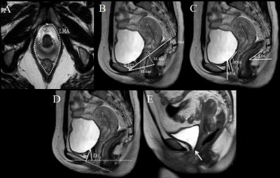
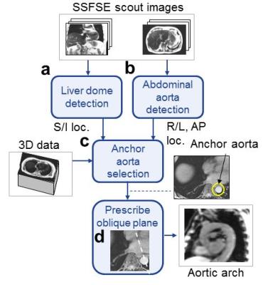
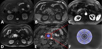
Phase Contrast Magnetic Resonance Imaging using Non-contrast-enhanced Magnetic Resonance Angiography using Balanced Steady-State Free-Precession Sequence and Time-Spatial Labeling Inversion Pulse: Measuring Left Gastric Vein Flow Velocity to Predict Esophageal Varices Development and Rupture - permission withheld
Akihiro Furuta, Hiroyoshi Isoda, Shigeshi Kohno, Koji Tokunaga, Ayako Ono, Rinpei Imamine, Rikiya Yamashita, Shigeki Arizono, Aki Kido, Naotaka Sakashita, Kaori Togashi
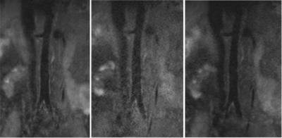
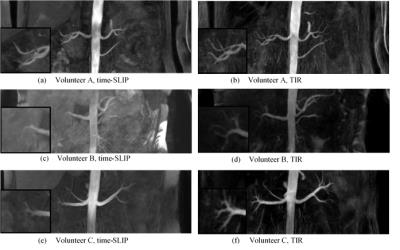
LGV flow velocity is clinically important to foresee esophageal varices development and rupture. But it is difficult to measure it's velocity exactly by echo or only phase contrast MRI (PC-MRI). To measure LGV flow velocity, 2D PC-MRI were set perpendicularly across vessel segments in the cross-sectional slice position determined from 3D selective visualized LGV using non-contrast-enhanced MRA with balanced steady-state free-precession sequence and time-spatial labeling inversion pulse. LGV flow velocity of all subjects could be measured exactly. This method is useful to measure LGV flow velocity.


4973.   
56 
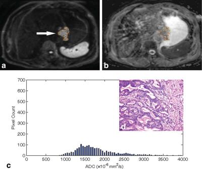
Assessment of Histological Differentiation in Gastric Cancers Using Whole-Volume Histogram Analysis of Apparent Diffusion Coefficient Maps
Zhengyang Zhou, Song Liu, Jian He, Weibo Chen
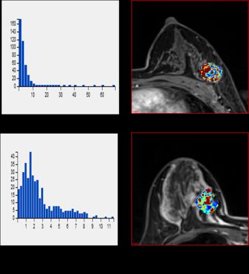
Seventy-eight patients with gastric cancer were underwent MRI to investigate whether the histogram analysis of the entire tumor volume in ADC maps could differentiate between histological grades. A series of histogram parameters were calculated and correlated with the histological grade of the surgical specimen. There were significant differences in the 5th, 10th, 25th, and 50th percentiles, skew, and kurtosis between poorly and well-differentiated gastric cancers. There were correlations between the degrees of differentiation and histogram parameters, including the 10th percentile, skew, kurtosis, and max frequency. Histogram analysis of the ADC maps can be useful in differentiating between histological grades.


4974.   
57 
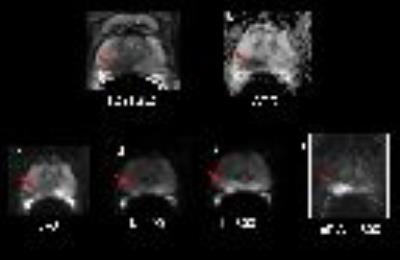
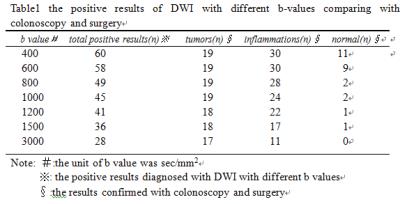
Diffusion-weighted MR Enterography imaging of the ileocecal segment: optimizing b-value for visually differentiating inflammatory and neoplastic lesions
Hao Yu, Daoyu Hu, Yaqi Shen, Zhen Li, Jianjun Li, Zi Wang, Yanchun Wang
To evaluate the ability of conventional MR Enterography (MRE) including coronal and axial T1/T2 weighted imaging and Diffusion-weighted imaging with different b-values(b=400,600,800,1000,1200,1500,3000 sec/mm2)to visually illustrate inflammatory lesions and neoplastic lesions in the ileocecal region comparing with colonoscopy or surgical results.As a result,MRE and DWI were capable of revealing the lesions in the ileocecal segment. DWI was superior to detect lesions especially inflammations comparing with conventional MRE, and the optimal b value of DWI for MRE was 800 sec/mm2 at 3T. Hyperintensity of ileocecal lesion on DWI with high b(?1000 sec/mm2) value wes more favor for tumor-like lesion.


4975.   
58 
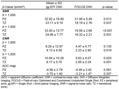
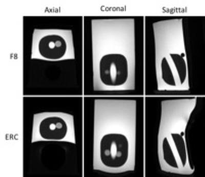
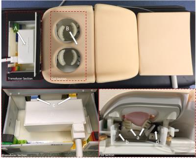
Ultra-fast abdominal imaging with high parallel-imaging factors:  Comparative study of a 60-channel receiver coil with the standard coil set-up
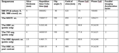
Ahmed Othman, Petros Martirosian, Wilhelm Horger, Jakob Weiss, Jana Taron, Karsten Jahns, Konstantin Nikolaou, Mike Notohamiprodjo
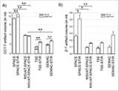
In this study, we evaluated a novel 60-channel coil setup for ultra-fast abdominal imaging using high PAT factors in a phantom, in healthy volunteers and in patients. We found that the 60-channel coil-setup is superior to a conventional 30-channel coil-setup yielding higher SNR and superior image quality and enabling ultra-fast image acquisition with diagnostic image quality.


4976.   
59 
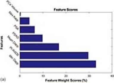
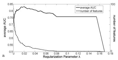
Radiomics Model for Preoperative Prediction of Lymph Node Metastasis in Rectal Cancer after Neoadjuvant Chemoradiatherapy Therapy
Haitao Zhu, Xiaoyan Zhang, Xiaoting Li, Yanjie Shi, Huici Zhu, Yingshi Sun
Preoperative evaluation of lymph node metastasis in locally advanced rectal cancer remains a problem especially after neoadjuvant chemoradiatherapy treatment (NCT). This study proposed a MRI-based radiomics method to predict lymph node involvement in rectal after NCT. Beside the features from the tumor, features from the lymph nodes were also included for the construction of the radiomics model to increase the accuracy of prediction. 10-fold cross-validation among 300 patients produced ROC with average AUC=0.78. Independent validation with 118 patients produced ROC with AUC=0.81.


4977.   
60 
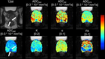
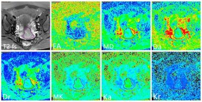
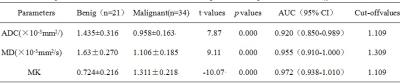
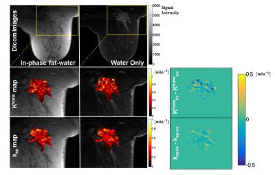
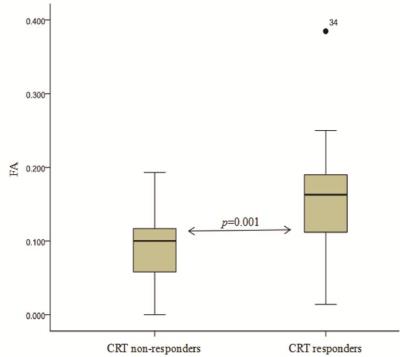
Pretreatment diffusion kurtosis imaging for predicting the response of locally advanced rectal cancer to neoadjuvant chemoradiation therapy
Hongliang Sun, Yanyan Xu, Kaining Shi, Wu Wang
Diffusion kurtosis imaging (DKI) is an emerging technique, which could reflect restricted water diffusion within the complex microstructure of most tissues based on non-Gaussian diffusion model. It has been reported that DKI was used in central system diseases, tumor grade, even assessment of treatment response. However, there is limited research reported about the clinical application of DKI in rectal cancer, and the value of DKI in monitoring rectal cancer treatment was still unclear.


4978.   
61 
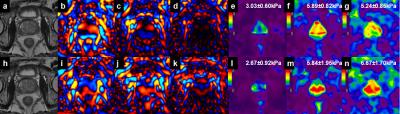
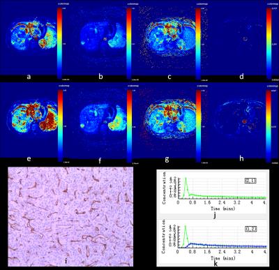
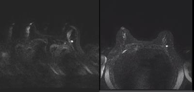
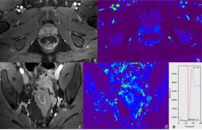
Dynamic Contrast-Enhanced Imaging of the Rectum Using Golden-Angle Radial Sparse Parallel MRI (GRASP): Initial Experience and Comparison to a Conventional Approach Using Time-resolved Angiography With Interleaved Stochastic Trajectories (TWIST).
Daniel Hausmann, Jing Liu, Philipp Riffel, Johannes Budjan, Robert Grimm , Tobias Block, Stefan Schoenberg, Ulrike Attenberger
MR perfusion images to discriminate between normal rectal wall and rectal cancers with less variance of perfusion values and superior image quality compared to conventional TWIST-Angiography can be generated using time-resolved free-breathing MRI with continuous golden-angle radial sampling and iterative reconstruction (GRASP). Additional morphologic assessment (“one-stop-shop”) with high spatial resolution, artifact-insensitive, multiphase, contrast-enhanced imaging may increase accuracy and diagnostic confidence of the examination.


4979.   
62 
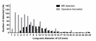
The Limitation in Predicting Lymph Nodes Stage by Using Magnetic Resonance Imaging on the Criterion of Size with Histopathological Analysis as Reference - permission withheld
Caizhen Feng, Jin Cheng, Jing Wu, Gongwei Wang, Yingjiang Ye, Yi Wang
In spite of LN status is critical to the prognosis of patients with gastric cancer, MDCT and MRI cannot accurately assess metastatic LNs prior to surgery.In our study, 802 LNs of 30 patients with gastric carcinoma were harvested.during D2 lymphadenectomy. Only 36.7% (295/802) LNs were detected on preoperative MRI.. 31.5% (217/688) LNs (<8mm) were identified as malignant by pathology, whereas, 44.7% (51/114) LNs (≥8mm) were defined as metastatic. Forty-one metastatic LNs (19%, 41/215) with(?3mm) were found in 7 patients (23.3%, 7/30) and caused N stage upstaging in 3 patients, which could not be detected by MRI.


4980.   
63 
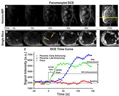
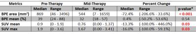
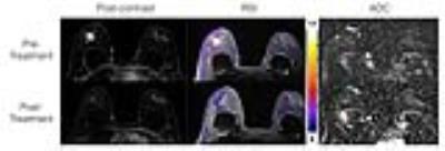
Dynamic contrast enhanced MR imaging for therapeutic response assessment after neoadjuvant Chemoradiotherapy in patient with local advanced rectal cancer - video not available
Yanfen Cui, Xiaotang Yang , Ning Huang
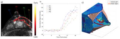
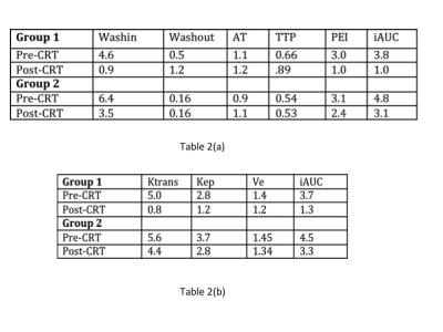
The pre-CRT Ktrans value and the percentage decrease in the Ktrans after CRT could be helpful to predict good therapeutic response to CRT for LARC. This may allow for personalized treatment-options in rectal cancer patients.  


4981.   
64 PET/MRI for rectal cancer staging: Longer PET acquisition times result in increased identification of nodal metastatic disease.
Colin Burke, Thomas Hope, Michael Ohliger, Zhen Wang, Katherine Van Loon, Madhulika Varma
Rectal cancer nodal staging guides the decision to whether neo-adjuvant chemoradiation is needed prior to surgical resection and is a predictor of survival and recurrence. However, staging based on size and morphologic criteria alone is limited.  Our data suggests that increased PET acquisition times with PET/MRI increases the identification of nodal metastatic disease in rectal cancer, particularly in small nodes sized 5 mm or less.


4982.   
65 
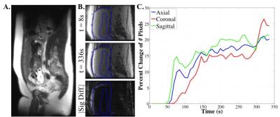
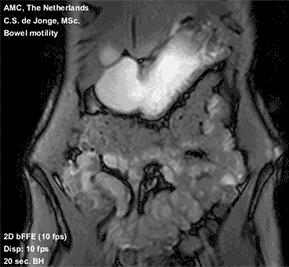
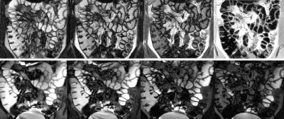
Dynamic MRI For Bowel Motility Imaging – How Fast And How Long?
C.S. de Jonge, R.M. Gollifer, A.J. Nederveen, D. Atkinson, S.A. Taylor, J. Stoker, A. Menys
Dynamic (cine) MRI of bowel motility is now routinely performed in clinical practice and advances in post-processing have enabled robust quantitation of this data facilitating numerous research applications. Generally, motility sequences are acquired in a 20 second breath hold at a temporal resolution of 1 fps.   In this study, we investigate these core assumptions and provide guidance information for future studies.  In summary, we show that a temporal resolution of at least 1 fps is necessary for a scan duration of at least 10 seconds. This is consistent with the majority of small bowel motility studies to date.


4983.   
66 
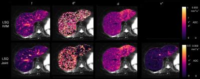
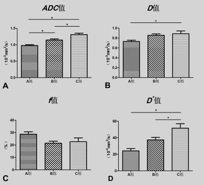
Optimization of high b values for intravoxel incoherent motion imaging of rectal cancer : a pilot study - video not available
Yankai Meng, Chongda Zhang , Hongmei Zhang, Chunwu Zhou
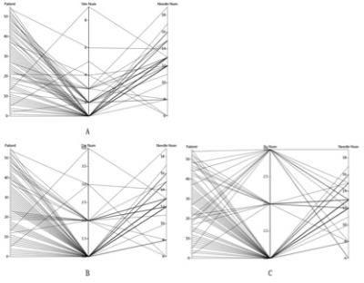
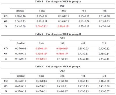
To optimize the high b values (>200s/mm2) for intravoxel incoherent motion imaging of rectal cancer and to observe the effect of high b values variation on IVIM parameters. Three groups (A group with all 16 b values: 0,10,20,30,40,60,80,100,150,200,400,800,1000,1200,1500,2000, B group with 14 b values: 0,10,20,30,40,60,80,100,150,200,400,800,1000,1200 and C group with 12 b values: 0,10,20,30,40,60,80,100,150,200,400,800) were selected respectively for measurement by a radiologist. The average values of each measurement were used for statistical analysis. One-way analysis of variance (ANOVA) and post-hoc test were performed on the mean values of IVIM parameters in groups A, B, and C, with a significance level of P<0.05. The p values of ANVOA results in ADC?D?D* values were less than 0.05, the differences were statistically significant. The p values of Bonferroni post-hoc test in D?D*?f values were not statistically significant differences in group A and B. With the number of high b values decrease, the values of ADC?D?D* values and standard error were increased, while of f values was not changed significantly. In our study, the reproducibility of the IVIM parameters caused by high b value variation was not significant. The value of selected b > 1500 need to be further studied.


4984.   
67 
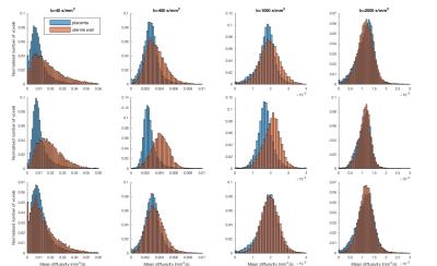
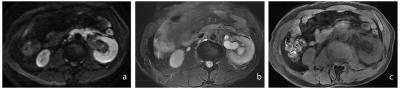
Optimized ROI size on ADC measurements of normal pancreas, pancreatic cancer and mass-forming chronic pancreatitis
Chao Ma, Jing Li, Mbaiaourer Bouka, Panpan Yang, Li Wang, Luguang Chen, Li Su, Yong Zhang, Jianxun Qu, Shiyue Chen, Qiang Hao, Jianping Lu
The effect of ROI size on ADC measurements in normal pancreatic tissue or pancreatic lesions have rarely been studied. This study investigated the influences of ROI size in ADC measurements for the differentiation between normal pancreas (NP), mass-forming chronic pancreatitis (MFCP) and pancreatic ductal adenocarcinoma (PDAC).


4985.   
 
68 
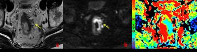
Diffusion kurtosis imaging for differentiating tumor KRAS mutation status in rectal cancer
Yanyan Xu, Hongliang Sun, Kaining Shi, Wu Wang
Diffusion kurtosis imaging (DKI), which is a non-Gaussian diffusion-weighted model proposed by Jensen et al 1, has the potential to characterize both normal and pathologic tissue 1-3, meanwhile, providing a new option for tumor garde 4 and assessment of treatment response 5-7. Previous studies 1-3, 8 found that DKI could better account for restricted water diffusion within the complex microstructure of most tissues. To our knowledge, however, no study has included evaluation of DKI characteristic in rectal cancer, especially in the aspect of KRAS mutant, which associated with clinical treatment and prognosis of colorectal cancers 9. 


4986.   
69 The utilization of DDC value in detecting the status of LVI in rectal cancer patients at 3.0T MRI
Guangwen Zhang, Jinsong Zhang
In this study, we aimed to investigate the value of DDC in assessing the status of lymphovascular invasion in patients with rectal cancer. Ninety-eight patients with rectal adenocarcinoma underwent DWI with 16 b-values at 3.0T MR system. We found there was an significant difference in DDC value between the LVI presence group (DDC=0. 893±0.151×10⁻³mm2/s, n=46) and the LVI absent group (DDC=0. 825±0.127×10⁻³mm2/s, n=52), (P=0.018). We speculate that DDC value derived from multi-b value DWI could be a useful functional parameter in detecting the status of LVI in rectal cancer patients. 


4987.   
70 
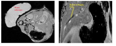
Measuring T1 and T2 of the small bowel wall at 3T
Hannah Williams, Penny Gowland, Luca Marciani, Robert Scott, Guruprasad Aithal, Caroline Hoad
Available techniques to measure in-vivo bowel permeability are inadequate for stratifying patients to identify those at risk of complications from increased bowel permeability. T1 and T2measurements could potentially be indicators of changes in bowel wall structure and thus permeability. We have measured the T1 and T2 of the bowel wall to be 1.68±0.57 s and 0.08 ±0.02 s respectively. We found significant variations between and within subjects. However it is currently unknown whether some of these variations are real and some due to errors in the measurement process. 


4988.   
71 
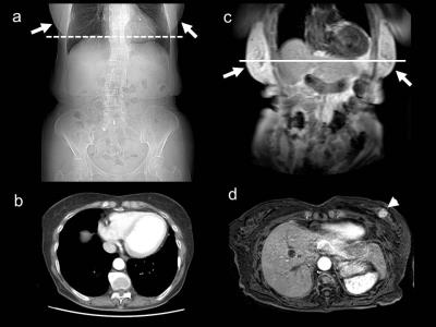
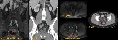
MR versus CT Imaging for Identifying the Etiology of Abdominal Pain in Emergency Department Patients
Michael Repplinger, Perry Pickhardt, Rebecca Bracken, Douglas Kitchin, Jessica Robbins, Timothy Ziemlewicz, Scott Reeder
Our study aimed to evaluate the diagnostic accuracy of MR versus CT for identifying the etiology of abdominal pain in emergency department patients. This is a prospective study that included patients ≥12-years-old who were being evaluated for possible appendicitis. All patients underwent both MR and CT; images were interpreted by three radiologists who were blind to the patient’s outcome. There were 113 instances of acute abdominal processes (15 different diagnoses). The overall accuracy of NC-MR, CE-MR, and CT was 77%, 83%, and 90% for individual reads and 82%, 84%, and 94% for consensus reads.


4967.   
50 Analysis of motility in apparently normal small bowel – relationship to Crohn’s symptoms
Ruaridh Gollifer, Alex Menys, Jesica Makanyanga, Carl Puylaert, Frans Vos, Jaap Stoker, David Atkinson, Stuart Taylor
Crohn’s disease (CD) patients often suffer abdominal symptoms even when their disease is apparently in remission with no identifiable active inflammation.1 Ongoing aberrant gut motility has been postulated as a cause, and this can now be quantified using MRI.2 This study tested the association between abdominal symptoms based on the Harvey-Bradshaw Index (HBI) and MRI derived motility metrics in morphologically healthy small bowel in CD patients.  An inverse association was found between reduced motility spatial variation across the small bowel and symptoms, particularly diarrhoea.  This association was strongest when HBI scores were higher.


4968.   
51 Feasibility of Performing Dynamic and Delayed Enhancement and Magnetization Transfer Ratios in pediatric patients undergoing clinically-indicated MRE: pilot study to assess image quality for quantitative evaluation
Mary-Louise Greer, Susan Shelmerdine, Kedar Patil, Claire Cuscaden, Debra Drossman, Logi Vidarsson
Purpose: Assess feasibility of applying magnetization transfer (MT) and dynamic and delayed enhancement (DCE) sequences during MR Enterography(MRE) in children. 

Methods: REB approved, in this prospective study, patients  =/< 18 years undergoing MRE for suspected or proven inflammatory bowel disease were consented for application of MT and DCE sequenced in addition to standard clinical sequences. These were assessed and  prospectively recruited and imaging sequences applied. Imaging was subjectively analysed by two radiologists or a radiologists and physicist in the first arm by consensus for sequence modification.

Results: Inter and intra-reader analysis was undertaken.

Conclusion: DCE is robust, MTR requires further modification.


4969.   
52 
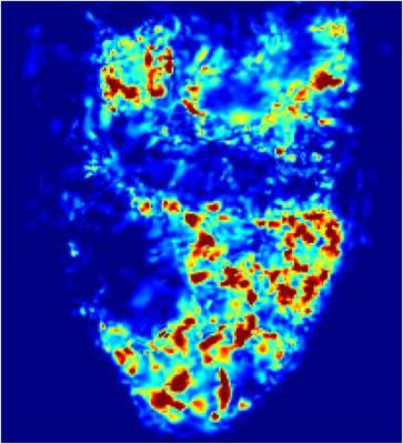
Magnetization transfer MRI for evaluating bowel fibrosis and inflammation in patients with stricturing Crohn’s disease - video not available
Xuehua Li, Zhuangnian Fang, Siyun Huang, Li Huang, Zhongwei Zhang, Xu Yan, Xiaolei Zhu, Jinjiang Lin, Mengchen Zhang, Mengjie Jiang, Shiting Feng, Canhui Sun, Ziping Li
This study aimed to assess the efficacy of Magnetization Transfer MRI (MTI) for evaluating bowel fibrosis and inflammation in patients with stricturing Crohn’s Disease (CD). Bowel wall MTR with normalization to skeletal muscle was calculated and correlated to histologic fibrosis and inflammation as well as amount of type I collagen and vessel density. The results showed that normalized MTRs correlated with histologic fibrosis and type I collagen scores, but did not correlate with inflammation scores or vessel densities. Thus, MTI can accurately detect and distinguish varying degrees of bowel fibrosis with or without coexisting inflammation in human CD. 


4970.   
53 
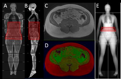
Comparison of Measurement of Abdominal Visceral Adipose Tissue in Men and Women by MRI vs. DXA
Cherie Shook, Bret Goodpaster, Heather Cornnell
Visceral adipose tissue (VAT) has been identified as a significant contributing factor to the metabolic complications of obesity and cardio-metabolic disease, thus its precise measurement is becoming more clinically relevant. Both MRI and DXA were used to measure different components of body composition including VAT, and these results were compared by gender. Both scan acquisitions took similar amounts of time, but DXA results were calculated automatically while MR data processing was completed offline, thus took more time. The results from this study indicate that DXA is a precise measure of only a portion of VAT while MRI can give a more accurate measurement of total VAT across the entire abdomen, potentially avoiding gender bias.


4971.   
54 
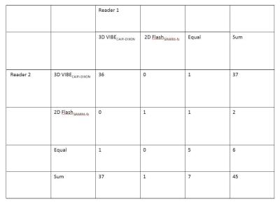
Clinical application of 3D VIBECAIPI-DIXON for enhanced imaging of the small intestine
Yang Yu, Lu Liang, Tao Jiang
The abstract discussed the clinical application of a fast 3D VIBE sequence with Dixon fat saturation and CAIPIRINHA acceleration techniques (3D VIBECAIPI-DIXON) by compare to a standard 2D FLASH sequence with spectral fat saturation and conventional GRAPPA acceleration technique (2D FlashGRAPPA-fs) for enhanced imaging of the small intestine


4966.   
49 
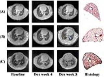
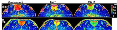
Metabolic Imaging of ß3-adrenoreceptor Activated BAT and its Systemic Effect on Abdominal Fat in Diet Induced Obese Model
Jadegoud Yaligar, Sanjay Kumar Verma , Venkatesh Gopalan , Anantharaj Rengaraj, Tian Xianfeng, Anna Ulyanova, Bhanu Prakash K.N, Suresh Anand Sadananthan, Navin Michael, S. Sendhil Velan
Imbalance in dietary intake and energy expenditure are associated with obesity, diabetes and metabolic disorders. Adipocyte size and expansion of adipose tissue plays a critical role towards the progression of diet induced obesity. Brown adipose tissue (BAT) plays a critical role in modulating different fat depots in the body. BAT can be functionally activated by administering the β3-adrenergic agonist. Understanding the mechanisms associated with BAT activation and the possibility of reversing insulin resistance and its impact on whole body metabolism is of current clinical interest for combating diabetes. In the current study, we investigated the fat partitioning in high fat diet induced obese rodent model by β-adrenergic-mediated BAT activation.


4989.   
72 
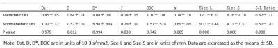
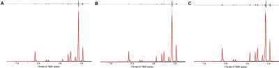
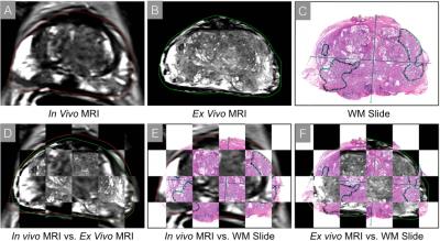
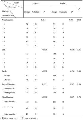
Chemical Shift Effect Predicting Lymph Node Status in Rectal Cancer using High-Resolution MR Imaging with Node-for-node Matched Histopathological Validation - video not available
chongda zhang, hongmei zhang, feng ye, yuan liu, chunwu zhou
To evaluate the value of chemical shift effect (CSE), as well as other criteria for the prediction of lymph node status. Lymph nodes harvested from transversely whole-mount specimens were compared with in vivo and ex vivo images to obtain MR characteristics including CSE, as well as other predictors of 255 benign and 35 metastatic nodes. Our results revealed that CSE is a reliable predictor for differentiating benign from metastatic lymph nodes. Other predictors of nodal location, border, signal intensity and minimum distance to rectal wall were also proved to be useful for the diagnosis.
 

The International Society for Magnetic Resonance in Medicine is accredited by the Accreditation Council for
Continuing Medical Education to provide continuing medical education for physicians.