ISMRM 25th Annual Meeting & Exhibition • 22-27 April 2017 • Honolulu, HI, USA

Electronic Poster Session: Cardiovascular
4752 -4775 Image Processing
4872 -4893 Preclinical CV Imaging
 
Image Processing
Electronic Poster
Cardiovascular

 
Wednesday, 26 April 2017
Exhibition Hall  13:45 - 14:45


    Computer #

 
4758.   
7 Automated 4D Flow Conservation Utilizing Adjacency Matrices
Carson Hoffman, Gabe Shaughnessy, Oliver Wieben
4D flow magnetic resonance imaging (MRI) can provide comprehensive information on vessel anatomy and hemodynamics for complex vessel system. Adjacency matrices are often used in computer science to help simplify complex graphs into a binary encoded matrix. The adaptation of adjacency matrices to 4D flow MRI can help reduce the complexity for analysis by structuring the data into an efficient binary matrix. One application of this new analysis method allows for flow conservation to be completed for complex volumes at all junctions. The conservation of flow at every junction can then be used to find segments of potential erroneous measurements.   


4752.   
1 Optimized PC-MRA using a New Processing Workflow for 4D Flow MRI Data
Lingzi Tashakkor, Susanne Schnell, Alex Barker, Kelly Jarvis, Emilie Bollache, Michael Markl
A new workflow was developed based on the information from 4D flow MRI to optimize the phase contrast MR angiogram. Five new PC-MRA algorithms were tested on 15 patients, and compared with and without the proposed pre-processing. Histogram equalization/remapping was applied to improve the dynamic signal range for easier segmentation and reduced user interaction. Results showed higher-quality PC-MRAs when the proposed pre-processing was applied.


4753.   
2 Slice Realignment for Motion-Corrupted Stacks of Short-Axis Cine Cardiac MR Images based on 3D Probabilistic Edge Maps
Giacomo Tarroni, Ozan Oktay, Andreas Schuh, Wenjia Bai, Antonio de Marvao, Declan O'Regan, Stuart Cook, Daniel Rueckert
Short-axis cine cardiac MR image stacks are acquired during multiple breath-holds, which often causes a misalignment of several slices. We propose a technique for in-plane spatial realignment of motion-corrupted short-axis slices which uses probabilistic edge maps of the myocardium (generated with decision forests) as input to image registration. The proposed technique was quantitatively tested on a dataset of motion-free stacks artificially corrupted by in-plane motion. Overlap measures such as the Dice coefficient - computed on myocardial masks segmented respectively on motion-free, motion-corrupted and motion-corrected stacks - suggest that the proposed technique is able to correctly compensate for slice misalignment.


4754.   
3 Semi-Automatic Ejection Fraction Calculation from Cardiac Low-Rank Tensor Images Based on Unsupervised Machine Learning
Zihao He, Anthony Christodoulou, Hua Guo, Debiao Li
Calculation of the ejection fraction from cardiac cine MR images requires segmenting multiple images of the left ventricle. This process, which is often performed manually, is time-consuming and observer-dependent. In this work, an unsupervised machine learning algorithm, combining hidden Markov random field and optical flow, has been proposed to perform semi-automatic tissue segmentation on T1/T2-weighted low-rank tensor images that have a built-in feature space due to low-rank factorization performed during image reconstruction. The segmentation results then allow automatic EF calculation. Demonstrated results have higher efficiency and similar accuracy compared with manual segmentation, and were stable with respect to different initializations.


4755.   
4 Non-rigid Groupwise Image Registration for Myocardial Strain Quantification from High-Resolution 3D Tagging
Valery Vishnevskiy, Christian Stoeck, Sebastian Kozerke
Cardiac motion can be efficiently assessed using 3D-tagged MR sequences for diagnostic purposes. However, there is a lack of robust post-processing tools to derive regional motion and strain data. Current registration methods tend to underestimate radial strain in the left ventricle. In order to provide accurate strain estimates we leverage temporal smoothness of displacements and low-rank structure of aligned images. The herein proposed method is 38% more accurate for radial strain, 27% for circumferential strain and 25% for longitudinal strain estimation than state-of-the-art registration implemented in Elastix while increasing computational speed by a factor of six.


4756.   
5 Cardiac image acquisition by SENSE combined compressed sensing with three dimensional quantification
Jong-Hyun Yoon, Pan-ki Kim, Young-Joong Yang, Jinho Park, Jin-Soo Kim, Byoung Wook Choi, Chang-Beom Ahn
SENSE combined compressed sensing technique is applied to multi-slice cardiac CINE imaging with breath holds. As to the compressed sensing, ITSC is used, which truncates small transformed coefficients in r-f domain to make data sparse, and it restores the measured data in k-t domain iteratively until the reconstructed images converge. Variation of ejection fraction (EF) is measured for two set of experiments, one from regenerated data set by resampling the original data set, and the other from real measurements.  Using the variation of EF and normalized mean square error clinical usefulness of the technique is demonstrated.


4757.   
6 Motion tracking in cardiac MRI: Cine Watermark tracking of myocardial strain
Davis Vigneault, Ronald Beyers, Chia Liu, Alison Noble, Thomas Denney, David Bluemke
Quantification of regional cardiac function is of central importance in cardiology, but has yet to be adopted into clinical practice due to limitations of the current techniques.  Here we present a method requiring minimal human intervention for tracking “cine watermark” images, in which features have been encoded into the phase image of a cardiac cine series.  The method employs nonlinear least squares optimization, which allows the sum of squared wrapped phase differences between patches in successive frames to be minimized globally across all frames, while regularizing over physically-motivated metrics.  Preliminary results in healthy human volunteers show robust tracking.


4759.   
8 MRI vessel slice tool visualization with an interactive 3-D display
Carson Hoffman, Eric Schrauben, Oliver Wieben
The comprehensive information on vessel anatomy and hemodynamics presented by 4D Flow MRI can be difficult to visualize. We introduce a new viewing mode using a ‘slice tool’. The use of this display algorithm can provide benefits for scalar visualization by preserving spatial location and avoiding ambiguities in cases of overlapping vessels. This novel approach can thus offer an improved understanding of complex hemodynamics within the body when used in conjunction with previously existing visualization methods eg. MIP images, pathline, and streamline visualizations.


4760.   
9 SUPER-RESOLUTION RECONSTRUCTION OF LATE GADOLINIUM ENHANCEMENT CARDIOVASCULAR MAGNETIC RESONANCE IMAGES USING A RESIDUAL CONVOLUTIONAL NEURAL NETWORK
Archontis Giannakidis, Ozan Oktay, Jennifer Keegan, Veronica Spadotto, Inga Voges, Gillian Smith, Iain Pierce, Wenjia Bai, Daniel Rueckert, Sabine Ernst, Michael Gatzoulis, Dudley Pennell, Sonya Babu-Narayan, David Firmin
Late gadolinium enhancement cardiac magnetic resonance (LGE-CMR) has enabled the accurate myocardial tissue characterization. Due to practical considerations, the acquisition of anisotropic two-dimensional (2D) stack volumes, with low through-plane resolution, still prevails in the clinical routine. We propose a deep learning-based method for reconstructing a super-resolved three-dimensional LGE-CMR data-set from a low resolution 2D short-axis stack volume. The method directly learns the residuals between the high and low resolution images. Results on clinical data-sets show that the proposed technique outperforms the state-of-the-art with regard to image quality. The fast speed of our model furthers facilitates its adoption for practical usage.


4761.   
10 Fully automatic bullseye analysis on short-axis MOLLI mapping: LV segmentation and AHA 17 parcellation - permission withheld
Yun-Wen Wang, Chun-Yu Huang, Hsiao-Hui Huang, Teng-Yi Huang
The free-breathing MOLLI (FB-MOLLI) presented in our previous study allowed T1 mapping in vivo without breath-hold. In this study, we attempted to implement unsupervised reconstruction for FB-MOLLI data sets and used a deformable method for image registration to improve the reliability of free-breathing T1 mapping. The results supported that the method improved the image alignments of the FB-MOLLI data sets and thus increased the quality of the T1 map. The variations of the repeated T1 measurements were significantly reduced in the anterolateral of the LV walls.


4763.   
12 Automatic detection of corrupted frames in cardiac DTI with machine learning
Francesca Cavallo, Pedro Ferreira, Zohya Khalique, Andrew Scott, Sonia Nielles-Vallespin, David Firmin
In vivo cardiac DTI is capable of probing the microstructure of the myocardium and its dynamics throughout the cardiac cycle. The typical cardiac DTI scan data will contain corrupted frames due to cardiac and respiratory motion. Currently an experienced observer identifies corrupted frames by means of a visual assessment and manually removes them. In this work we show that machine learning can be used to accurately assess DTI corrupted frames, reducing the user input, accelerating analysis and removing human subjectivity.


4764.   
13 A Novel 4D Semi-automatic Segmentation Algorithm for Whole-heart 3D Cine Magnetic Resonance Imaging
Ruizhi Liao, Danielle Pace, Andrew Powell, Polina Golland, Mehdi Hedjazi Moghari
Three-dimensional (3D) time-resolved (cine) whole-heart magnetic resonance imaging promises to greatly facilitate comprehensive and evaluation of cardiac function and morphology. We present here a robust semi-automatic 4D segmentation algorithm, using patch-based volumetric segmentation and a temporal image registration method, to reduce the segmentation time of 3D cine datasets to less than 30 minutes and to enable wide clinical use. Resulting volumetric measurements of the left ventricle and right ventricle are aligned with measurements from the current clinical routine. By visualizing the anatomy and dynamics of the heart, we show that 3D cine datasets promise to enhance surgical planning for patients with complex congenital heart disease.


4765.   
14 Non-iterative model for synthetic image-based registration of MOLLI cardiac T1 mapping images
Laura Saunders, Neil Stewart, David Kiely, Martin Graves, Andy Swift, Jim Wild
Cardiac T1 maps rely upon the acquisition of spatially aligned images. When patients fail to maintain breath hold, respiratory motion can cause T1 map inaccuracies due to poor image alignment. In the method demonstrated here, images are registered by co-registration to synthetic images, which are created via a non-iterative, automatic, model-based method. This method is compared to an iterative registration method using an energy minimisation process for quantitative registration accuracy and speed. Both methods were found to significantly improve image registration. The resultant registrations from both methods did not significantly differ, however the non-iterative model-based method reduced processing time by 1/5th. 


4766.   
15 An MRI based High-Resolution Geometrical Framework to Quantify Left Ventricular Remodeling in Murine Model of Myocardial Infarction - permission withheld
Siamak Ardekani, Geoffrey Gunter, Jiadi Xu, Robert Weiss, Laurent Younes
Computational models of left ventricular (LV) geometry and function that characterize regional cardiac response to injury can provide valuable diagnostic and predictive information. We have developed a mathematical tool to non-rigidly match a high-resolution surface mesh of the LV geometry to a set of LV epi and endocardial contours that are extracted from cardiac MR. We have applied our algorithm on murine model of myocardial infarction to quantify cardiac remodeling process. This approach enables us to perform statistical analysis of LV 3D geometry and function using only sparse sets of 2D plane contours, therefore facilitating cross-subject examination of shape variation.


4767.   
16 Texture analysis of native T1 mapping in dilated cardiomyopathy
Xiaoning Shao, Yingjie Sun, Yong Zhang, Jingliang Cheng, Shaoyu Wang
MR native T1 mapping technique was found have the ability of detecting diffuse myocardial fibrosis which was found in histological examination of dilated cardiomypathy (DCM). Texture analysis of T1 mapping had been proved to be able to provide more information besides T1 value. We found several histogram parameters (statistical class of texture analysis) were significantly higher in DCM than that of healthy volunteers. The areas under the receiver operating characteristic curve of histogram parameters were calculated. The results indicate that texture analysis of T1 mapping may be helpful for the diagnosis of DCM.


4768.   
17 Improving Precision of Arrhythmia-Insensitive Rapid Cardiac T1 Maps using a Non-Local Means Filter
Suvai Gunasekaran, KyungPyo Hong, Jeremy Collins, James Carr, Daniel Kim
Compared with other investigations, images produced from arrhythmia-insensitive rapid (AIR) cardiac T1 mapping pulse sequences contain significant random noise which limits the accuracy of quantitative measurements. Therefore, denoising filters were used to reduce noise present in the images. From our analysis, the non-local means filter was best able to reduce the amount of noise while still maintaining accurate T1 measurement data. 


4769.   
18 Validation of dMRI-Derived Fiber Orientations Using PLI in Human Fetal Hearts - permission withheld
Feng Yang, YueMin Zhu, Gabrielle Michalowicz, Yves Usson, Laurent Fanton, Magalie Viallon, Patrick Clarysse, Pierre-Simon Jouk, Pierre Croisille
To validate to what extent the fiber orientations derived from diffusion MRI (dMRI) reveal anatomical reality, polarized light imaging (PLI) allowing the fiber orientations of the human heart to be physically measured with high spatial resolution was used. The dMRI and PLI orientation measurements of the same hearts are then compared using a multimodal registration-based framework. Experimental results show that dMRI and PLI have similar variation patterns of elevation or azimuth angles, except that dMRI introduced a decrease of about 24º in transmural elevation angle range. No significant differences were observed on azimuth angle in both modalities.


4770.   
19 Regularized Curve Fitting Improves T1 Bias in Cardiac T2 Mapping - video not available
Galen Reed, Kenneth Johnson, Michelle Nystrom, Okai Addy, Reeve Ingle, Bob Hu, Juan Santos, William Overall
Bias error is studied in T2-prepared single shot SSFP cardiac T2 mapping. Phantom experiments showed that although centric phase encode ordering reduces bias from T1 relaxation during single shot readouts, a residual bias of approximately 20% remains in the 1000 ms T1 regime. A regularized, three parameter exponential fitting model reduces this bias and generates T2 maps with low variance using 4 echo times. 


4771.   
20 The Myocardial Feature Tracking of LA, LV and RV: Age-related in Normal Chinese
Junping Peng, Mingwu Lou, Lei Zhao, Zhanming Fan, Xiaohai Ma, Liang Zhong, Xiaodan Zhao, Hui Chen, Zheng Wang, Shuang Leng
To quantify the age-related global cardiac deformation, GLS and GCS derived from feature-tracking cardiovascular magnetic resonance were measured using post-processing software in 81 healthy Chinese volunteers. It was found that age significantly influenced GLS of LV and LA. This is an important indicator for further research of quantitation MR Myocardial Feature Tracking.


4772.   
21 Workflow Improvement with a Cardiac MRI Positioning Assist Function
Kensuke Shinoda, Shuhei Takemoto, Shuhei Nitta
In this study, we evaluated how the Cardiac MRI positioning assist function can reduce the operation steps and times in CMRI. When using the assist function, the total number of the operation steps was reduced to about 25% for both the experts and the beginners. The operation time was also reduced to about 25%. The mean and SD of the elapsed time for the CMRI procedure performed by experts were reduced from 42m17s±10m53s to 32m46s±3m29s, and those for coronary angiography were reduced from 20m42s±8m5s to 15m22s±2m49s.


4773.   
22 4D Flow MRI based aortic wall shear stress analysis using two different software tools and datasets from MRI scanners of two different appliance brands
Michael Beldoch, Thekla Oechtering, Victoria Schultz, Peter Hunold, Joerg Barkhausen, Alex Frydrychowicz
Wall shear stress (WSS) is an increasingly used vessel wall parameter derived from 4D Flow-MRI data. There is no reference standard for evaluating WSS and different software approaches are available. However, their comparability is not known. Hence, it was the aim of this study to compare two available software tools (GTFlow and FlowTool). To achieve this goal, data from 21 healthy volunteers scanned on MRI scanners of different vendors were analyzed with respect to average and segmental WSS. Results showed good agreement between tools.


4762.   
11 Fully automatic myocardial ECV mapping: deformable image registration, LV segmentation and a cloud computing pipeline
Yun-Wen Wang, Chiao-Ning Chen, Teng-Yi Huang, Ming-Ting Wu
In this study, we developed an automatic pipeline and segmentations of left ventricle walls for mapping myocardial extracellular volume fraction (ECV) and calculating global ECV values. The system combined with user-friendly web-based user interface was packaged into a software container. We used this system to analyze myocardial T1 images obtained from subjects with tetralogy of Fallot (TOF). The ECV values of the TOF data sets were significantly higher than those of the normal subjects. This automatic pipeline could be a practical tool for clinical evaluations of myocardial fibrosis with MR MOLLI T1 images.


4774.   
23 Free-Breathing cardiovascular imaging: comparing central k-space amplitude vs. phase based self-gating approaches
Feifei Qu, Uday Krishnamurthy, Brijesh Yadav, Ewart Haacke, Jaladhar Neelavalli
Two self-gating approaches to reconstruct free-breathing cardiovascular image generated by Golden Angle radial trajectory MRI were compared. The results show that both central k-space magnitude and phase self-gated image have comparable quality with breathhold Cartesian CINE image, and the phase self-gating signal is less sensitive to the coil sensitivity compared to the magnitude based self-gating signal.


4775.   
24 Evaluation of three-dimensional magnetic resonance imaging of autopsied human heart specimens for computational modeling of congenital heart diseases - permission withheld
Yoshiaki Morita, Wataru Ueki, Ryo Haraguchi, Takaaki Matsuayama, Yoshiaki Watanabe, Tatsuya Nishii, Atsushi Kono, Naoaki Yamada, Tetsuya Fukuda
This study aimed to compare the visibility of a formalin-fixed heart using various 3D MRI sequences as well as to determine the optimal sequence for computational modeling of congenital heart diseases. Our results demonstrated that MPRAGE showed the best contrast with good image quality for imaging of the myocardium and the vascular structure when surrounded by normal saline in a plastic container. We believe that computational cardiac modeling of human autopsied heart specimens using MPRAGE plays a critical role in education and/or research.
 
Preclinical CV Imaging
Electronic Poster
Cardiovascular

 
Wednesday, 26 April 2017
Exhibition Hall  14:45 - 15:45


    Computer #

 
4872.   
1 
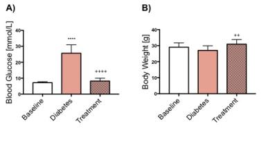
Reversible Changes in Cardiac Function and Metabolism in an Inducible Mouse Model of Type 1 Diabetes
Dragana Savic, Maria Rohm, Vicky Ball, Mary Curtis, Lisa Heather, Frances Ashcroft, Damian Tyler
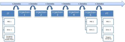
Heart disease is the leading cause of death in Type 1 diabetic patients, however the mechanistic link has not been fully established. In this study an inducible and reversible mouse model of Type 1 diabetes was used. Depression of cardiac function and the increase in blood glucose occur in combination with suppression of pyruvate to bicarbonate conversion. By reversing diabetes with Glibenclamide, cardiac function and blood glucose concentration was restored. This study demonstrated changes that occur alongside the development of a reversible model of Type 1 diabetes and how the action of Glibenclamide can affect metabolism and function of the heart. 


4874.   
3 
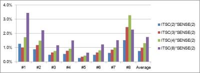
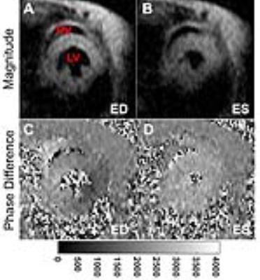
Repeatability and User Variability of Myocardial Tissue Phase Mapping in Mice
Nivedita Naresh, Cynthia Yang, Sol Misener, Bradley Allen, Michael Markl, James Carr, Daniele Procissi
Mouse models can help investigate the molecular mechanisms underlying complex cardiovascular diseases. Assessment of myocardial regional wall motion plays a very important role in the diagnosis and management of several cardiovascular diseases and can be linked to many underlying biological processes. In this study, we evaluated the repeatability and inter-user variability of the myocardial tissue phase mapping method in mice. We found that myocardial tissue phase mapping can be performed with good repeatability and little user variability in mice to reliably quantify both global and regional myocardial velocities.


4875.   
4 
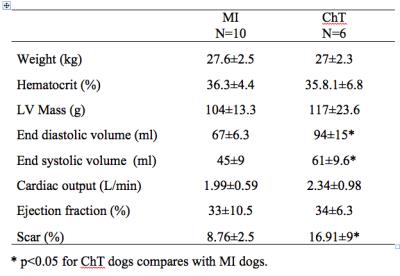
MRI/PET of Myocardial Extracellular Volume in a Canine model of Chemotherapy
Chia-Ying Liu, Cynthia Davies-Venn, Comfort Elumogo, Rolf Symons, Victoria Hoffmann, Kelly Rice, Roberto Maass-Moreno, Veit Sandfort, Stefan Zimmermann, Amir Pourmorteza, Mark Ahlman, David Bluemke
We used simultaneous MR/FDG PET to measure the myocardial extracellular volume (ECV) and glucose metabolism (estimated by standard uptake value, SUV) in a canine model of chemotherapy (ChT); comparison was made to ECV in relationship to a myocardial infarct (MI) models.  MRI ECV in the ChT group was elevated by 16% and 23% compared to the MI group in the remote and adjacent myocardial segments, respectively. PET SUV in the ChT group was reduced by 49% and 41% compared to the MI group in the remote and adjacent myocardial segments, respectively. Difference was also observed in the MRI partition coefficient but not in the native T1.


4873.   
2 
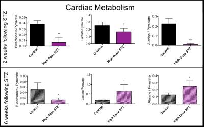
Type 1 Diabetic Hearts show Unexpected Biphasic Metabolic and Functional Progression as Evaluated with Hyperpolarised [1-13C]Pyruvate and CINE MR.
Dragana Savic, Vicky Ball, Carolyn Carr, Lisa Heather, Damian Tyler
Type 1 diabetes patients are insulin deficient resulting in hyperglycaemia. Diabetic patients have a higher risk of cardiovascular diseases. In this study the progression of cardiac metabolic and functional decline was followed in a streptozotocin (STZ) induced Type 1 diabetic model.  Flux through pyruvate dehydrogenase was significantly decreased at 2 and 6 weeks post STZ injection. Interestingly, the incorporation of the 13C-label from pyruvate into lactate and alanine was decreased at 2 weeks, but significantly increased at 6 weeks. Cardiac output was normalized after 6 weeks. Such studies will allow a better understanding of the interactions between metabolism and function in the diabetic heart.


4876.   
5 
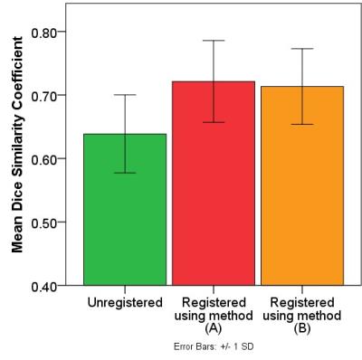
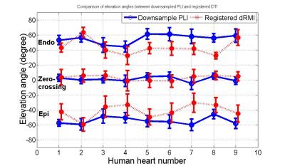
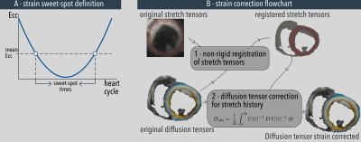
Evaluation of the impact of strain correction on the secondary eigenvector of diffusion with in vivo and ex vivo porcine hearts
Pedro Ferreira, Sonia Nielles-Vallespin, Ranil de Silva, Andrew Scott, Daniel Ennis, Daniel Auger, Jonathan Suever, Xiaodong Zhong, Bruce Spottiswoode, Dudley Pennell, Andrew Arai, David Firmin
Myocytes have a laminar organization, where sheets of myocytes interleave with collagen-lined shear layers. Cardiac diffusion tensor imaging is capable of probing sheet dynamics with secondary diffusion directions, although questions remain about cardiac strain being a possible confounder. Here we study the validity of strain-correcting cardiac diffusion tensor data by directly comparing in vivo DTI data without and with strain correction, to ex vivo DTI data of the same porcine hearts arrested in a diastolic or systolic conformation. Results show that the current strain correction model exaggerates the contribution of microscopic strain to diffusion resulting in an over-correction. 


4877.   
6 
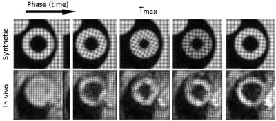
Simultaneous Cardiac and Renal oblique-slice T1-Mapping Differentiates Contrast Agent Activity in Normal and Doxorubicin-treated Rats
Ronald Beyers, Dean Schwartz, Tessa Hutchinson, Meghan Ward, Nouha Salibi, Christian Goldsmith, Thomas Denney
We developed a cardiac multi-oblique-slice T1-mapping sequence, called Tmax, for simultaneous in vivo cardiac and multi-region T1-mapping in rats.  We validated Tmax with gadolinium contrast agent (CA) scans then applied it to support our concurrent development of a reactive oxygen species activated T1-shortening agent, called H4qtp2, in doxorubicin-treated (Dox) rats.  The new Tmax sequence performed excellent at simultaneously quantifying gadolinium T1 effects in cardiac and renal regions.  However, application of Tmax with low dose levels of H4qtp2 CA in Dox rats gave marginal results from too low dose of H4qtp2 to sufficiently affect the T1 and quantify Dox-induced pathology


4878.   
7 
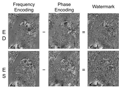
TWO-DIMENSIONAL PHASE-CONRAST MAGNETIC RESONANCE IMAGING AS TOOL TO DETERMINE HEPATIC HEMODYNAMICS IN RATS WITH  A HEALTHY, FIBROTIC OR CIRRHOTIC LIVER
Denise Schaffner, Dominik von Elverfeldt, Peter Deibert, Irmgard Merfort, Adhara Lazaro, Lisa Lutz, Manfred Baumstark, Wolfgang Kreisel, Wilfried Reichardt
In this work we wanted to test a Magnetic Resonance (MR) scanning protocol as a non-invasive tool to determine hepatic hemodynamics and to assess the liver fibrosis degree in an animal model of liver fibrosis and cirrhosis. MR rat liver images provided a good discrimination of healthy from diseased liver, but for the assessment of liver fibrosis degree histology is indispensable. The results show that portal and aortal flow patterns for a cardiac cycle could be measured with high reliability in Conclusion, this MR scanning protocol presents a reliable non-invasive tool to determine hepatic hemodynamics in healthy and diseased rats


4879.   
8 Feasibility of 4D-Flow Imaging of Uterine Blood Flow in the Pregnant Rhesus Macaque
Philip Corrado, Jacob Macdonald, Sydney Nguyen, Kevin Johnson, Chris Francois, Ronald Magness, Scott Reeder, Ian Bird, Dinesh Shah, Thaddeus Golos, Oliver Wieben
4D-Flow MRI is introduced as an alternative to Doppler velocimetry in monitoring blood flow to the placenta in pregnancy. Our 3D radially undersampled PC-VIPR technique provided volumetric coverage of uterus and relevant vasculature with a 10-minute scan. Uterine arteries and ovarian veins were visualized in a 0.83mm isotropic resolution angiogram and flow rates and vessel sizes were measured retrospectively. Repeated scans of four rhesus macaques on subsequent days showed reproducibility of flow rate and cross sectional area measurements in vessels of interest, demonstrating the potential for 4D-Flow MRI for assessing utero-placental vascular health.


4880.   
9 
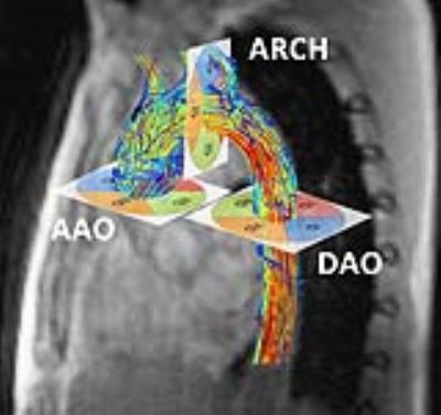
4D flow MRI measurements in ex vivo beating pig hearts as a testing platform for transcathether aortic valve implantation
Eva Peper, Alberto Leopaldi, Sjoerd van Tuijl, Nicky de Jonge, Gustav Strijkers, Arend de Weger, Aart Nederveen, Henk Marquering, Pim van Ooij
In this 4D flow MRI study, ex vivo beating pig heart models in an MR-compatible setup mimicking human physiological conditions were used to investigate flow alterations after implantation of artificial valves designed for transcathether aortic valve implantations (TAVI). Two pig hearts had implanted TAVI valves (one with and one without attachment to the aortic sinuses) and were compared to five pig hearts without valve implantation. For both pig hearts with implanted valves, substantial aortic backflow, and thus paravalvular leakage, was observed. The ex vivo set-up presented is thus suitable for cardiac flow experiments.


4881.   
10 
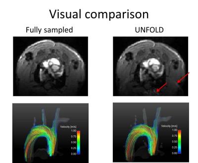
Evaluation of accelerated preclinical 4D-flow imaging with UNFOLD
Moritz Braig, Axel Krafft, Jochen Leupold, Juergen Hennig, Marius Menza, Dominik von Elverfeldt
There is growing interest in preclinical imaging and analysis of complex flow patterns. For example, 4D flow imaging of mouse models with vascular plaques or aortic constriction could provide insights into the development and pathogenesis of cardiovascular diseases. Unfortunately, 4D-flow MRI is often compromised by a trade-off of reasonable acquisition durations and achievable spatial resolution. Our work evaluates an undersampling strategy, namely UNFOLD, for preclinical 4D flow MRI, which has the potential to decrease measurement time by 50%. Artifacts emerging from the undersampling are removed by unaliasing in the temporal domain using a lowpass filter after fourier transformation.


4882.   
11 Quantitative gated and non-gated rat phase contrast MRI: optimized analysis of blood flow and wall shear stress.
Chen-You Huang, Chiun-Wei Huang, Shao-Chieh Chiu, Wu-Chung Shen, Shin-Lei Peng
Goals of this study are to test effects of gated/non-gated, velocity encoding (VENC) and spatial resolution on blood flow, wall shear stress (WSS) and artery area when performing phase-contrast (PC) MRI for rat common carotid artery (CCA). Results show the usage of gated instrument can provide more reproducible results. VENC has insignificant influences on flow, WSS and artery area. To compromise the trade-off between accuracy and time-consuming, the resolution of 0.21 mm is suggested for extracting hemodynamic information about rat CCA.  


4883.   
12 Right ventricular myocardial strain in rats
Emil Espe, Jan Magnus Aronsen, Lili Zhang, Ivar Sjaastad
The function of the right ventricle (RV) is closely linked to clinical outcome in many cardiovascular diseases. Experimental heart disease in rodents play an irreplaceable role in modern cardiovascular research, but no in vivo method exists offering robust measurements of RV myocardial function in small animals.

We used phase-contrast MRI to measure RV strain in rats. We found that RV strain and ejection fraction were closely related, and confirmed that high RV afterload is linked to reduced RV strain.

We show, for the first time, that it is possible to accurately measure myocardial function in the RV in rodents.


4884.   
13 Cardiac MRI measurement of right ventricular strain using feature tracking in a model of embolic pulmonary hypertension.
Zachary Borden, Donald Benson, Alejandro Roldan, Heidi Kellihan, Ashley Mulchrone, Naomi Chesler, Christopher Francois
Right ventricular strain was assessed using an MRI tissue tracking algorithm on bSSFP axial sequences in both acute and chronic embolic pulmonary hypertension canine models.  Strain values were heterogeneous in the acute population with statistically significant decreases in acute radial and longitudinal strain rate and chronic radial and longitudinal strain and strain rate values.  Findings suggest MRI cardiac strain measurement is a promising technique in the clinical evaluation of post embolic pulmonary hypertension patients. 


4885.   
14 
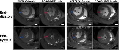
Cardiac Magnetic Resonance for Characterizing a Spontaneous Hypertrophic Cardiomyopathy Mouse Model
Min-Chi Ku, Till Huelnhagen, Andreas Pohlmann, Thoralf Niendorf
HCM is the most common inherited heart disease. The two most frequently seen mutated genes are MYH7 and MYBPC3 which account for nearly 80% of familiar HCM. In this study we hypothesized that these gene variants will affect both LV and RV function. By in-vivo CMR we detected LV hypertrophy in a mouse strain DBA/2J bearing the two gene variants. Interestingly there is no defected LV function found but changes in RV function as both male and female DBA/2J mice had declined RVEF. Our results provide new insights into the correlation of genetic alteration and HCM phenotype.


4886.   
15 
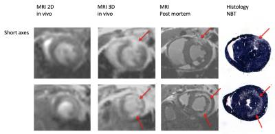
4D Cine Strategy for Assessment of Mouse Cardiac Function and Infarct Size in a Single Acquisition Optimized for a Clinical 3T MR System
Lindsey Crowe, Fabrizo Montecucco, Federico Carbone, Iris Friedli, Anne-Lise Hachulla, Vincent Braunersreuther, Francois Mach, Jean-Paul Vallée
Small cardiac imaging on clinical 3T machines is an important, cost effective translational step for new contrast media and sequence validation. We developed for the first time a new 4D strategy tailored for 3T to simultaneously assess function and infarct in mice, validated against 2D cine, post mortem and histology. Isotropic 3D cardiac cine of mice on a clinical 3T system improved coverage and reduced flow artifacts with higher spatial and temporal resolution for more accurate quantification of cardiac function. Infarct volume enhancement was quantifiable from the same 3D cine acquisition.


4887.   
16 
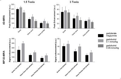
Gadobutrol- vs. gadoterate meglumine-enhanced multi-phase 3D-MRA and 4D-MRA at 1.5T and 3T: an intra-individual quantitative and qualitative comparison of macrocyclic contrast agents in minipigs - permission withheld
Dariusch Hadizadeh, Gregor Jost, Vera Keil, Christian Marx, Maximilian Rauch, Frederic Schmeel, Hubertus Pietsch, Hans Schild, Winfried Willinek
In an animal model bolus kinetics and image quality of the macrocyclic contrast agents (CA) gadobutrol (standard and half-dose) and gadoterate meglumine (standard-dose) were investigated intra-individually in multi-phase 3D- (MP3D) MRA and in 4D-MRA at 1.5T and 3T. Standard dose gadobutrol provided significantly higher signals in both MP3D- and 4D-MRA at both field strengths. Differences were most prominent in venous imaging phases. At 3T, arterial first pass peaks were truncated in 7/8 minipigs using standard dose CA. Image quality analysis of MP3D confirmed higher image quality in venous phases with standard-dose gadobutrol compared to gadoterate meglumine at both field strengths. 


4888.   
17 
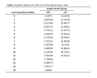
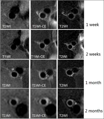
Histological validation of loading lag of heart iron with respect to liver iron in a rabbit model
xiaodong chen, Zipan Chen, Heng Lv, Ziliang Cheng, Qihua Yang, Zuoquan Zhang, Zebin Luo, Jiaji Mao, Queenie Chan, Yingjie Mei, Jingwen Huang, Wubiao Chen, Biling Liang, Hua Guo
Whether there exists a lag in the iron loading between liver and heart in transfusion dependent patients has clinical significance in prevention and treatment of heart iron overload. We performed this study to verify this time lag on a rabbit model. The result shows that the iron loading, measured with pathology, MRI and atomic absorption spectrophotometer, was much faster and heavier in the liver than that in the heart. Therefore, there existed a time lag between heart and liver iron overload on the rabbit model. This may lead to improved clinical guidelines for cardioprotection.


4889.   
18 Multiparametric CMR protocol and analysis methods for the detection of early cardiotoxicity remodelling in the mini-swine.
Delphine Perie, Clémence Balosetti, Hélène Héon, Nagib Dahdah, Farida Cheriet, Matthias Friedrich, Daniel Curnier
Some Cardiovascular magnetic resonance (CMR) studies investigated the long term effects of cancer treatments, but were never applied to the detection of early changes during cardiotoxicity remodelling. The CMR parameters we investigated in the miniature swine therapeutic model with doxorobucin was able discriminate treated animals from controls. Differences were detectible earlier than onset of classical echocardiographic changes. Translating these observations to personalized medicine approach could be the premise for the oncologist to know accurately when the treatment just starts to have deleterious effect on myocardium instead of just observing that the heart was damaged by doxorubicin.


4890.   
19 
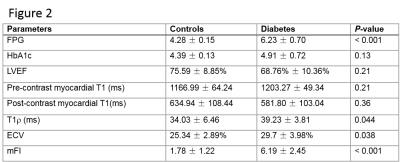
A spontaneous type 2 diabetic rhesus monkey model for cardiac MR study - permission withheld
Yu Zhang, Jie Zheng, Fabao Gao
The aim of this study was to provide a human-like diabetic monkey model for cardiovascular research. Myocardial function and tissue characterizations were measured by CMR in 14 diabetic and 5 control monkeys. In addition to diastolic dysfunction, minor and moderate diffuse fibrosis was shown in cardiac T1, T1r, ECV, and non-contrast fibrosis index images in all the hearts of all diabetic monkeys, confirmed by histopathology finding in one diabetic monkey.  


4891.   
20 
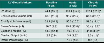
Early Prediction of Chronic Infarct Size by Acute Strain: A Cardiac MRI Study of Myocardial Infarction in Pigs
Sarayu Parimal, Smita Sampath, Ibrahim Mazlan, Grace Croft, Teresa Totman, Yvonne Tay Wei Zheng, Elaine Manigbas, Michael Klimas, Jeffrey Evelhoch, Dominique Kleijn, Chih-Liang Chin
We characterized cardiac structure and function longitudinally in myocardial infarcted pigs, induced by permanent ligation of left circumflex artery of left ventricle (LV), to identify early strain biomarkers that are predictive of late stage remodeling. Pigs were imaged pre- and post-surgery at 1-wk and 4-wk. Reduction of percentage of infarct was observed at basal antero-lateral and mid infero-lateral regions at 4-wk post-surgery. Decreased peak circumferential strain was observed at infarcted areas showing compromised contractility. In addition, ROC analyses revealed that acute strain at 1-wk and early strain change from baseline can predict chronic infarct size suggesting that LV strain could potentially serve as early biomarker for novel therapies.


4892.   
21 Aortic Wall MR Imaging for Assessing Rosuvastatin Therapy in Atherosclerotic Animal Model
Juan Huang, Yan Song , Mingmei Li, Xiaotao Deng, Sheng Jiao, Jingying Yu, Min Chen, Xiaoqi Wang
Here we present the high-resolution MRI imaging for assessing therapy of titrated rosuvastatin for atherosclerosis in rabbit models. This study shows that abdominal aorta wall in both control and treated rabbits get thicker with prolonged continuous feeding of cholesterol, aortic plaques grows bigger with this progression. The MRI vascular wall imaging measured atherosclerotic plaques with well correlation of histological classifications. Statical regression showed that the group with rosuvastatin treatment reduced the aortic plaques comparing to the control rabbits.


4893.   
22 
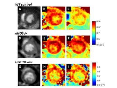
T1 Mapping with Nitric Oxide Synthase (NOS) Inhibition Detects Impaired Coronary Endothelial Function in Mice Fed a High Fat Diet
Sophia Cui, Frederick Epstein
Endothelial nitric oxide synthase (eNOS)-mediated production of NO regulates the microvasculature, controlling both vessel diameter and permeability.  We hypothesized that T1 mapping of the healthy heart during NOS inhibition would detect increased water content resulting from increased coronary microvascular permeability, while a blunted change in T1 between baseline and NOS inhibition would indicate coronary eNOS dysfunction. Using these methods, we detected an increase in myocardial T1 of 113±15 ms due to NOS inhibition in control mice, but no change in eNOS-/- or HFD mice, demonstrating the eNOS mechanism and detection of coronary endothelial dysfunction in a model of heart disease. 
 

The International Society for Magnetic Resonance in Medicine is accredited by the Accreditation Council for
Continuing Medical Education to provide continuing medical education for physicians.