Reconstruction & Post-Processing
Electronic Poster
Acquisition, Reconstruction & Analysis
Wednesday, 26 April 2017
| Exhibition Hall |
16:15 - 17:15 |
| |
|
Computer # |
|
5061.
 |
97 |
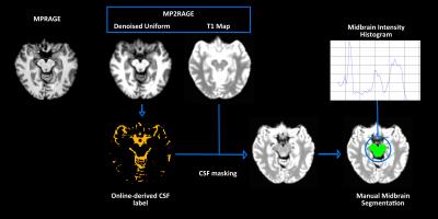
Semi-automated identification of Substantia Nigra in healthy controls and patients with Parkinson's Disease: a feasibility study using MP2RAGE - video not available
Maria Eugenia Caligiuri, Gaetano Barbagallo, Tobias Kober, Umberto Sabatini, Aldo Quattrone, Andrea Cherubini
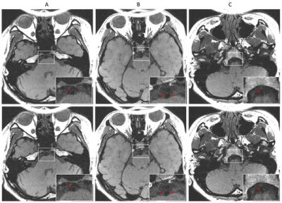
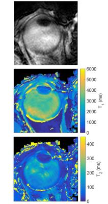
Reliable in vivo assessment of human substantia nigra (SN) requires highly trained operators and different MRI sequences. Advanced techniques have recently facilitated SN identification, but their acquisition in routine clinical practice may not be feasible. MP2RAGE allows for quantitative T1 mapping with an acceptable acquisition time (< 10 minutes). Moreover, SN can be seen on T1 maps, but not on standard MPRAGE. In this study, we tested the feasibility of semi-automated SN identification on MP2RAGE-derived T1 maps by using a thresholding approach, and compared SN volume and T1 values between healthy controls and patients with Parkinson's disease.
|
|
5062.
 |
98 |
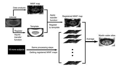
Myelin water atlas for cervical spinal cord: A template for spinal cord pathway myelin microstructure 
Hanwen Liu, Emil Ljungberg, Erin MacMillan, Laura Barlow, Shannon Kolind, John Kramer, Cornelia Laule
In-vivo microstructural information of myelin in the spinal cord is desirable for studying spinal cord injury and neurodegenerative diseases. We used myelin water imaging combine with Spinal Cord Toolbox to create a standard microstructure template specific to myelin content, so-called myelin water atlas, for healthy cervical spinal cord. The resulting atlas is able to distinguish myelin content in 7 different spinal cord pathways and agrees with well-known anatomical characteristics. Our work shows the potential of using a myelin water atlas as a microstructure reference to visualize demyelination in spinal cord injuries or diseases.
|
|
5063.
 |
99 |
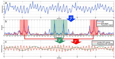
Visualization of Cardiac and Respiratory Pressure Gradient of Cerebrospinal Fluid Based on Asynchronous Two-Dimensional Phase Contrast Imaging 
Saeko Sunohara, Satoshi Yatsushiro, Mitsunori Matsumae, Kagayaki Kuroda
To visualize the distribution of the pressure gradients of the cardiac- and respiratory-driven cerebrospinal fluid (CSF), asynchronous two-dimensional phase-contrast velocity imaging was performed in 9 healthy subjects. The pressure gradients were calculated by the Navier–Stokes equations after the total CSF motion was classified into either cardiac or the respiratory components in the frequency domain. In the prepontine, the pressure gradients in the caudal-to-cranial direction were 14.9 ± 3.17 Pa/m for cardiac components and 1.28 ± 0.46 Pa/m for respiratory components; the cardiac pressure gradient was also significantly larger than the respiratory pressure gradient in other regions.
|
|
5064.
 |
100 |
On the profile ordering of Golden Angle radial Simultaneous Multi-Slice imaging 
Pim Borman, Rob Tijssen, Bas Raaymakers, Chrit Moonen, Clemens Bos
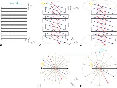
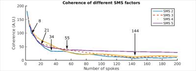
Golden angle radial sampling allows for flexible sliding window reconstructions while minimizing motion sensitivity and undersampling artifacts. Simultaneous MultiSlice (SMS) can be used to increase the spatial coverage without decreasing the framerate. Here we show that the golden angle profile ordering is in principle not compatible with the in-plane phase cycling scheme required by SMS. It follows that these are only compatible for a certain number of spokes, namely when the number of spokes is part of the Fibonacci sequence and the SMS factor is a divisor of this number.
|
|
5065.
 |
101 |
Non-rigid Groupwise Registration for Intravoxel Incoherent Motion Imaging of the Heart 
Valery Vishnevskiy, Georg Spinner, Christian Stoeck, Constantin von Deuster, Sebastian Kozerke
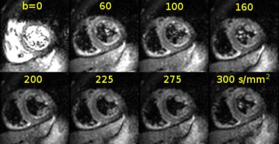
Intravoxel Incoherent Motion (IVIM) imaging is an attractive approach for contrast-agent free in vivo perfusion measurement in the heart. Since the data is acquired during free breathing, nonrigid respiratory motion is considerable and needs to be corrected before estimation of IVIM parameters. In order to provide accurate image registration, an approach with parametric total variation regularization of displacements and low-rank structure of aligned images stack is presented. The proposed method allows for robust IVIM parameter estimation and improves mean squared residuals of the IVIM model by 24% on average compared to state-of-the-art non-rigid registration methods.
|
|
5066.
 |
102 |
A supervised automated segmentation strategy for renal DCE-MR images 
Wenjian Huang, Hao Li, Jue Zhang, Xiaoying Wang, Jing Fang
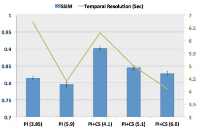
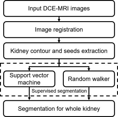
A supervised DCE-MR images classification strategy is proposed in this study. First, the training set was obtained by an automated seeds extraction procedure. Subsequently, support vector machine (SVM) and random walk algorithms were employed as two separate classification approaches to achieve image segmentations, respectively. The automated segmentations and a repeated manual segmentation were compared quantitatively with a reference manual segmentation. The average similarity indexes for SVM, random walker and repeated manual segmentation were 0.78, 0.76 and 0.72, respectively. The results indicate that the proposed strategy yield a satisfied similarity with manual segmentation and is more stable than the manual segmentation.
|
|
5067.
 |
103 |
Improving Accuracy in MR Fingerprinting by Off-Resonance Deblurring - permission withheld
Peter Koken, Thomas Amthor, Mariya Doneva, Holger Eggers, Karsten Sommer, Jakob Meineke, Peter Börnert
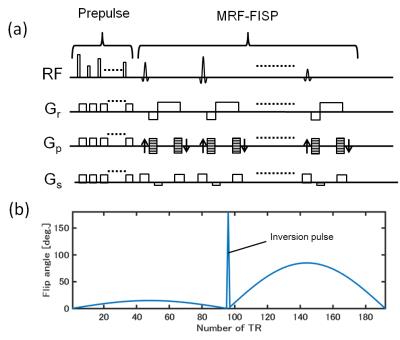
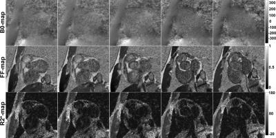
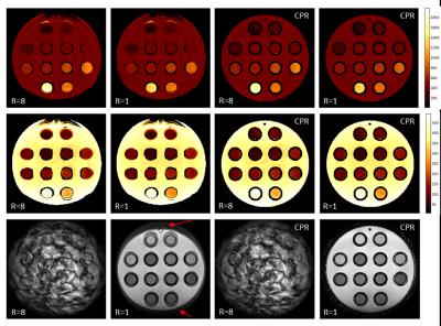
Efficient, highly under-sampled spiral acquisition is preferred in magnetic resonance fingerprinting (MRF). However, although the spiral is very efficient in terms of sampling, it is sensitive to all kinds of off-resonance effects resulting in signal blurring. This effect leads to geometric distortion and matching errors, reducing accuracy significantly. To overcome these limitations, the present work proposes to combine spiral-based MRF with field map-based deblurring, e.g. by conjugate phase reconstruction (CPR). The basic feasibility of this approach for under-sampled MRF is shown in phantom and in-vivo experiments, underlining the effectiveness of this simple correction approach paving the way for even more efficient MRF sampling.
|
|
5068.
 |
104 |
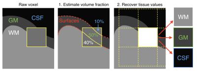
Partial volume effect correction for surface-based cortical mapping 
Camille Van Assel, Gabriel Mangeat, Benjamin De Leener, Nikola Stikov, Caterina Mainero, Julien Cohen-Adad
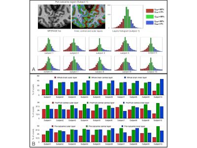
Partial Volume Effect (PVE) hampers the accuracy of studies aiming at mapping MRI signal in the cortex due to the close proximity of adjacent white matter (WM) and cerebrospinal fluid (CSF). The proposed framework addresses this issue by disentangling the various sources of MRI signal within each voxel, assuming three classes (WM, gray matter, CSF) within a small neighbourhood. MRI scans of 17 healthy subjects suggest robust estimations of PVE, allowing accurate extraction of MRI metrics using surface-based analysis. This method can be particularly useful for probing pathology in outer or inner cortical layers, which are subject to strong PVE with adjacent CSF or WM.
|
|
5069.
 |
105 |
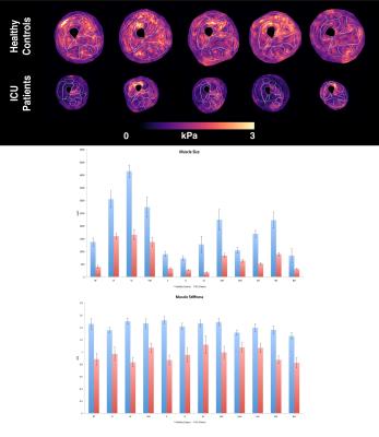
Muscle Change Associated with Time in Intensive Care Unit (ICU) 
Michael Perrins, Lucy Hiscox, Calum Gray, Scott Semple, Lucy Barclay, Rachael Kirkbride, Lisa Salisbury, Colin Brown, Timothy Walsh, Edwin van Beek, Neil Roberts, David Griffith
Muscle wasting is common during critical illness. In this study, thigh muscles of previously mobile patients surviving an episode of severe critical illness were imaged by Magnetic Resonance Imaging (MRI) during convalescence and compared to healthy controls. We present preliminary findings of the first clinical study using Magnetic Resonance Elastography (MRE) to measure muscle stiffness (kPa) and muscle cross-sectional area (mm2) for Intensive Care Unit (ICU) patients. A statistically significant reduction in muscle area and muscle stiffness in patients was found when compared to the healthy control group. There was a significant cross-sectional muscle area increase following ICU patient discharge.
|
|
5070.
 |
106 |
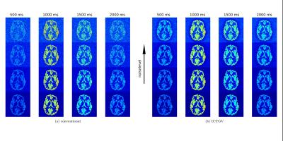
Improved Denoising of Dynamic Arterial Spin Labeling with Infimal Convolution of Total Generalized Variation Functionals (ICTGV) 
Matthias Schloegl, Stefan Spann, Christoph Aigner, Martin Holler, Kristian Bredies, Rudolf Stollberger
Dynamic arterial spin labeling MRI provides important quantitative information about blood arrival time and perfusion. However, the inherently low signal-to-noise ratio requires repeated measurements to achieve a reasonable image quality. This leads to long acquisition times and hence increases the risk of motion artifacts, which impedes clinical applicability. To overcome this limitation we propose to reconstruct the dynamic ASL data employing ICTGV regularization from a reduced number of averages. The performance of the method is evaluated on synthetic and in-vivo ASL data.
|
|
5071.
 |
107 |
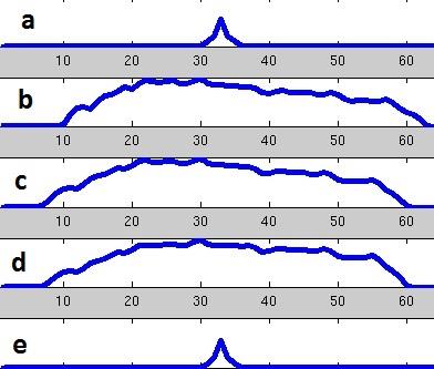
Fast Non-iterative Image Reconstruction for O-space Imaging 
Maolin Qiu, Yuqing Wan, R. Todd Constable
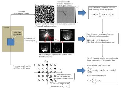
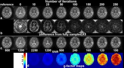
MRI with non-linear spatial encoding magnetic fields (SEM) can provide high quality MR images, but there images are usually calculated using iterative optimization procedures. Such reconstruction methods can take a long time to converge, and the results may depend on the initial iteration parameters. We propose a fast, non-iterative image reconstruction method for O-space imaging based on the local K-space and local SEMs and demonstrate its effectiveness for image reconstruction of nonlinear O-space imaging data.
|
|
5072.
 |
108 |
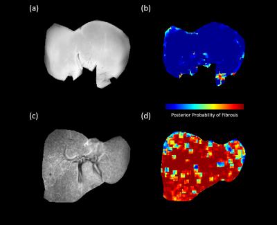
Wavelet based Texture Analysis of Liver Fibrosis in Delayed Phase Gadolinium-Enhanced T1-weighted in vivo Images 
Lavanya Umapathy, Jonathan Brand, Jean-Philippe Galons, Lars Furenlid, Diego Martin, Maria Altbach, Ali Bilgin
Non-invasive imaging techniques that can identify early structural changes due to fibrosis in vivo are of high clinical importance. In this work, a five-level wavelet decomposition of biopsy confirmed normal and fibrotic ex vivo liver tissues is performed and histogram-based features are extracted from the wavelet subbands. A linear classifier is trained using the top 10 features and applied to classify liver fibrosis in Gadolinium-enhanced delayed phase T1-weighted in vivo images. The results show that normal samples yield low posterior probabilities for fibrosis whereas these values are very high for fibrotic samples.
|
|
5073.
 |
109 |
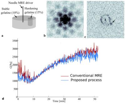
An alternative to phase image-based Magnetic Resonance Elastography (MRE) using k-space data processing 
Nadège Corbin, Elodie breton, Michel de Mathelin, Jonathan Vappou
MR Elastography (MRE) requires substantial data processing involving phase image reconstruction, wave enhancement and inverse problem solving. The objective of this study is to propose an alternative reconstruction method based on direct k-space data processing, particularly adapted to applications requiring fast MRE measurements such as the monitoring of elasticity changes. Elastograms are directly reconstructed from raw MR data without prior phase image reconstruction, circumventing thereby the delicate step of phase unwrapping. The k-space MRE method shows promising results by providing elasticity values similar to the ones obtained with conventional MRE in phantoms and in vivo in porcine liver.
|
|
5074.
 |
110 |
Texture Analysis for Evaluating Image Registration 
Vikas Kotari, Vasiliki Ikonomidou
Accurate image registration is essential for both cross-sectional and longitudinal MR studies. In longitudinal studies aligning same contrast intra-subject images, which are the focus of our work, registration is assumed to be a rigid body problem. This assumption is questionable due to global and local changes in brain volume either due to hydration or atrophy. Consequently, misregistration at the voxel level may occur in these studies, which might lead to subject data being discarded. This misregistration is evident particularly around the cortex. Visual inspection of images is used to determine registration accuracy. While this approach is suitable for assessing alignment of landmark structures, it fails to capture the millimteric or sub-millimetric misregistrations. Automatic metrics can precisely estimate the overall performance of a given registration algorithm by employing an evaluation database. However, to estimate the registration accuracy of a given pair of images, such metrics are unsuitable. In this work we propose texture analysis of a subtraction image to evaluate the registration accuracy of a given pair of same contrast, intra-subject images. Once registered, images are intensity normalized, blurred and subtracted. In the event of registration errors, or violation of the rigid body assumption, the subtraction images have artifacts. The texture features of these artifacts are different from the artifact-free (clean) areas of the subtraction images. Using a texture-based classifier, artifact areas in the subtraction images that indicate failed registration are identified. In addition to determining if the registration has failed, our approach can identify the specific locations of misregistration, which can be corrected, leading to a more inclusive subject data.
|
|
5075.
 |
111 |
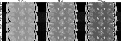
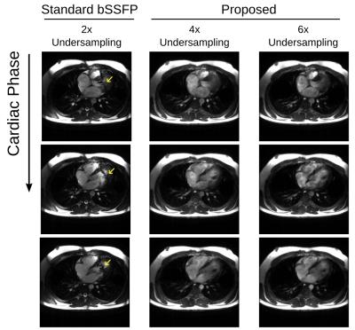
Dynamically Phase-Cycled bSSFP Cardiac Cine in a Single Breathhold with Phase-Cycle Consistency Regularization 
Corey Baron, Anjali Datta, Dwight Nishimura
At high field strengths, cardiac cine acquisitions acquired with balanced SSFP can suffer from banding artifacts. To mitigate this issue, dynamically phase-cycled cardiac cine was acquired in a single breathhold with undersampling rates of 4 and 6, and images were reconstructed with a phase-cycle banding profile regularization that exploits redundancy between phase-cycles.
|
|
5076.
 |
112 |
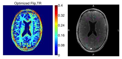
Myelin Water Fraction Estimation from Optimized Steady-State Sequences using Kernel Ridge Regression 
Gopal Nataraj, Jon-Fredrik Nielsen, Jeffrey Fessler
This work introduces a new framework for myelin water fraction (MWF) estimation. We use a novel scan design approach to construct a sequence a fast steady-state sequences and optimize corresponding flip angles and repetition times for precise MWF estimation. We quantify MWF and five other parameters per voxel using a novel method based on kernel ridge regression. We obtain MWF maps in vivo that are comparable to those reported in literature, with possibly shorter overall scan time.
|
|
5077.
 |
113 |
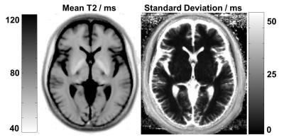
A T2 Template Map from a Healthy Cohort to Identify Localized Anomalies in Single Subjects 
Tom Hilbert, Alexis Roche, Cristina Granziera, Guillaume Bonnier, Kieran O’Brien, Tony Stöcker, Pavel Falkovskiy, Reto Meuli, Jean-Philippe Thiran, Gunnar Krueger, Tobias Kober
We construct a database of normal T2 values by spatially normalizing quantitative maps from healthy subjects into a common space. A low standard deviation across all subjects demonstrates good reproducibility of the T2 values in white matter and deep grey matter. Additionally we adopt a standard voxel-based procedure that compares the quantitative T2 map of a patient to the database and test it on three multiple sclerosis datasets. The obtained z-score maps show that white matter lesions can be detected in the limits of the available resolution and the applied smoothing.
|
|
5078.
 |
114 |
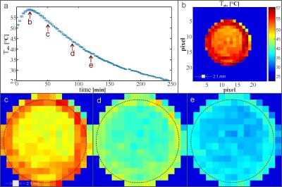
Temperature Mapping of Fluorinated (19F) Gas in a Cool Down Experiment 
Tobias Hoh, Eduardo Coello, Jorge Carretero Benignos, Axel Haase, Rolf Schulte
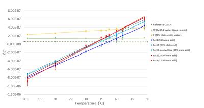
Magnetic resonance imaging (MRI) temperature mapping of gases is challenging due to limited sensitivity. In this proof of concept study, proton thermometry methods from tissue temperature monitoring were transferred to 19F gas MRI. A phase-dependent 19F resonance frequency shift temperature mapping is proposed for fluorinated gas at 3T based on a spiral readout to overcome limitations of ultra-short relaxation times.
This work demonstrates the feasibility of 2D thermometry in a canonical setup with inert fluorinated gases.
|
|
5079.
 |
115 |
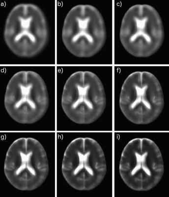
Improved 23Na MRI Quantification of the Human Brain at 3T Using Partial Volume Correction Techniques 
Tie-Qiang Li, Elaine Lui, Patricia Desmond
In this study we have focused on the development and application of PVC techniques for improving the quantification accuracy of STC with 23Na MRI at 3T. Although PVEs are known to induce errors in quantification, it has not been widely used in 23Na MRI. While different PVC algorithms have been proposed in the PET literature, each method has its limitations and relies on simplified assumptions. The proposed hybrid methods, such as GTM+RBV, aimed to overcome such limitations are most robust.
|
|
5080.
 |
116 |
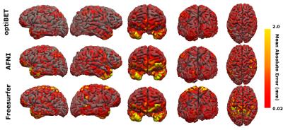
Optimization of brain extraction increases global cortical thickness accuracy 
Antonio Carlos Senra Filho, Gareth Barker, Luiz Otávio Murta Junior, Flavio Dell'Acqua
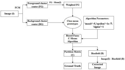
Reconstruction of the cortical surface is of great importance as a biomarker for many brain diseases, and in recent years a number of advances in image processing and analysis have been made in the cortical reconstruction process. However, despite the scientific community employing a range of advanced surface reconstruction algorithms in order to improve quantitative accuracy, cortical thickness measurement is still a challenge. Here, we address the question of whether a better brain extraction procedure can improve the cortical surface reconstruction. Our analyses suggest that a more accurate brain mask directly affects the global cortical thickness estimate, reducing its quantitative uncertainty.
|
|
5081.
 |
117 |
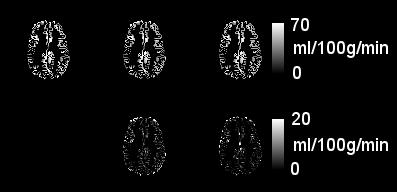
ADRIMO: Anatomy-DRIven MOdelling of spatial correlation to improve analysis of arterial spin labelling data 
David Owen, Andrew Melbourne, David Thomas, Joanne Beckmann, Jonathan Rohrer, Neil Marlow, Sebastien Ourselin
Arterial spin labelling (ASL) offers valuable measurements of perfusion in the brain and other organs. However, ASL data have low SNR and are prone to partial volume effects. We present a Bayesian model of anatomically-derived spatial correlation in ASL data (ADRIMO), which improves the accuracy of perfusion estimates and hence improves the analysis of ASL data. The method is assessed experimentally by examining ASL images from a cohort of 130 preterm-born adolescents.
|
|
5082.
 |
118 |
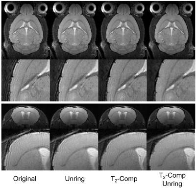
Reduction of ringing artifacts for high resolution 3D RARE imaging at high magnetic field strengths 
Martin Krämer, Karl-Heinz Herrmann, Silvio Schmidt, Otto Witte, Jürgen Reichenbach
High resolution 3D-RARE imaging at 9.4T can be very challenging due to increased artifacts caused by strong T2- and Gibbs ringing. In this work, we present the combination of two correction algorithms, local subvoxel-shift unringing and T2-compensation, to reduce effectively both types of artifacts. For this purpose, the local subvoxel-shift algorithm has been extended to the third spatial dimension and evaluated in healthy mice.
|
|
5083.
 |
119 |
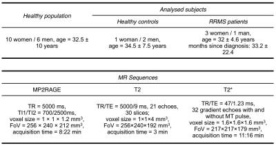
Personalized map to assess diffuse and focal brain damage - permission withheld
Guillaume Bonnier, Alexis Roche, Tom Hilbert, Gunnar Krueger, Cristina Granziera, Tobias Kober
We propose a new methodology, which is based on the quantification of region-specific brain tissue-properties to provide personalize maps of brain damage in a single patient. To achieve this aim, we used T1, T2, T2* and MTI maps and applied the method to detect brain abnormalities in multiple sclerosis patients.
|
|
5084.
 |
120 |
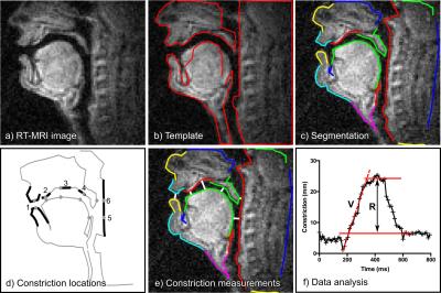
Test-retest repeatability of human speech biomarkers from static and real-time dynamic magnetic resonance imaging 
Johannes Toger, Tanner Sorensen, Krishna Somandepalli, Asterios Toutios, Sajan Goud Lingala, Shrikanth Narayanan, Krishna Nayak
This study presents a test-retest repeatability framework for quantitative speech biomarkers from static MRI and real-time MRI (RT-MRI), and applies the framework to healthy volunteers (n=8). Repeatability was quantified using intraclass correlation coefficient (ICC) and mean within-subject standard deviation (σe). Inter-study agreement was strong to very strong for static anatomical biomarkers, (ICC: min/median/max 0.71/0.89/0.98, σe: min/median/max 0.90/2.20/6.72 mm), poor to very strong for dynamic RT-MRI biomarkers of articulator motion range (ICC: 0.26/0.75/0.90, σe: 1.6/2.5/3.6 mm) and poor to very strong for velocity (ICC: 0.26/0.56/0.93, σe: 2.2/4.4/16.7 cm/s). The introduced framework can be used to guide future development of speech biomarkers.
|
|

