Tuesday, 25 April 2017
| Room 313A |
16:15 - 18:15 |
Moderators: Myriam Chaumeil, Afonso Silva |
Slack Channel: #s_neuro
Session Number: O01
16:15
|
0590.
 |
Investigation of the role of the venous system and the glymphatic system in brain waste clearance 
Yimin shen, Quan Jiang, Guangliang Ding, Nicholas Guys, E. Mark Haacke, Jiani Hu*
The recently discovered glymphatic system has become an exciting area of research because of its broad implications in both normal neurophysiological activities and neurological disorders. However, the exact relationship between the vascular system and the glymphatic system in terms of waste clearance for the brain is unclear . In addition to the glymphatic system, our preliminary MRI results suggest that the venous (but not the arterial) system also directly participates in waste removal. Fully elucidating the roles of the venous and glymphatic systems in waste removal from the brain is important for understanding the influence of waste clearance on neurological diseases.
|
16:27
 |
0591.
 |
Anterograde manganese transport in donor optic nerve following whole eye transplantation
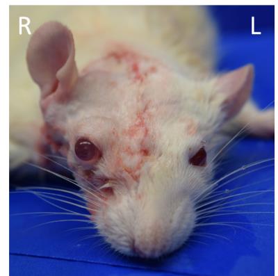
Yolandi van der Merwe, Chiaki Komatsu, Lin He, Maxine Miller, Ian Rosner, Huamin Tang, Joel S. Schuman, Jose-Alain Sahel, Michael B. Steketee, Kia M. Washington, Kevin C. Chan
Approximately 39 million people worldwide suffer from irreversible blindness. Our recently established whole eye transplant model (WET) gives the opportunity to provide an intact optical system that could restore lost vision. In this study we use manganese-enhanced MRI to examine the anterograde transport of the transplanted and recipient visual pathways following WET. Our results show comparable manganese enhancement between donor and naïve intraorbital optic nerves, suggesting the presence of anterograde manganese transport in the donor optic nerve. This in vivo imaging model system may allow future examinations of neuroregenerative approaches for connecting between the transplanted eye and the recipient’s brain.
|
16:39
|
0592.
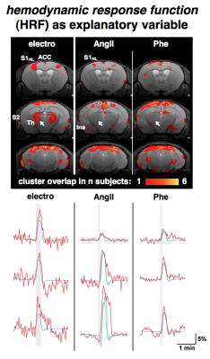 |
Detailing the Origin of BOLD fMRI in Mice: Somatosensory Stimulation versus Pharmacologically Induced Blood Pressure Alterations
Henning Reimann, Mihail Todiras, Till Huelnhagen, Michael Bader, Andreas Pohlmann, Thoralf Niendorf
The combination of somatosensory fMRI (sfMRI) and mouse genomics holds great potential to unravel the underlying mechanisms of chronic pain. Transient stimuli were shown to induce unspecific BOLD patterns in mice. It is known that increases in mean arterial blood pressure (MABP) can mimic BOLD activations in rats. To detail the origin of the unspecific BOLD patterns evoked in the murine brain we performed sfMRI along with pharmacologically induced blood pressure alterations while monitoring MABP. We compared MABP changes, BOLD signals and BOLD patterns between the types of stimuli and observed confounding effects of MABP on murine BOLD fMRI.
|
16:51
|
0593.
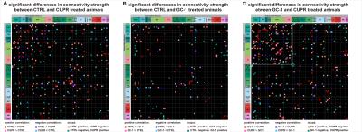 |
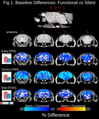
Remodeling of resting state functional connectivity following thyromimetic induced remyelination in the mouse brain - permission withheld
Neele Hübner, Annika Hammerschmitt, Thiago Marques De Melo, Thomas Bienert, Jürgen Hennig, Dominik von Elverfeldt, Laura Harsan
Characterization of the brain network architecture and its alterations in pathologies are essential for a better understanding of mechanisms of action in health and disease and paves the way for the development of targeted therapeutic strategies. In the field of demyelinating disorders, thyromimetic treatment provides a promising remyelinating strategy inducing oligodendrogenesis in vitro. We tested the potential of sobetirome (GC-1), a thyroid hormone analogue, to induce remyelination in vivo in the cuprizone demyelinated mouse model and we investigated its action on the resting state (rsfMRI) mouse brain functional connectivity.
|
17:03
|
0594.
 |
Genetic rescue of brain morphometry in a mouse model of neurodevelopmental disorder
Rylan Allemang-Grand, Jacob Ellegood, Leigh Spencer Noakes, Brian Nieman, Jason Lerch
In this study, we scanned a mouse model of Rett syndrome before and after reactivation of Mecp2, the gene strongly implicated in the disorder. We found that reactivation of Mecp2 at three different time points in adulthood lead to drastic growth of the neuroanatomy across many regions of the cortex, cerebellum and medulla. Our findings demonstrate that the developmental delayed brain retains an innate plasticity that can be recruited to restore neuroanatomical structure in adulthood.
|
17:15
|
0595.
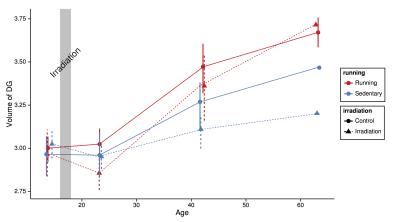 |
Can mice outrun the deleterious impacts of radiation to the brain?
Kamila Szulc, Shannon Egan, Elizabeth de Guzman, Aidin Arbabi, Donald Mabbott, Brian Nieman
Pediatric cancer patients who receive cranial radiation therapy (CRT) exhibit cognitive deficits later in life. These deficits are often accompanied by brain structure abnormalities, especially prevalent in the white matter and hippocampus. The objective of this study was to explore the potential of physical exercise to mitigate some of the deleterious effects of CRT on the brain, using a mouse model and high-resolution MRI as a measure of brain structure. We found that irradiated mice housed in cages with access to running wheels showed a remarkable recovery of a number of CRT-induced brain volume deficits, most notably in the hippocampus.
|
17:27
|
0596.
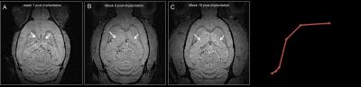 |
MRI visualization of brain-like tissue formation following implantation of neural precursors into in cerebrospinal fluid
Nikorn Pothayee, Dragan Maric, Kathryn Sharer, Jung-Hwa Tao-Cheng , Stephen Dodd, Alec Calac, James Pickel, Alan Koretsky
Neural stem cell transplantation has been hailed as a promising approach for treatment of neurological diseases. While most in vivo studies have implanted cells into specific sites in brain tissue, little is known whether the cerebrospinal fluid (CSF) provides a permissive environment in cultivating tissue growth. Here, using MRI, we investigate whether early neural precursor cells could initiate a large-scale formation of new brain tissue in the CSF of adult rat.
|
17:39
|
0597.
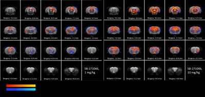 |
Use of Pharmacological MRI (phMRI) to understand the mechanisms leading to convergent procognitive effects of 5-HT6 serotonergic receptors agonist (EMD-386088) and antagonist (SB-271046).
Willy Gsell, Rachel Asselot, Nicolas Delcroix, Uwe Himmelreich, Valentine Bouet, François Dauphin
Either blocking or activating 5HT6 receptors (5-HT6R), respectively with antagonists or agonists, exert beneficial cognitive effects. Through phMRI, we demonstrated for the first time the similarities and discrepancies in the brain activation induced by an agonist and an antagonist of 5-HT6R. Both drugs similarly activate cortices and hippocampus. SB-271046 positively activates a network including the medio-dorsal raphe while EMD-386088 activates the rostral dorsal raphe. The different patterns of activation elicited in brain regions such as the habenula and the MG/DG nuclei (for SB-271046), or the amygdala and substantia nigra (for EMD-386088) supports different interactions with polysynaptic pathways.
|
17:51
|
0598.
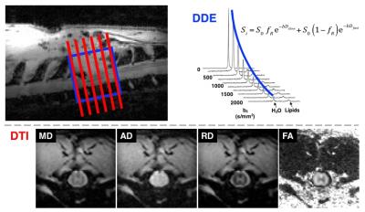 |
Predicting Functional and Histological Outcomes in Spinal Cord Injury: Comparing Double Diffusion Encoding and Diffusion Tensor Imaging in the Rat
Nathan Skinner, Shekar Kurpad, Brian Schmit, Natasha Beucher, Kyle Stehlik, L. Muftuler, Matthew Budde
Using a rat model of spinal cord injury (SCI), diffusion tensor imaging (DTI) is compared to double diffusion encoding (DDE) at the acute and chronic stages after injury. Acute DDE measurements show a strong relationship with chronic functional outcomes whereas DTI has poor prognostic sensitivity. On the other hand, during the chronic stage, DTI outperforms DDE as a marker of functional status. The differences reflect evolving pathologies that must be considered for the appropriate application and interpretation of DTI and DDE. The results also highlight the prognostic potential of DDE in acute SCI.
|
18:03
|
0599.
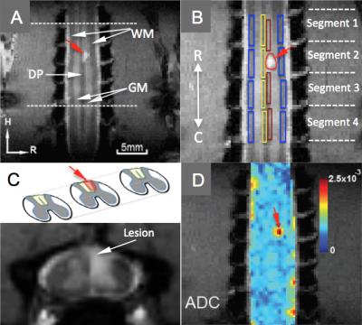 |
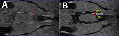
Spinal DTI Parametric Changes Following Traumatic Injury in Monkeys
Arabinda Mishra, Feng Wang, Li Chen, John Gore
The proposed DTI study aims at quantification of the effect of experimentally induced dorsal column lesion at upper cervical level (C4/5) in squirrel monkeys. Diffusion parametric changes based on the directionality, and mobility of water molecules in the cellular environment, characterizes the spinal cord. Change in diffusion parameters on specific white matter tracks above and below the SCI location were compared between the lesioned and normal side of the spinal cord. A systematic group analysis of the inter-ROI changes along the white matter pathways can therefore be used to correlate the loss and recovery of sensory/motor functions over time.
|
|