Tuesday, 25 April 2017
| Room 320 |
08:15 - 10:15 |
Moderators: Nivedita Agarwal, Gary Miller |
Slack Channel: #s_neuro
Session Number: O08
08:15
|
0412.
 |
SPINAL CORD SODIUM AND AXONAL LOSS IN MULTIPLE SCLEROSIS 
Bhavana Solanky, Ferran Prados , Carmen Tur, Sebastien Ourselin, Xavier Golay, Olga Ciccarelli, Claudia Gandini Wheeler-Kingshott
Increased sodium concentration in the normal appearing brain of multiple sclerosis (MS) patients has recently been reported, which may be a consequence of axonal loss or an accumulation of intracellular sodium. Here we investigated spinal cord total sodium concentrations in MS patients and healthy controls. Spinal cord atrophy, as a result of axonal loss, often presents in MS, therefore we also tested for the association of sodium with SC area. We found increased total sodium levels in the SC, but no correlation with the decrease in SC cross sectional area in the MS cohort.
|
08:27
 |
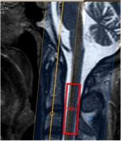
0413.
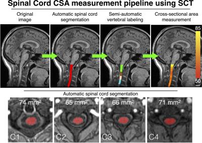 |
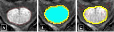
Repeatability and reproducibility of spinal cord atrophy measurements in a multiple sclerosis population using the Spinal Cord Toolbox
Benjamin De Leener, Tobias Granberg, Katharina Fink, Nikola Stikov, Julien Cohen-Adad
Spinal cord atrophy is a major determinant of physical disability in multiple sclerosis (MS) and other diseases with neurodegeneration. The upper spinal cord cross-sectional area (CSA) is therefore a clinically important measurement reflecting global spinal cord atrophy. New image analysis software enable semi- and fully-automatic quantification of spinal cord atrophy. This study characterizes the repeatability and reproducibility of semi-automatic CSA measurements of the spinal cord in healthy subjects and in patients with multiple sclerosis, using the Spinal Cord Toolbox (SCT). Results demonstrated the high repeatability and reproducibility of CSA measures using SCT in both healthy persons and in MS.
|
08:39
|
0414.
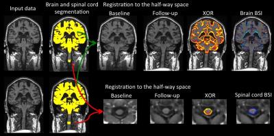 |
Boundary shift integral to compute brain and cervical spinal cord longitudinal volume changes using the same 3DT1w volumetric scans in multiple sclerosis
Ferran Prados, Özgür Yaldizli, Manuel Jorge Cardoso, Marios Yiannakas, Francesco Grussu, Esther Ruberte, Jens Wuerfel, Frederik Barkhof, Claudia Gandini Wheeler-Kignshott, Sebastien Ourselin
Brain atrophy is considered to be the net accumulative irreversible disease burden as the ultimate consequence of different pathological processes found in the multiple sclerosis brain. A recent cross-sectional study demonstrated the possibility to assess atrophy of the spinal cord using 3DT1w brain volumetric scans. However, to date, no unified technique has been presented to longitudinally assess brain and spinal cord atrophy using the same MRI acquisition. Here we present a proof-of-concept data from a pipeline that uses the boundary shift integral to compute both brain and cervical spinal cord atrophy rates using the same 3DT1w brain volumetric scans.
|
08:51
|
0415.
 |
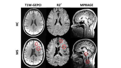
Improved Visualization of the Spinal Cord Lesions in Multiple Sclerosis using MP2RAGE at 3T
Jennifer Lefeuvre, Sunil Patil, Gunnar Krueger, Tobias Kober, Stéphane Lehéricy, Daniel Reich, Govind Nair
Spinal cord lesions are known to be prevalent and contribute to disability in multiple sclerosis (MS). We explored the use of the MP2RAGE sequence for better delineation of spinal cord lesions in a clinical setting, and compared it with standard sequences used in MS. A more accurate measurement of lesion load and better visualization of the lesion location within the spinal cord can help improving MS diagnosis and may be developed to an imaging biomarker of disease burden.
|
09:03
|
0416.
 |
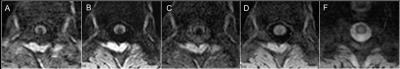
Outer spinal cord rim visualization using magnetization-prepared 3D T1w TFE at 3T: Application to multiple sclerosis.
Marios Yiannakas, Torben Schneider, Matthew Clemence, James Fairney, Ferran Prados, Hugh Kearney, Sebastien Ourselin, David Miller, Claudia Gandini Wheeler-Kingshott
Neuropathological studies in multiple sclerosis (MS) have suggested that meningeal inflammation in the brain and spinal cord (SC) may be associated with disease progression. However, the use of structural imaging protocols to depict meningeal tissues has not been reported. Herein, we investigate the feasibility of obtaining sufficiently high image contrast to resolve the SC rim that we speculate corresponds to the SC pia mater (SCPM) using the magnetization-prepared 3D T1w TFE sequence. Preliminary results show that it is possible to resolve SCPM, and that significant differences in signal intensity values within SCPM exist between healthy controls and people with MS.
|
09:15
|
0417.
 |
On the Relationship between Tissue Damage in Cervical Spinal Cord and Brain in Multiple Sclerosis.
Biao Xiang, Jie Wen, Amber Salter, Anne Cross, Dmitriy Yablonskiy
We examined quantitative relationships between tissue damage in the cervical spinal cord (characterized by cross-sectional area (CSA)) and cortical gray matter (GM) (characterized by thickness and tissue specific R2* values) in multiple sclerosis (MS) patients, and determined relative contributions of these to neurologic disability. We found correlations between CSA and GM R2* values and thickness of several cortical regions. Compared with cortical R2* and thickness, cervical spinal cord CSA correlated better with neurological impairment status. CSA, thickness and age-corrected R2* values all differentiated MS subjects from healthy Controls. CSA and GM thickness could further distinguish MS clinical subtypes.
|
09:27
|
0418.
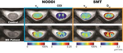 |
Investigation of Advanced Diffusion Models in the Spinal Cord: Comparison of NODDI and SMT in MS Patients
Samantha By, Junzhong Xu, Bailey Box, Seth Smith
NODDI and Spherical Mean Technique (SMT) are multi-compartmental diffusion models that have demonstrated promise in the brain, but have never been applied to the in vivo spinal cord in multiple sclerosis (MS) patients. These models estimate axonal volume fractions, which can be indicative of microstructural integrity. We apply NODDI and SMT in healthy controls and MS patients to evaluate feasibility of both models in the human spinal cord. Results indicate that NODDI (p=0.004) and SMT (p=0.030) can characterize disparity in axonal volume fraction in MS patients, suggesting potential utility of advanced diffusion models in the spinal cord.
|
09:39
|
0419.
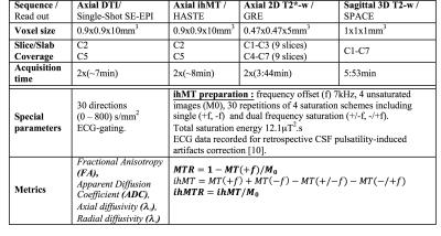 |
Column-specific demyelination in Spinal Cord Normal Appearing White Matter occurring in Multiple Sclerosis: A preliminary study using inhomogeneous Magnetization Transfer and DTI
Henitsoa RASOANANDRIANINA, Bertrand AUDOIN, Olivier GIRARD, Guillaume DUHAMEL, Sarah DEMORTIERE, Maxime GUYE, Jean-Philippe RANJEVA, Jean PELLETIER, Virginie CALLOT
This preliminary study investigates the added value of the inhomogeneous Magnetization Transfer (ihMT) technique when studying Spinal Cord (SC) tissue demyelination occurring in Multiple Sclerosis (MS). DTI and MT/ihMT data collected in 9 MS patients, analyzed in specific normal appearing white matter regions, indicated significant demyelination in corticospinal and posterior sensory tracts as compared to age-matched healthy controls, with a very high sensitivity of ihMTR that appears very promising for further objective therapeutic evaluation or topographic quantification of progressive SC demyelination.
|
09:51
|
0420.
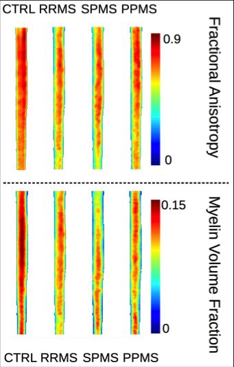 |
Comparative Myelin Imaging and Multi-Shot Diffusion Tensor Imaging in Multiple Sclerosis Cervical Spinal Cord
Hagen Kitzler, Hannes Wahl, Gesine Pach, Judith Eisele, Katja Thomas, Sean Deoni, Tjalf Ziemssen, Jennifer Linn
Spinal cord myelin imaging holds potential to quantify cervical spinal cord tissue myelin loss, and hence, the subtle spinal burden of demyelinating diseases, invisible for conventional magnetic resonance imaging. We present the approach of combined spinal 3D double-inversion recovery imaging with diffusion tensor imaging and myelin sensitive multicomponent driven equilibrium single pulse observation of T1 and T2, with subsequent normal spine space registration and group comparison in different multiple sclerosis courses. Whereas no significant differences were found between the different disease courses using fractional anisotropy, relapsing-remitting and primary-progressive multiple sclerosis were discriminated based on differences in relative cord myelin content.
|
10:03
|
0421.
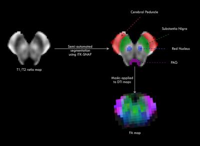 |
DTI abnormalities in the midbrain in pediatric-onset multiple sclerosis.
Ritobrato Datta, Sophia Ly, Christine Till, Elisea De Somma, Nadine Akbar, Sudipto Dolui, Douglas Arnold, Sridar Narayanan, Brenda Banwell
Pediatric-onset MS (POMS) is characterized by a high frequency of brainstem lesions early in the disease. It is unknown whether normal-appearing tissue in this region is involved at this early time point. Using diffusion tensor imaging (DTI) at 3T, we evaluated fractional anisotropy (FA) changes in midbrain substructures in POMS patients and age- and sex-matched healthy controls. Mean FA of midbrain was significantly reduced in the MS group, a difference that remained significant even after removing any focal midbrain lesions. Our results indicate a widespread disruption in the midbrain that exceeds lesional tissue disruption alone.
|
|