Monday, 24 April 2017
| Room 320 |
13:45 - 15:45 |
Moderators: Nivedita Agarwal, Yukio Miki |
Slack Channel: #s_neuro
Session Number: O09
13:45
 |
0212.
 |
Metabolic counterparts of sodium accumulation in Multiple Sclerosis: A whole brain 1H-MRSI and 23Na-MRI study 
Maxime Donadieu, Adil Maarouf, Yann Le Fur, Soraya Gherib, Elisabeth Soulier, Lauriane Pini, Stanislas Rapacchi, Sylviane Confort-Gouny, Maxime Guye, Jean Pelletier, Bertrand Audoin, Wafaa Zaaraoui, Jean-Philippe Ranjeva
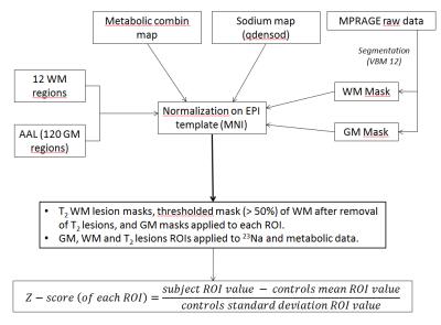
To determine the metabolic counterparts of cerebral total sodium accumulations in patients with Multiple Sclerosis, we acquired fast 3D-1H-EPSI and Density-adapted 3D-UTE 23Na MRI at 3 Tesla covering the whole brain in 21 patients and 20 volunteers. Patients showed increased 23Na and decreased NAA, Glx and Cho levels. Stepwise analyses highlights association of 23Na accumulations with i) decreased NAA and Glx levels and increased Cho levels within GM, ii) with decreased NAA and increased Cho levels within NAWM and T2 lesion compartments. Clinical status of patients assessed by MSFC was correlated to GM and NAWM 23Na, NAA and Glx levels.
|
13:57
|
0213.
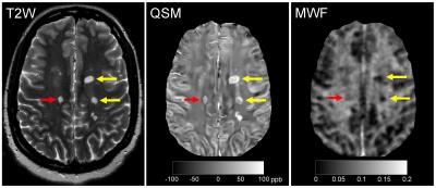 |
MS lesions demonstrating a QSM hyperintense-rim have more myelin loss compared to those without a QSM hyperintense-rim
Yihao Yao, Thanh Nguyen, Sneha Pandya, Sandra Hurtado Rúa, Amy Kuceyeski, Yi Wang, Susan Gauthier
Iron causes proinflammatory activation of microglia near the rim of white matter MS lesion. This is chronic inflammation with associated myelin tissue damage. We propose to use quantitative susceptibility mapping (QSM) to assess chronic inflammation, as hyperintense rim on QSM can be unequivocally interpreted as iron. We use myelin water fraction (MWF) to measure myelin. We have found that MS lesions with hyperintense rims on QSM have lower MWF and higher susceptibility compared to lesions without hyperintense rims on QSM (p<0.01). Hyperintense rim on QSM may provide a biomarker for tissue injury due to iron associated chronic inflammation.
|
14:09
|
0214.
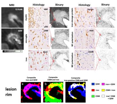 |
Combining QSM and MWF in multiple sclerosis: a marker for the inflammatory state of MS lesions?
Carsten Stueber, Alexey Dimov, Kofi Deh, Thanh Nguyen, Yi Wang, David Pitt
Multiple sclerosis (MS) is a demyelinating disease of the central nervous system. In particular, excess iron is considered to play an essential role in lesion activity. In this study, we combine iron-reflecting quantitative susceptibility mapping (QSM) and myelin water fraction (MWF) with histology in post-mortem tissue. Our results show that elevated iron concentrations at the lesion rim reflect pro-inflammatory microglial activity, suggesting to use QSM to determine levels of lesion inflammation and MWF for detecting ongoing demyelination.
|
14:21
 |
0215.
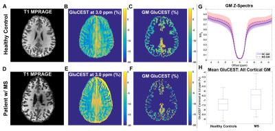 |
Glutamate-Sensitive CEST in Cortical Gray Matter: Application to Cognitive Impairment in Multiple Sclerosis
Kristin O'Grady, Adrienne Dula, Bailey Lyttle, Benjamin Conrad, Bailey Box, Siddharama Pawate, Francesca Bagnato, Seth Smith
Altered glutamate regulation in gray matter (GM) has been implicated in the pathogenesis of cognitive impairment in multiple sclerosis (MS), but such pathology in GM is subtle and difficult to detect using conventional MRI techniques. In this work, we apply a quantitative, glutamate-sensitive chemical exchange saturation transfer (GluCEST) MRI technique at 7.0T to gain new insights into molecular changes underlying GM pathology and their relationship to cognitive impairment in MS. We found significant differences in cortical GM GluCEST contrast between healthy controls and patients with MS, and in some cortical regions, GluCEST contrast correlates significantly with measures of cognitive impairment.
|
14:33
|
0216.
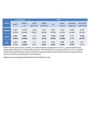 |
Using myelin water and diffusion basis spectrum imaging to differentiate demyelination, inflammation, oedema and axonal damage in subjects with multiple sclerosis
Irene Vavasour, Peng Sun, Shannon Kolind, David Li, Alex MacKay, Sheng-Kwei Song, Robert Carruthers, Anthony Traboulsee
This study compared myelin water fraction (MWF), intra/extracellular water geometric mean T2 (ieGMT2) and diffusion basis spectrum imaging (DBSI)-derived measures in multiple sclerosis (MS) lesions and normal appearing white matter. 14 MS subjects were scanned with 48-echo T2 relaxation and DBSI sequences. Significant correlations were found for MWF vs radial diffusivity, MWF vs fiber fraction, and ieGMT2 vs restricted fraction. Lesions showed changes consistent with decreased myelin and axons. Enhancing lesions also showed increased oedema. By quantitatively distinguishing and tracking inflammation, axon and myelin injury, DBSI and myelin water imaging can inform us of the pathological processes involved in MS.
|
14:45
|
0217.
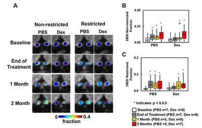 |
Corticosteroid Treatment Fails to Prevent Long-term Axonal Loss Assessed by Diffusion Basis Spectrum Imaging
Tsen-Hsuan (Abby) Lin, Jie Zhan, Chunyu Song, Michael Wallendorf, Peng Sun, Anne Cross, Sheng-Kwei Song
Glucocorticoids are commonly used to treat acute optic neuritis. Herein, we employed longitudinal diffusion basis spectrum imaging (DBSI) to examine and compare optic nerve integrity in EAE with PBS or Dexamethasone treatment. Our results indicate that anti-inflammatory treatment with corticosteroids alone is not sufficient to prevent eventual axonal loss in mice, and may have relevance for treatment of MS exacerbations with corticosteroids. DBSI could serve as an outcome measure to monitor longitudinal disease progression and to help stratify treatments.
|
14:57
|
0218.
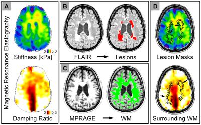 |
Multiple sclerosis lesions are softer than surrounding white matter: An MR elastography study
Curtis Johnson, Christian Thompson, Brian Sandroff, Thomas Edwards, Elizabeth Hubbard, Rachel Klaren, Hillary Schwarb, Bradley Sutton, Lara Pilutti, Robert Motl
Mechanical properties of the brain measured with magnetic resonance elastography (MRE) have proven sensitive to tissue health in neurological conditions, including multiple sclerosis (MS). In this study, we use high-resolution MRE to examine the mechanical properties of focal lesions in subjects with MS to determine if they exhibit viscoelastic signatures that differ from surrounding white matter. In a sample of fourteen subjects, we found that lesions are significantly softer than surrounding white matter. This finding suggests MRE is sensitive to tissue disruption localized to focal lesions, and may provide novel measures of tissue health in the assessment of MS.
|
15:09
|
0219.
 |
Investigation of outer and inner cerebellar MTR abnormalities in different MS clinical subtypes
Rebecca Samson, Manuel Cardoso, Nils Muhlert, Varun Sethi, Özgür Yaldizli, Maria Ron, Ferran Prados , Sebastian Ourselin, David Miller, Claudia Gandini Wheeler-Kingshott, Declan Chard
Histopathology has demonstrated extensive grey matter (GM) damage in MS, and an association with meningeal inflammatory factors has previously been suggested. We applied a method to subdivide the cerebellar GM (CGM) into inner and outer regions, and investigated for magnetization transfer ratio (CGM-MTR) abnormalities in MS subtypes compared to healthy controls (HC). Outer was lower than inner CGM-MTR in all groups including HC. Outer and inner CGM-MTR reductions were observed in progressive MS subtypes. Stronger correlations of outer than inner CGM-MTR with clinical scores were observed, suggesting that outer CGM-MTR may reflect more clinically relevant pathology, particularly in progressive MS.
|
15:21
|
0220.
 |
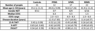
High Spatial Resolution Mapping of Trans-Capillary Water Exchange in Progressive Multiple Sclerosis
Ian Tagge, Manoj Sammi, Rebecca Spain, Dennis Bourdette, Randy West, John Grinstead, Katherine Powers, Xin Li, Charles Springer, William Rooney
DCE-MRI data were acquired from 14 healthy control (HC) and 16 secondary progressive multiple sclerosis subjects on a 7T MRI instrument to investigate difference in brain blood vessel properties. The Shutter-Speed Paradigm was used to map blood volume fraction and trans-capillary water exchange kinetics. Our finding suggest abnormalities in brain blood vessel properties suggestive of impaired metabolism in secondary progressive MS.
|
15:33
|
0221.
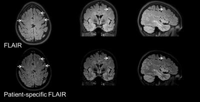 |
Improving white matter lesion conspicuity in multiple sclerosis using patient-specific optimization of 3D FLAIR
Refaat Gabr, Amol Pednekar, Koushik Govindarajan, Xiaojun Sun, Roy Riascos, María Ramírez, Khader Hasan, John Lincoln, Flavia Nelson, Jerry Wolinsky, Ponnada Narayana
Fluid-attenuated inversion recovery (FLAIR) imaging is widely used in multiple sclerosis (MS) scan protocols for its good lesion to tissue contrast. Optimization of 3D FLAIR acquisition parameters for the individual patient could further improve lesion conspicuity. In this work, tissue contrast between lesions and white matter for 3D FLAIR was optimized in the same scan session based on fast measurement of the relaxation times and proton density. Results on 16 MS patients show ~30% improved lesion contrast with the patient-specific acquisition parameters compared to the fixed-parameter 3D FLAIR sequence.
|
|