Tuesday, 25 April 2017
| Room 314 |
13:45 - 15:45 |
Moderators: Salil Soman, Meiyun Wang |
Slack Channel: #s_neuro
Session Number: O14
13:45
|
0506.
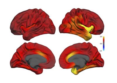 |
Structural brain changes after Electroconvulsive therapy are broadly distributed. 
Leif Oltedal, Ute Kessler, Donald Hagler, Vera Erchinger, Dominic Holland, Ketil Oedegaard, Anders Dale
Major depression is the leading cause of disability in the world. Electroconvulsive therapy (ECT) which is used in major depression when other treatments are ineffective, has been shown to cause increased volume of multiple specific subcortical and cortical regions. A sample of 19 patients with T1-weighted 3D volumes acquired before and after ECT was analyzed by using nonlinear registration and unbiased methods for quantification of regional anatomical change (Quarc). The effect sizes of ECT-induced brain changes are large, and the changes are more broadly distributed than previously thought. The results suggest a global effect, probably modulated by the stimulation parameters.
|
13:57
 |
0507.
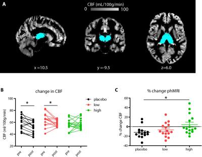 |
Dose-dependent effects of citalopram on serotonergic function assessed with SPECT and pharmacological MRI
Anouk Schrantee, Henk Mutsaerts, Jan Booij, Liesbeth Reneman
Serotonin transporter (SERT) imbalances are involved in the pathogenesis of a wide range of neuropsychiatric diseases, including depression. SERT blockers, like citalopram, decrease radioligand binding to the SERT in a dose-dependent manner, as measured with SPECT. In addition to replicating this finding, we show that pharmacological MRI (phMRI) can also detect differences in SERT occupancy with different oral doses of citalopram; higher citalopram plasma levels are associated with lower subsequent changes in the phMRI signal upon an intravenous citalopram challenge. This is important as this non-ionizing technique allows longitudinal assessment of the serotonin system.
|
14:09
|
0508.
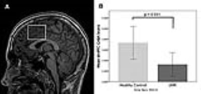 |
Relationship Between Prefrontal GABA Levels and Hippocampal Resting Activity in Subjects at Ultra High Risk of Psychosis: A Combined MRS-pCASL study
Gemma Modinos, Fatma Simsek, Jamie Horder, Matthijs Bossong, Carly Samson, Matilda Azis, Beverly Quinn, Ilaria Bonoldi, Paul Allen, Philip McGuire, James Stone
Converging evidence from preclinical studies indicates that dysfunction of the gamma-aminobutyric acidergic (GABAergic) neurotransmitter system plays a major role in the pathophysiology of schizophrenia. Despite the improved methods and reliability of neuroimaging measurements, which have recently facilitated testing predictions from animal models in humans, the extent to which GABAergic neurotransmission is altered in patients with psychosis is less clear. Furthermore, although preclinical evidence suggests that decreased cortical interneuron function leads to hippocampal activity overdrive, no study has explicitly investigated the relationship between neurotransmission and neurophysiology in humans. Here we show that prefrontal GABA function is reduced in individuals at ultra high risk of developing psychosis and that this reduction is related to hippocampal (and nominally to prefrontal) resting activity. These findings shed light on the pathophysiology of vulnerability for schizophrenia by showing that alterations in GABAergic systems have downstream effects on hippocampus before the onset of psychosis.
|
14:33
|
0509.
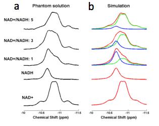 |
“Brain Rust” in Schizophrenia Revealed by in vivo Redox (NAD+/NADH) Measurement
Fei Du, Sang-Young Kim, Bruce Cohen, Xi Chen, Scott Lukas, Ann Shinn, Cagri Yuksel, Tao Li, Dost Ongur
A growing body of evidence suggests that an “immuno-oxidative” pathway including redox dysregulation associated with oxidative stress, mitochondrial dysfunction, neuroinflammation, and cell-mediated immune response may contribute to disruptions in brain activity in schizophrenia (SZ). The aim of this study is to assess possible redox imbalance in SZ patients by using a novel in vivo 31P-MRS technique to measure NAD+ and NADH. Our results revealed a ~40% decrease of NAD+/NADH ratio compared to healthy individuals of similar age, indicating higher levels of oxidative stress in patients with schizophrenia. This work may lead to new strategies to protect the brain from oxidative stress and improve brain function in schizophrenia or the other brain disorders.
|
14:45
 |
0510.
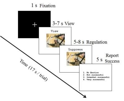 |
Effective Connectivity Network of Emotion Regulation in Soldiers with Trauma
D Rangaprakash, Michael Dretsch, Thomas Daniel, Thomas Denney, Jeffrey Katz, Gopikrishna Deshpande
Conscious regulation of emotions is essential for sound functioning of an individual, while its disruption leads to several severe symptoms observed in psychiatric disorders like posttraumatic stress disorder (PTSD) and mild-traumatic brain injury (mTBI). While the brain regions activated in emotion regulation have been elucidated in prior works, an understanding of the underlying network has been elusive. Employing an emotion regulation task, we discovered the network of emotion regulation in healthy soldiers, and dysregulation in soldiers with comorbid PTSD/mTBI (N=59). Our work is significant given that we present, for the first time, the evidence for the network of emotion regulation/dysregulation.
|
14:57
|
0511.
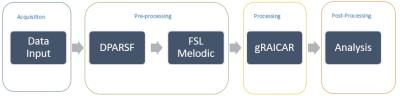 |
Investigating Brain Connectomic Alterations in PTSD and PCS using the Reproducibility of Independent Components obtained from Resting-State Functional MRI Data
Mohammed Syed, D Rangaprakash, Michael Dretsch, Thomas Denney, Jeffrey Katz, Gopikrishna Deshpande
Posttraumatic stress disorder (PTSD) and Post-concussion syndrome (PCS) are heterogeneous neurological disorders where fMRI connectivity metrics derived from them may not be highly reproducible, leading to poor generalizability and consequently lower classification accuracies. We present a method that characterizes the reproducibility of networks using ‘generalized Ranking and Averaging Independent Component Analysis by Reproducibility’ (gRAICAR) algorithm followed by unsupervised clustering to discriminate between the groups based on functional brain networks that are most reproducible within PTSD, PCS, and healthy control groups separately. We identify dorsolateral prefrontal cortex, inferior parietal lobule, caudate and medial prefrontal cortex as regions within the most reproducible independent components.
|
15:09
|
0512.
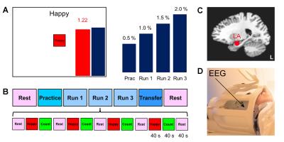 |
Real-time fMRI Neurofeedback of the Amygdala Enhances Amygdala-orbitofrontal Connectivity and Lateralized EEG Coherence in Veterans with Combat-related PTSD - permission withheld
Vadim Zotev, Raquel Phillips, Masaya Misaki, Chung Ki Wong, Brent Wurfel, Matthew Meyer, Frank Krueger, Matthew Feldner, Jerzy Bodurka
We have performed a study of emotion regulation training in veterans with combat-related PTSD using real-time fMRI neurofeedback (rtfMRI-nf) with simultaneous EEG. Eighteen PTSD patients learned to upregulate their left amygdala activity using rtfMRI-nf during a positive emotion induction task based on retrieval of happy autobiographical memories. Enhancement in the amygdala-orbitofrontal functional connectivity during the rtfMRI-nf task showed positive correlation with severity of PTSD symptoms. Enhancement in left-lateralized upper alpha EEG coherence also positively correlated with PTSD severity. These results suggest that the rtfMRI-nf of the amygdala has the potential to correct the functional connectivity deficiencies specific to PTSD.
|
15:21
|
0513.
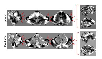 |
The Cerebellum and Brainstem Together Increase Classification Accuracy for Autism Spectrum Disorder over the Whole Brain
Muriel Bruchhage, Leon Aksman, Andre Marquand, MRC AIMS Consortium, EU TACTICS Consortium, Jan Buitelaar, Declan Murphy, Steven Williams
Autism spectrum disorder (ASD) has been linked to cerebellar and brainstem dysfunction and abnormal development, but it remains unclear whether these regional abnormalities can help classify the disorder. Performing machine learning based classification using Jacobian determinant based features on two independent male ASD cohorts (adult and paediatric) of different sizes and age range, we demonstrated a consistently higher classification accuracy by up to 15% using the cerebellum and brainstem as regions of interest classifiers over the whole brain. In both cohorts, classification was driven by regional differences in the posterior lateral cerebellum.
|
15:33
|
0514.
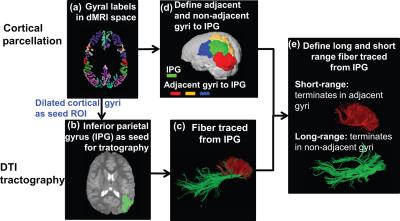 |
Atypical maturation of short-range fibers connecting higher-order brain regions in children with autism aged 2-7 years
Minhui Ouyang, Jennifer Muller, Hua Cheng, Yun Peng, J. Edgar, Timothy Roberts, Hao Huang
A pattern of local or short-distance “over-connectivity” and long-range under-connectivity is frequently hypothesized in individuals with autism spectrum disorder (ASD). Little is known about the spatiotemporal characterization of structural short-distance connections in typically developing (TD) children or children with ASD. We hypothesized that altered trajectories of short-range association fibers (SAF) are not uniform across the brain regions, with abnormal maturation primarily observed in higher-order but not in primary sensory brain regions in children in ASD. Here, we quantified SAF with a novel index defined as normalized SAF (NSAF) based on diffusion MRI tractography, and characterized its trajectories across brain regions.
|
|