Wednesday, 26 April 2017
| Room 312 |
13:45 - 15:45 |
Moderators: René Botnar,
Xihai Zhao |
Slack Channel: #s_cv
Session Number: O20
13:45
 |
0796.
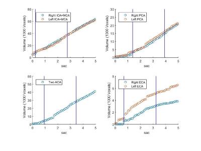 |
Volumetric blood flow measurement with territorial segmentation of time-resolved contrast enhanced magnetic resonance angiography of the brain 
Oren Geri, Shelly Shiran, Jonathan Roth, Moran Artzi, Liat Ben-Sira, Dafna Ben Bashat
We propose a method for territorial segmentation and volumetric flow rate (VFR) measurement based on time-resolved contrast enhanced MR-angiography. Eight territories: right/left internal carotid arteries; the two anterior cerebral arteries (combined); the right/left external carotid arteries; the right/left posterior cerebral arteries; and the vertebrobasilar territory, were segmented using an iterative region-growing algorithm based on the bolus-arrival-time with increased temporal resolution. VFR was measured based on the territorial volume as a function of time. Healthy subjects' VFR results were similar to literature values. The clinical potential of this method is demonstrated on one patient with Moyamoya before and after surgery.
|
13:57
 |
0797.
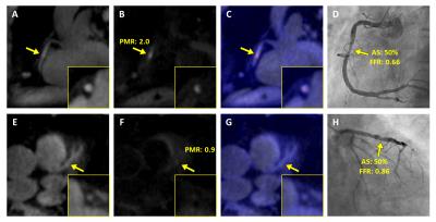 |
Coronary Plaque Hyper-Intensity on Dark-Blood T1-Weighted MRI Helps Identify Lesion-Specific Ischemia: Insights from the Comparison Study with Invasive Fractional Flow Reserve (FFR)
Yibin Xie, Young-Jin Kim, Sang-Eun Lee, Jianing Pang, Anthony Christodoulou, Qi Yang, Zixin Deng, Daniel Berman, Hyuk-Jae Chang, Debiao Li
Coronary Atherosclerosis T1-weighted Characterization (CATCH) is an accelerated MR technique for detecting high-risk atherosclerotic lesions. However, the relationship between plaque signal and lesion-specific ischemia is still unclear. In this study we applied CATCH in a patient cohort undergoing invasive FFR and discovered the association between plaque hyper-intensity and hemodynamic functional significance. The results presented here support the potential clinical utility and added value of MR coronary plaque characterization as a “gate-keeper” for invasive and costly coronary procedures.
|
14:09
|
0798.
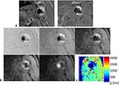 |
Multi-contrast Acquisition in a Single Scan (MASS) for Three-dimensional Quantitative T1 Mapping of Carotid Atherosclerosis
Haikun Qi, Huiyu Qiao, Shuo Chen, Zechen Zhou, Xinlei Pan, Yishi Wang, Chun Yuan, Huijun Chen
Intra-plaque hemorrhage (IPH) is a dynamic process and change of the IPH MR signal was found to be correlated with plaque developments. So quantitative T1 mapping of plaque is essential to monitor plaque progression. In this study, we proposed an IR prepared 3D golden angle radial sampling sequence, enabling multiple T1 contrasts acquisition in a single scan (MASS) with application to carotid artery T1 mapping. The accuracy and feasibility of MASS was demonstrated in phantom studies and in vivo imaging experiments on healthy volunteers and carotid atherosclerosis patients. MASS may be a one-stop solution to carotid atherosclerotic plaque imaging.
|
14:21
|
0799.
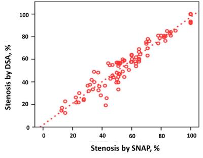 |
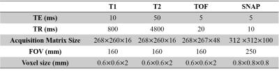
Assessment of Carotid Atherosclerotic Disease Using 3D Simultaneous Non-contrast Angiography and Intraplaque Hemorrhage (SNAP) imaging: Comparison with Digital Subtraction Angiography
Huilin Zhao, Jianrong Xu, Xiaosheng Liu, Beibei Sun, Weibo Chen, Chun Yuan, Xihai Zhao
3D fast Simultaneous Non-contrast Angiography and intraPlaque hemorrhage (SNAP) imaging was recently proposed as a technique for joint MRA and intraplaque hemorrhage (IPH) imaging. This study sought to determine the accuracy of this technique at quantifying carotid atherosclerosis disease compared to conventional intra-arterial digital subtraction angiography (DSA) in patients with at least 50% carotid stenosis. We found that 3D SNAP imaging had excellent agreement with DSA in measuring luminal stenosis and identification of ulceration in carotid arteries. Our findings suggest that, SNAP imaging might be a potential candidate technique for comprehensive evaluation of carotid high-risk atherosclerotic disease.
|
14:33
|
0800.
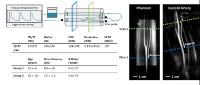 |
Measuring Local Pulse Wave Velocity in the Carotid Arteries using a Compressed Sensing reconstruction for high temporal resolution (4 ms) 2D PC CINE MRI
Eva Peper, Wouter Potters, Abdallah Motaal, Pim van Ooij, Aart Nederveen, Gustav Strijkers, Bram Coolen
Measuring PWV with MRI would provide a useful tool to measure arterial stiffness locally. However, the limitation of MRI for this implementation is temporal resolution. This study validates a technique to measure local PWV at up to only 5 cm of the carotid arteries using 2D PC CINE MRI data of a high temporal resolution compressed sensing (CS) reconstruction. The method is validated using a pulsatile flow phantom and two groups of 10 elderly and 10 younger healthy volunteers. A significant difference between age groups was found.
|
14:45
|
0801.
 |
Automatically Identify Plaque Components in Carotid Artery using Simultaneous Non-Contrast Angiography and intraPlaque hemorrhage (SNAP) imaging
Qiang Zhang, Huiyu Qiao, Shuo Chen, Zhensen Chen, Xihai Zhao, Chun Yuan, Huijun Chen
The purpose of this study is to develop an automatic method to identify plaque components using a single 3D Simultaneous Non-Contrast Angiography and intraPlaque hemorrhage (SNAP) acquisition. Using artifact neural network classifier with the intensities of multiple images generated from SNAP and the morphology information, the automatic identified components area has a high correlation with manual segmentation on 2D multi-contrast MR images: 0.82 (necrotic core), 0.79 (calcification) and 0.88 (fibrous tissue). This study further enhanced ability of 3D SNAP sequence in plaque components identification, suggesting SNAP would be a practical clinical solution for carotid atherosclerotic plaque evaluation.
|
14:57
|
0802.
 |
Longitudinal Analysis of Vascular Inflammation and Intraluminal Thrombus Composition using High Resolution MRI in Abdominal Aortic Aneurysms
Chengcheng Zhu, Bing Tian, Joseph Leach, David Saloner, Michael Hope
Clinical management of abdominal aortic aneurysm (AAA) disease is based on the maximal diameter. Vascular inflammation and intraluminal thrombus (ILT) composition have been explored as novel imaging markers of progressive AAA disease, but studies to date have been limited by short follow-up time (~6 month). We followed 37 patients for an average of 4 years using CT/CTA and high resolution black-blood MRI. Our results show that inflammation identified by delayed ultrasmall superparamagnetic iron oxide (USPIO) particle uptake was strongly associated with AAA growth and/or intervention, whereas ILT composition was not. Imaging of vascular inflammation may improve AAA patient risk stratification.
|
15:09
|
0803.
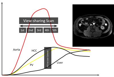 |
Simultaneous acquisition of MR angiography and diagnostic images on contrast-enhanced view-sharing multi-arterial phases
Yoshifumi Noda, Satoshi Goshima, Kimihiro Kajita, Hiroshi Kawada, Nobuyuki Kawai, Hiromi Koyasu, Masayuki Matsuo, Tomohiro Namimoto, Norihiro Shinkawa, Masataka Nakagawa, Toshinori Hirai, Yasuyuki Yamashita
While magnetic resonance angiography (MRA) is clinically used to evaluate vascular anatomy, whereas it needs independent scan leading to a decreased throughput. We generated MRA using early phase images in the contrast-enhanced multi-arterial phase images with view-sharing technique. Aortic branches and anatomical anomalies were clearly visualized on MRA without significant differences in contrast effect and conspicuity regardless of contrast material with different r1 value (gadoterate meglumine and gadobutrol).
|
15:21
|
0804.
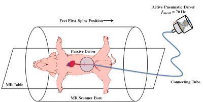 |
In Vivo Magnetic Resonance Elastography of Abdominal Aortic Aneurysm in A Porcine Model
Huiming Dong MS, Matthew Joseph BS, Prateek Kalra MS, Xiaokui Mo PhD, Richard White MD, Rizwan Ahmad, Arunark Kolipaka PhD
Abdominal aortic aneurysm (AAA) can result in life-threatening aortic rupture. Although AAA diameter is utilized for assessing rupture risk clinically, it is a poor indicator of rupture potential. Aortic stiffness is an important biomechanical property that can provide critical information about the overall mechanical integrity of AAA and thus results in more accurate rupture risk evaluation.Therefore, the aim of this study is to utilize non-invasive in vivo MRE to estimate aortic stiffness in AAA-induced animal models, and compare it with the stiffness obtained from ex vivo mechanical testing as well as AAA diameters.
|
15:33
|
0805.
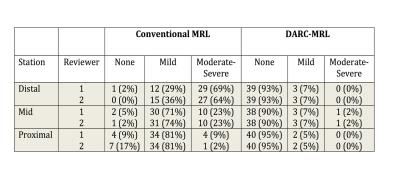 |
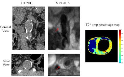
Robust MR Lymphangiography using DARC-MRL: Evaluation of Venous Suppression and Strategies for More Efficient Clinical Examinations
Jeffrey Maki, Beth Ripley, Neeraj Lalwani, Noah Briller, Peter Neligan, Gregory Wilson
Dual Agent Relaxation Contrast MR Lymphangiography (DARC-MRL) effectively eliminates venous enhancement through the up-front i.v. injection ferumoxytol (USPIO causing marked blood T2* shortening) combined with obtaining MRL datasets at prolonged, precisely determined echo times. Echo time prolongation does cause an approximately 45% loss of lymphatic signal intensity, however with excellent lymphatic-to-tissue contrast, there was no clinically significant lymphatic signal loss. Data regarding the time course of lymphatic enhancement progression demonstrate most MRL enhancement can be fully captured in only two time points, allowing for a more efficient, faster examination. Multi-echo DARC offers to add further robustness and visualization capability.
|
|