Monday, 24 April 2017
| Room 312 |
16:15 - 18:15 |
Moderators: Jeremy Collins, Tino Ebbers |
Slack Channel: #s_cv
Session Number: O21
16:15
|
0287.
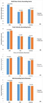 |
Multi-year 4D flow MRI Follow-Up Study of Bicuspid and Tricuspid Aortic Valve patients and Association between Wall Shear Stress and Aortic Diametric Growth 
Ozair Rahman, Alex Barker, Carmen Blanken, Emilie Bollache, Michael Rose, Pim Van Ooji, Jeremy Collins, James Carr, Chris Malaisrie, Patrick McCarthy, Michael Markl
Patients with Bicuspid Aortic Valve (BAV) are at increased risk of developing aortopathy compared to Tricuspid Aortic Valve (TAV) patients. However, there is imited data presenting the development of pathophysiologic changes taking place over multi year time period. Our study attempts to quantify the changes that take place from baseline and follow-up scans to help us better understand this process.
|
16:27
|
0288.
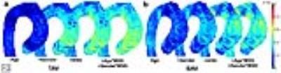 |
3D Linear Regression Analysis Reveals Relationships of 4D flow MRI-derived Aortic Dimensions with Age, Gender and Wall Shear Stress in Patients with Aortopathy
Pim van Ooij, Jeremy Collins, Paul Fedak, Aart Nederveen, James Carr, Michael Markl, Alex Barker
In two groups of patients with bicuspid valves (BAV) and with tricuspid valves with dilated aortas (TAV), 3D correlation coefficient r maps were created to investigate linear relationships between 3D aortic diameter maps and 3D wall shear stress maps (WSS), with age and gender as co-variables. The dependence of diameter on gender was higher for TAV, whereas the dependence of diameter on age was higher for BAV patients. With the addition of WSS to the model, r increased slightly for both groups. In general, r was significantly higher for TAV: BAV mediated aortopathy is suspected to have genetic associations.
|
16:39
 |
0289.
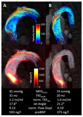 |
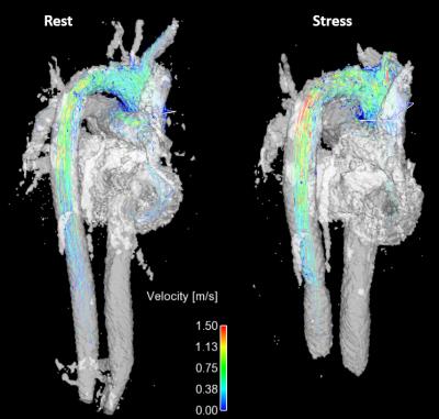
Multiparametric Assessment of Patients with Aortic Stenosis using Multipoint 4D Flow MRI - Correlation with Cardiac Biomarkers - permission withheld
Alexander Gotschy, Christian Binter, Robert Manka, Sebastian Kozerke
Various flow characteristics, such as Turbulent Kinetic Energy (TKE), flow displacement or jet angle, derived from 4D Flow MRI, have been used to investigate the hemodynamic effects of aortic stenosis (AS). However, the predictive value of these flow parameters is still unknown. Therefore, we investigated the correlation between multiple flow parameters and cardiac biomarkers which are known to provide prognostic information on the progression and outcome of AS. Our results revealed that MRI-based TKE and peak velocity significantly correlate with NT-proBNP, implying potential relevance of these imaging parameter for future risk stratification of AS patients.
|
16:51
|
0290.
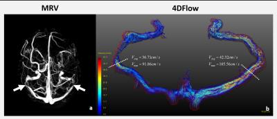 |
Blood flow characterization in sigmoid-sinus using 4D-Flow MR among patients with pulsatile tinnitus
Yunduo Li, Le He, Xiangyu Cao, Xianling Wang, Shubin Chen, Huijun Chen, Rui Li, Chun Yuan
Pulsatile tinnitus (PT) is suspected to be associated with abnormal hemodynamics in sigmoid-sinus. In this study, we used 4D-Flow MRI to characterize blood flow in sigmoid-sinus among patients with pulsatile tinnitus and demonstrated that high blood velocity in sigmoid-sinus might be an authentic marker of PT. This study may provide more information for diagnosis and treatment of pulsatile tinnitus, especially for patients with PT of venous origin.
|
17:03
|
0291.
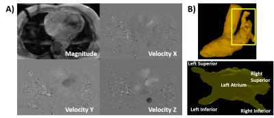 |
4-Dimensional Phase-Contrast Magnetic Resonance Imaging of Left Atrial Stasis in Patients with Paroxysmal Atrial Fibrillation: A Comparative Study of Patients Pre- and Post-Ablation
Julio Garcia, Michael Bristow, Carmen Lydell, Andrew Howarth, Bobby Heydari, Frank Prato, Maria Drangova, Rebecca Thornhill, Pablo Nery, Stephen Wilton, Allan Skanes, James White
This study may be of interest for clinicians and clinical researchers who study atrial diseases. This study demonstrates that 4D flow-derived LA 3D stasis is clinically feasible and it may be useful for characterize differences between pre- and post-ablation patients.
|
17:15
|
0292.
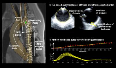 |
4D flow MRI based quantification of regional stiffness in the thoracic aorta in stroke patients compared to transesophageal echocardiography
Thomas Wehrum, Felix Günther, Anja Hennemuth, Johann Drexl, Hanieh Mirzaee, Andreas Harloff
Our purpose was to quantify regional stiffness in the aorta in stroke patients using 4D flow MRI based pulse-wave-velocity quantification in comparison with stiffness quantification using parameters based on transesophageal echocardiography (TEE). MRI and TEE based stiffness parameters were highly correlated and increased stiffness as measured using 4D flow MRI and TEE was associated with presence of atherosclerosis. Accordingly, we were able to predict the presence of atherosclerotic lesions with high sensitivity and specificity using both, 4D flow MRI and TEE. Hence, especially non-invasive 4D flow MRI can be used in future longitudinal studies investigating early development of atherosclerotic lesions.
|
17:27
|
0293.
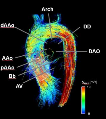 |
Increased aortic wall shear stress and wall shear stress gradient in patients with an anatomically shaped sinus prosthesis using 4D Flow MRI
Victoria Schultz, Thekla Oechtering, Malte Sieren, Michael Scharfschwerdt, Anja Hennemuth, Markus Hüllebrand, Hans-Hinrich Sievers, Jörg Barkhausen, Alex Frydrychowicz
Patients with anatomically shaped sinus prosthesis have been shown to have near physiological hemodynamics in the aortic bulb but altered flow characteristics distal to the prosthesis. The aim of this study was to compare the aortic wall shear of 12 patients with sinus prosthesis with 12 age-matched volunteers using 4D flow sensitive MRI. The wall shear stress analysis in 8 analysis planes revealed a tendency towards decreased WSS in the region of the prosthesis and increased WSS values distal to the prosthesis. Interestingly, the WSS gradient per plane and segmental WSS distal to the prosthesis were increased throughout the patients.
|
17:39
 |
0294.
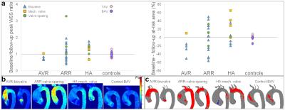 |
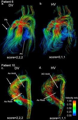
Post-surgical changes in aortic wall shear stress patterns in patients with aortopathy: a follow-up 4D flow MRI study
Emilie Bollache, Paul Fedak, Pim van Ooij, Ozair Rahman, Alex Hong, Eric Keller, S Chris Malaisrie, Patrick McCarthy, James Carr, Jeremy Collins, Michael Markl, Alex Barker
Our purpose was to follow up post-surgical changes in peak wall shear stress (WSS) and extent of at-risk tissue using 4D flow MRI in 34 aortopathy patients. Highly variable changes between pre- and post-surgery were found according to the intervention or replaced aortic valve type, while WSS patterns were unchanged in 20 other patients who did not undergo surgery. The reproducible 4D flow MRI WSS indices should be studied in larger cohorts and compared with patient outcome to potentially detect risk of future events in aortopathy patients, while optimizing the extent of resected aortic tissue.
|
17:51
|
0295.
 |
4D Flow MRI during Exercise in Prematurely Born Adults and Children
Jacob Macdonald, Arij Beshish, Kristin Haraldsdottir, Marlowe Eldridge, Oliver Wieben, Christopher Francois
Preterm birth can result in impaired development of lung airways and vasculature, but the long term implications are unclear. With this in mind, we performed 4D flow imaging in the aorta and pulmonary artery during exercise at 70% maximal power in both children and adults who had been born prematurely to identify any differences in flow characteristics relative to healthy controls. Although no statistically significant differences were identified between our groups, some preterm cohorts showed increased cardiac output and mean velocity. The significance of these trends should become apparent as we continue to recruit subjects and increase our statistical power.
|
18:03
|
0296.
 |
k-t Accelerated Dual-Venc 4D flow MRI for Improved Flow Visualization in Pediatric Patients with Congenital Heart Disease
Liliana Ma, Michael Rose, Ozair Rahman, Kelly Jarvis, Joshua Robinson, Cynthia Rigsby, Michael Markl, Susanne Schnell
This study explores the potential of using dual-velocity encoding 4D flow MRI for in-vivo assessment of complex blood flow patterns in patients with diverse presentations of congenital heart disease.
|
|