Tuesday, 25 April 2017
| Room 320 |
16:15 - 18:15 |
Moderators: Giulia Ginami, Sebastian Kozerke |
Slack Channel: #s_cv
Session Number: O22
16:15
 |
0630.
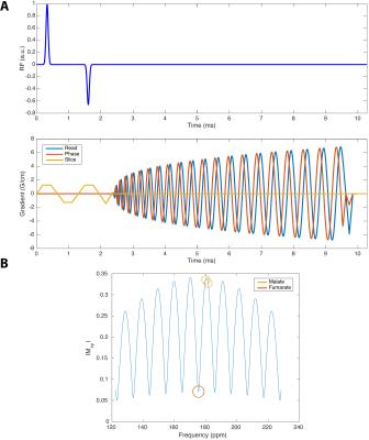 |
Hyperpolarized [1,4-13C2]Fumarate is a probe of necrosis in myocardial infarction 
Jack Miller, Angus Lau, Giles McMullen-Klein, Andrew Lewis, Vicky Ball, Carolyn Carr, Ferdia Gallagher, Damian Tyler, Marie Schroder
Previous work has shown that hyperpolarized [1,4-13C2]fumarate is a probe of cellular necrosis. We demonstrate here that the ratio of cardiac hyperpolarized malate to fumarate is increased by a factor of $$$\sim$$$82 one day after cryoinduced myocardial infarction in rats, decreasing to an $$$\sim$$$30-fold increase one week after injury. We additionally image this injury with a novel spiral multiband pulse sequence. Hyperpolarized fumarate therefore forms a sensitive probe of myocardial injury in vivo, and could form a clinical monitor of cellular damage and necrosis after infarction.
|
16:27
|
0631.
 |
Accelerated Cine Imaging of the Heart using Blipped Multiband SSFP
Anthony Price, Lucilio Cordero-Grande, Shaihan Malik, Joseph Hajnal
Multiband accelerated cine SSFP of the heart is demonstrated using blipped gradients for controlled aliasing of simultaneous slices. The benefit of using this method over the more complex phase cycling RF multiband pulse sets is that the frequency response, and thus banding artefacts, remain unchanged. Here we demonstrate up to MB4 acceleration of full short-axis stacks, sufficient to cover the left ventricle within a single breath-hold.
|
16:39
 |
0632.
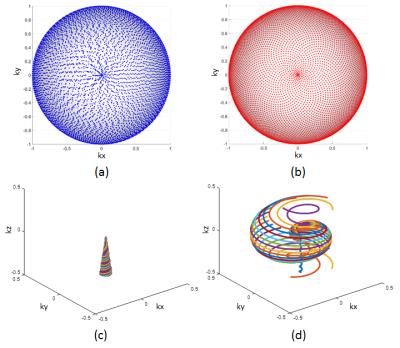 |
Improved Whole-Heart Coronary MR Angiography Using a 3D Cones Phyllotaxis Sequence
Mario Malavé, Corey Baron, Nii Addy, Joseph Cheng, Bob Hu, Phillip Yang, Dwight Nishimura
We have developed a 3D cones alternating-TR steady state free precession (SSFP) sequence for whole-heart coronary MR angiography (CMRA). In this study, we compare motion artifacts and coronary image quality between a sequential-cones and phyllotaxis-cones sequence. Coronary image quality was analyzed using qualitative scores obtained through blinded reading by two board-certified cardiologists, and the IEPA vessel sharpness metric. The results of the study show that the phyllotaxis-cones acquisition can alleviate motion artifacts, improve qualitative image quality scores and increase coronary vessel sharpness.
|
16:51
|
0633.
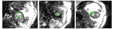 |
Automated Heartbeat Detection for Self-Gated Fetal Cardiac MRI
Robin Demesmaeker, Tobias Kober, Jérôme Yerly, Jérôme Chaptinel, Milan Prsa, Yvan Mivelaz, Leonor Alamo, Yvan Vial, Gregoire Berchier, Chantal Rohner, Matthias Stuber, Davide Piccini
Fetal cardiac cine MRI requires an MRI-based cardiac gating signal since recording a fetal ECG is fraught with significant challenges. Existing approaches usually extract the signal from real-time image series and mandate semi-manual user interaction. However, these give often inconsistent results or suffer from reduced spatio-temporal resolution. We propose a novel algorithm which automatically localizes the fetal heart on real-time low-resolution images, and provides a precise frequency estimate of the cardiac motion signal that can be used for gating. We show that this automated method leads to images with equal or better quality than those obtained with the manual approach.
|
17:03
|
0634.
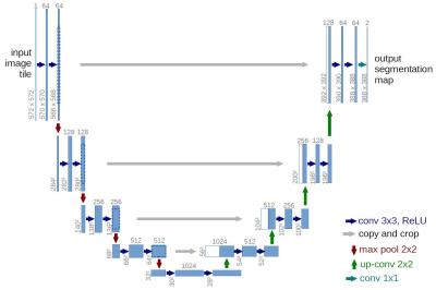 |
Automated Left Ventricular Volumetric Quantitation from Short-axis CMR Images with Machine Learning using a Deep Convolutional Neural Network - permission withheld
James Goldfarb, Jie Cao, Julian de Wit
Automatic segmentation of the LV bloodpool using deep learning with a convolutional neural network is a promising, accurate and efficient method for segmentation of cardiac MR images. Although there were a few cases with inaccurate results, "big fails", accuracy is high, R2=0.93 and ejection fraction error ~4%. In the future it may provide a customizable, fast and accurate method for comprehensive evaluation of cardiac MR images.
|
17:15
|
0635.
 |
DeepVentricle: A Fully Convolutional Neural Network for Automating Functional Measurements in Cardiac MR
Hok Kan Lau, Jesse Lieman-Sifry, Matthieu Le, Sean Sall, John Axerio-Cilies, Dominik Fleischmann, Aya Kino, Frandics Chan, Daniel Golden
We present DeepVentricle, an automated approach to ventricular segmentation in cardiac MR. DeepVentricle uses a fully convolutional neural network to simultaneously perform semantic segmentation of the left ventricle (LV) and right ventricle (RV) endocardium, and LV epicardium; segmentations are then used to estimate ejection fraction and myocardial mass. We show that the error rates of LV ejection fraction and mass are within the expected range of expert annotator inter-rater variation. This suggests that contours calculated using DeepVentricle could be useful on their own or as an initial estimate for clinicians as part of their semi-automated annotation workflow.
|
17:27
|
0636.
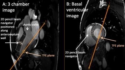 |
Using transient intrinsic torsional shear wave propagation to measure Left Ventricular Myocardial stiffness with a 2D pencil beam navigator at 0.5ms temporal resolution: Initial results from phantom studies and volunteers
Jessica Webb, Jurgen Runge, Jordi Martorell, Gerald Carr-White, Reza Razavi, David Nordsletten, Ralph Sinkus
Heart Failure with preserved Ejection Fraction is common, associated with high morbidity and mortality, and is challenging to diagnose. We have developed a novel patient friendly non-invasive technique to quantify myocardial stiffness using transient MR Elastography. Aortic valve closure results in a shear wave propagating through the myocardium. The torsional wave propagation can be visualised using a 2D pencil beam navigator positioned along the myocardium, using four breath holds each 15 seconds. With a temporal resolution of 0.5ms this technique can be used in all patients in sinus rhythm. Increased myocardial stiffness results in increased speed of shear wave propagation.
|
17:39
|
0637.
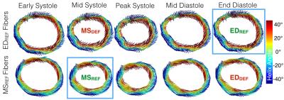 |
Time Resolved In Vivo Myofiber Orientations from Combined Cardiac DENSE and cDTI
Patrick Magrath, Luigi Perotti , Eric Aliotta , Ilya Verzhbinsky , Kévin Moulin, Daniel Ennis
Resolving the dynamics of cardiac myofiber orientations with cDTI could have significant value in the assessment of cardiac disease. Unfortunately, cDTI measurements are not reliable at all cardiac phases and would require extremely long acquisitions. Fusing cDTI with time-resolved displacement maps from cine DENSE could enable evaluation of the dynamics of myofiber orientations using cDTI obtained at a single cardiac phase. The objective of this study was to generate dynamic myocardial fiber maps and validate the approach with dual-phase cDTI measurements.
|
17:51
|
0638.
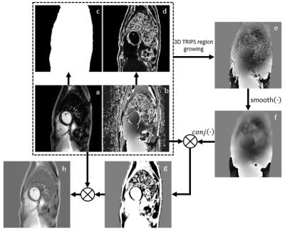 |
3D True Polarity Recovery with Independent Phase Estimation Using Multi-layer Stacks Based Region-Growing (3D-TRIPS)
Haining Liu, Gregory Wilson, Niranjan Balu, Jeffery Maki, Martin Gunn, Hiroko Watase, Daniel Hippe, Chun Yuan
3D whole heart phase sensitive late gadolinium enhanced (LGE) imaging can improve myocardial scar detection. Phase sensitive inversion recovery (PSIR) LGE is sub-optimal for clinical 3D application as it doubles the scan time with a fully sampled reference image. To address this, we develop 3D True Polarity Recovery with Independent Phase Estimation Using Multi-layer Stacks Based Region-Growing (3D-TRIPS) for direct reconstruction of 3D phase sensitive images without need for a separate reference scan. We demonstrate that 3D-TRIPS images shows good agreement and less artifacts compared with PSIR images. 3D-TRIPS will allow 3D-LGE imaging with half the scan time of PSIR.
|
18:03
|
0639.
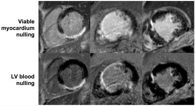 |
Dark-Blood Late Gadolinium Enhanced MRI: A Novel Method without Additional Magnetization Preparation for Improved Myocardial Scar Detection
Robert Holtackers, Amedeo Chiribiri, David Higgins, Rene Botnar
Late gadolinium enhanced (LGE) MRI often suffers from poor scar-to-blood contrast when used for detection of endocardial scar due to the bright signal of adjacent blood. We report a method that significantly reduces left ventricular blood signal by setting a shorter inversion time in combination with a phase-sensitive inversion recovery (PSIR) sequence. Nulling the left ventricular blood signal with PSIR significantly increases scar-to-blood contrast since blood signal and scar signal no longer have similar signal levels. As no additional magnetization preparation is used, clinical application on current MR systems is readily available without the need for software modifications or additional training.
|
|