Wednesday, 26 April 2017
| Room 313A |
16:15 - 18:15 |
Moderators: Martijn Froeling, David Sosnovik |
Slack Channel: #s_cv
Session Number: O24
16:15
 |
0922.
 |
Quantification of Increased Myocardial Stiffness in Patients with Hypertrophic Cardiomyopathy Using 3D High Frequency Cardiac Magnetic Resonance Elastography 
Shivaram Poigai Arunachalam, Arvin Arani, Ian Chang, Yi Sui, Phillip Rossman, Kevin Glaser, Joshua Trzasko, Kiaran McGee, Armando Manduca, Richard Ehman, Richard Ehman, Philip Araoz
Abnormal thickening of myocardium in patients with hypertrophic cardiomyopathy impairs the pump function, and in particular affects diastolic filling with a known increase in myocardial stiffness. The purpose of this work was to determine if 3D high frequency cardiac MR elastography (MRE) can quantitatively differentiate increased myocardial stiffness in HCM patients compared to healthy volunteers. 36 patients with clinical diagnosis for hypertrophic cardiomyopathy (HCM) and 47 healthy volunteers were studied. The myocardial stiffness of HCM patients (mean: 12.01 kPa) was found to be significantly stiffer (p < 0.01) than healthy controls (mean: 7.79 kPa).
|
16:27
|
0923.
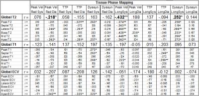 |
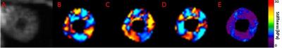
CMR Demonstrates Structure-Function Relationship in Patients after Heart Transplantation
Ryan Dolan, Amir Rahsepar, Julie Blaisdell, Kai Lin, Kenichiro Suwa, Allen Anderson, Kambiz Ghafourian, Esther Vorovich, Jonathan Rich, Jane Wilcox, Clyde Yancy, Jeremy Collins, James Carr, Michael Markl
Cardiac MRI demonstrates differences between heart transplant recipients and controls using tissue phase mapping (TPM), T2, and T1. Significant correlations between myocardial velocities and dyssynchrony obtained from TPM (myocardial function) and T2 and T1 (myocardial tissue structure) suggest a relationship between impaired structure and function among transplant recipients.
|
16:39
|
0924.
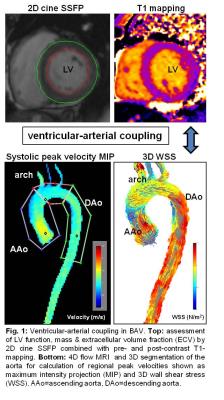 |
4D Flow MRI, Cardiac Function, and Myocardial T1-Mapping: Ventricular-arterial coupling in Patients with Bicuspid Aortic Valve (BAV)
Julia Geiger, Amir Rahsepar, Kenichiro Suwa, Alex Powell, Alex Barker, Jeremy Collins, James Carr, Michael Markl
BAV is the most prevalent congenital cardiovascular malformation. Its association with progressive ascending aortic dilatation and concomitant aortic valve stenosis or regurgitation with increasing age has a critical impact on patients’ morbidity. We applied a comprehensive CMR protocol in 50 BAV patients consisting of cine-imaging, T1-mapping and 4D flow MRI to simultaneously assess cardiac parameters and aortic hemodynamics. We observed significant relationships between LV mass and WSS as well as peak velocities in the AAo and arch, likewise in the sub-cohort with normal valve function, leading us to the hypothesis that there is proof for ventricular-aortic coupling in BAV patients.
|
16:51
|
0925.
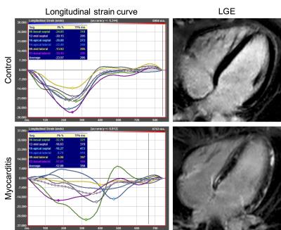 |
Feature tracking myocardial strain analysis in acute myocarditis: Diagnostic value and association with myocardial inflammation and edema
Julian Luetkens, Ulrike Schlesinger-Irsch, Daniel Kütting, Darius Dabir, Rami Homsi, Jonas Doerner, Frederic Schmeel, Alois Sprinkart, Claas Naehle, Hans Schild, Daniel Thomas
Cardiac magnetic resonance (CMR) can detect inflammatory myocardial alterations in patients with acute myocarditis. The addition of myocardial strain analysis might further broaden the diagnostic targets of CMR. We investigated myocarditis patients using multiparametric CMR including a feature-tracking analysis of myocardial strain parameters. We could demonstrate that myocardial strain measurements can reliable discriminate between diseased and healthy patients. Furthermore, strain measurements are associated with the extent of myocardial edema/inflammation. These findings indicate that CMR feature-tracking strain analysis adds important diagnostic information, and might serve as a new tool for the assessment of myocardial dysfunction in patients with acute myocarditis.
|
17:03
|
0926.
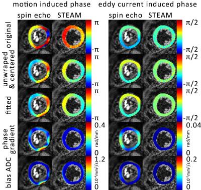 |
Impact of eddy-currents and cardiac motion in DTI of the in-vivo heart - a comparison of second-order motion compensated SE versus STEAM
Christian Stoeck, Constantin von Deuster, Robbert van Gorkum, Sebastian Kozerke
Motion-compensated (M2) spin-echo (SE) and stimulated-echo acquisition mode (STEAM) sequences have been proposed to generate diffusion contrast in in-vivo cardiac imaging. When comparing measured fractional anisotropy and mean diffusivity of cardiac tissue, marked differences have been reported between SE and STEAM. Cardiac motion, perfusion, different mixing times and eddy-currents have been discussed as potential source of discrepancies. In this study it is shown that signal dephasing due to eddy-currents play a minor role. While SE is more prone to motion-induced dephasing compared to STEAM, reported differences in mean diffusivity can only marginally be explained by motion-induced signal loss in SE.
|
17:15
 |
0927.
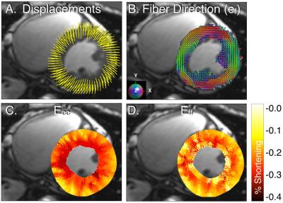 |
In Vivo Assessment of Cardiomyocyte Performance Using Combined Cardiac DENSE and cDTI
Patrick Magrath, Luigi Perotti , Eric Aliotta , Ilya Verzhbinsky , Kévin Moulin, Daniel Ennis
Circumferential strain (Ecc) derived from cardiac DENSE 3D displacement maps is a promising biomarker for diagnosing early stages of cardiac disease. Ecc is commonly evaluated only in the mid-wall where it is expected to align with the local myofiber direction. Our aim was to combine cDTI and DENSE displacement data to directly characterize strain along the fiber direction (Eff) throughout the heart. Across all healthy subjects (N=9), Eff had a smaller peak systolic transmural gradient than Ecc (-0.035±0.041 vs. -0.097±0.030 (unitless), p<0.001). Eff is a more spatially uniform measure of regional LV function in healthy volunteers and provides a microstructurally anchored measure of cardiomyocyte performance.
|
17:27
|
0928.
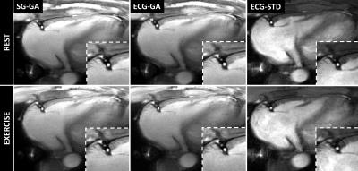 |
Self-Gated Golden Angle Spiral CINE MRI for Endothelial Function Assessment
Gabriele Bonanno, Allison Hays, Robert Weiss, Michael Schär
A novel self-gated 2D spiral CINE MRI method is proposed to assess coronary endothelial function (CEF) and was tested in healthy volunteers. Cardiac self-gating data were extracted from the k-space center and showed high correlation with simultaneously-recorded ECG. High coronary image quality and CEF measures, in good agreement with a standard ECG-triggered method, can now be obtained without the need for ECG.
|
17:39
|
0929.
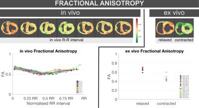 |
Characterisation of in-vivo and ex-vivo cardiac Diffusion Tensor Imaging scalar measures of cardiac microstructure in healthy swine - permission withheld
Sonia Nielles-Vallespin, Pedro Ferreira, Andrew Scott, Ranil de Silva, Philip Kilner, Daniel Ennis, Dudley Pennell, David Firmin, Andrew Arai
In-vivo cDTI was performed at several cardiac phases in healthy swine (N=16), followed by ex-vivo cDTI in two contractile states. The three eigenvalues (L1, L2, L3), MD, FA and Mode were compared. All trends between in-vivo diastole and systole matched those between ex-vivo relaxed and contracted states, except for MD, which decreased ~10% in-vivo from diastole to systole, with no significant differences ex-vivo between relaxed and contracted states. These results provide a useful baseline for future preclinical studies with cardiac disease models, and might contribute towards formulating a strain correction model that accounts for the microstructural constraints and deformations of the myocardium.
|
17:51
|
0930.
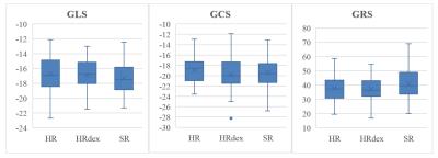 |
Strain analysis methods from CMR more sensitive than echocardiographic methods to small differences in cardiotoxicity remodeling between risk groups of cancer survivors.
Delphine Perie, Hadi Begdouri, Mohamed Aissiou, Farida Cheriet, Tarik Hafyane, Matthias Friedrich, Caroline Laverdière, Maja Krajinovic, Daniel Sinnett, Gregor Andelfinger, Daniel Curnier
The use of cardiac strain mapping may provide useful knowledge that may help in detecting doxorubicin-induced cardiotoxicity at a functional scale. Although the feasibility of CMR has been established, there are no standard acquisition protocols or processing pipelines to assess cardiac strain maps. Compared to echocardiography, strain analysis methods from CMR are more sensitive to small differences in cardiotoxicity between risk groups in cancer survivors. While strain mapping from echocardiography remains adequate to detect large differences between healthy volunteers and patients with diseases, our study highlighted the necessity to combine different strain mapping methods to fully describe small cardiac damages
|
18:03
|
0931.
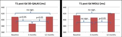 |
Longitudinal study of myocardial T1 and T2 relaxation times in aortic stenosis patients: before, and 3- and 12 months after aortic valve replacement
Sofia Kvernby, Mattias Rönnerfalk, Marcel Warntjes, Carl-Johan Carlhäll, Eva Tamás, Jan Engvall, Tino Ebbers
The purpose of this pilot study is to investigate whether myocardial relaxation times (T1 and T2), alter over time in patients with severe aortic valve stenosis, from pre-surgery to 12 months after aortic valve replacement. Myocardial relaxation times were measured pre surgery, and 3 months and 12 months post surgery with 3D-QALAS, T1-MOLLI and T2-GraSE. The results demonstrated significant changes in myocardial relaxation times over time after surgery in this patient group.
|
|