Oral
Body: Breast, Chest, Abdomen, Pelvis
Monday, 24 April 2017
| Room 320 |
16:15 - 18:15 |
Moderators: Sooah Kim, Bachir Taouli |
Slack Channel: #s_body
Session Number: O27
16:15
|
0317.
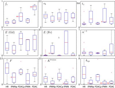 |
Physiologically-constrained Multiagent DCE-MRI for Pancreatic Cancer Imaging 
Matthias Schabel, Erin Gilbert, Alexander Guimaraes, Cory Wyatt
Physiologically-constrained multiagent pharmacokinetic modeling in pancreas using sequential injections of gadoteridol and ferumoxytol reveals differences between healthy pancreas in high-risk patients and both IPMN and pancreatic ductal adenocarcinoma.
|
16:27
|
0318.
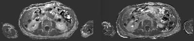 |
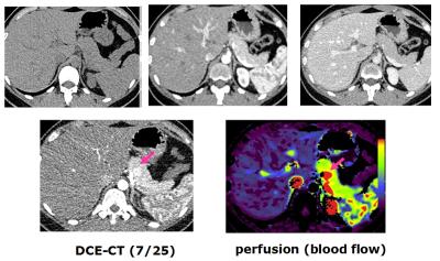
Improved Characterization of Contrast Uptake in Human Pancreatic Cancer
Douglas Kelley, Benjamin Yeh, Michael Ohliger, Eric Collisson, Zhen Wang
We present an improved method for quantifying contrast agent uptake in highly desmoplastic pancreatic tumors in human patients. The new method separates the T1 and T2* effects of the Gadolinium-based contrast agent; uses fast acquisition and non-rigid registration to minimize motion effects; and two point Dixon imaging to separate the water component on a pixel by pixel basis.
|
16:39
|
0319.
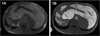 |
Prognostic value of hepatobiliary phase MRI in patients with primary sclerosing cholangitis – Assessment of clinical outcome and evaluation of surrogate parameters
Jennifer Schulze, Henrike Lenzen, Jan Hinrichs, Michael Manns, Frank Wacker, Kristina Ringe
In this prospective study we assessed the prognostic value of hepatobiliary phase (HBP) MRI in patients with primary sclerosing cholangitis (PSC). Relative enhancement (RE) in the HBP after gadoxetate disodium injection correlated significantly with clinical scores (MELD, Mayo Risk) established to estimate survival in patients with chronic liver disease and PSC. More importantly, a significant correlation with previously suggested surrogate parameters for clinical outcome as well as with solid clinical endpoints (development of tumor, liver transplantation, death) at follow-up could be observed. These promising results attest HBP MRI in patients with PSC a potential prognostic role and warrant further long-term evaluation.
|
16:51
|
0320.
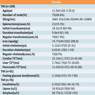 |
Early screening of pancreatic iron overload in thalassemia major with MRI T2* - permission withheld
Jingwen Huang, Qihua Yang, Jinglian Zhong, Xiaodong Chen, Ziliang Cheng, Taihui Yu, Yun Su, Biling Liang
Diabetes Mellitus is a serious complication of thalassemia major. Intensive chelation therapy in the early stage may avoid diabetes. So we aimed to determine the optimal timing age of pancreatic iron screening with MRI T2* technique. Early pancreatic hemosiderin was found in thalassemia major, with the youngest one of 5.3 years old. Early dysfunction of pancreatic exocrine and endocrine glands was found in thalassemia major, with the youngest one of 5.5 years old. Therefore, we suggest age of 5 to 6 years old as the optimal initial age for pancreatic T2* scanning.
|
17:03
|
0321.
 |
The Development And Prognostication Of Magnetic Resonance Elastography Thresholds In Primary Sclerosing Cholangitis
Kartik Jhaveri, Hooman Hosseini-Nik, Nima Sadoughi, Harry Janssen, Jordan Feld, Sandra Fischer, Ravi Menezes, Angela Cheung
Primary sclerosing cholangitis (PSC) is a chronic, progressive, cholestatic liver disease which causes bile duct structuring and eventually causes liver cirrhosis requiring liver transplantation. Due to heterogeneity of liver fibrosis distribution and lack of optimal method to assess disease severity accurate disease stratification is challenging. Magnetic resonance elastography (MRE) has shown very good results for quantification of hepatic fibrosis. MRE may provide a unique means of stratification and prognostication in PSC with its ability to assess a larger volume of hepatic tissue compared to biopsy or transient elastography(VCTE) and this what we explored in this prospective study.
|
17:15
|
0322.
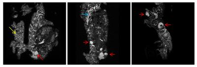 |
Diffusion imaging detects differences in disease trajectory between two mouse models of pancreatic cancer
Palamadai Venkatasubramanian, Matthew Smith, Jesse Yan, Brian Hallis, Emman Mascarinas, Andrew Diaz, Brian DeCant, Ron McKinney, Paul Grippo, Alice Wyrwicz
Multiparametric MR microimaging deteced differences in pancreatic microstructure between two mouse models of pancreatic cancer, EL-KRASG12D (EK) and p48-Cre/LSL-Kras (KC) mice, that overexpress mutant KRas via different mechanisms. MR signatures characteristic of acinar-ductal metaplasia, fibrosis, cystic neoplasms and precancerous lesions revealed different trajectories of disease development between the two genetically engineered mice.
|
17:27
|
0323.
 |
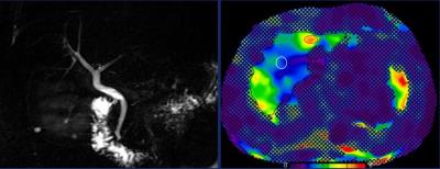
Insulinoma localization with cross-sectional imaging: head-to-head comparison of contrast-enhanced CT, volume perfusion CT and multi-parametric MR
Liang Zhu, Zhao-yong Sun, Hua-dan Xue, Tian-yi Qian, Zheng-yu Jin
This study aims to compare insulinoma localization with CECT, VPCT and multi-parametric MR (mp-MR) at 3T in the same patients, in a prospective manner. CECT, VPCT and mp-MR were performed in patients with suspected insulinomas. The presence/absence of tumor within the pancreatic head, neck, body and tail region was evaluated with 5-scale confidence levels. ROC analysis was performed. Surgical pathology served as reference standard. We found that VPCT and mp-MR showed improved diagnostic performance for insulinoma localization, compared to CECT. Both modalities could serve as problem-solving tools in difficult cases. mp-MR has the potential to replace CECT as the first-line examination.
|
17:39
|
0324.
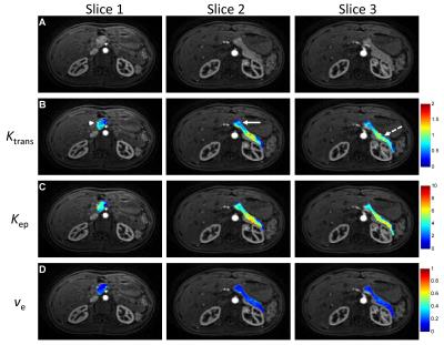 |
3D Pancreatic Perfusion MRI using Through-Time Spiral GRAPPA Acceleration
Yong Chen, Shivani Pahwa, Mark Griswold, Nicole Seiberlich, Vikas Gulani
In this study, free-breathing 3D pancreatic perfusion quantification was achieved using a rapid non-Cartesian parallel imaging technique. The method was applied to 11 asymptomatic subjects and the values are in good agreement with literature values. Significant differences in both Ktrans and Kep were noticed between different locations in the pancreas, but no significant difference was found in the volume of distribution (Ve).
|
17:51
|
0325.
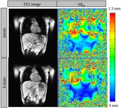 |
Geometric distortion due to B0 inhomogeneity in liver MR imaging under inhalation and exhalation breath-hold - permission withheld
Oi Lei Wong, Jing Yuan, Yihang Zhou, Siu Ki Yu, Kin Yin Cheung
Geometric accuracy is critical for radiotherapy and is one major concern in the application of MRI in radiotherapy. Since the geometric distortion and B0 inhomogeneity are related, we aim to evaluate the geometric distortion due to ΔB0 variation at inhalation and exhalation breath-hold. Based on our results, larger geometric distortion was noted during inhalation than exhalation.
|
18:03
|
0326.
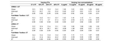 |
Simultaneous Iron and Fat Quantification Using an Auto Regressive Moving Average Model at 1.5T and 3T
Aaryani Tipirneni-Sajja, Axel Krafft, Brian Taylor, Ralf Loeffler, Ruitian Song, Nathan Artz, Jane Hankins, Claudia Hillenbrand
A major confounder of hepatic iron assessment by R2*-MRI is fat (e.g. steatosis) which introduces signal modulations. In this study, we systematically evaluate two signal modeling techniques, an autoregressive moving average (ARMA) model and the method provided by the ISMRM Fat-Water Toolbox for simultaneous iron and fat quantification in phantoms and in vivo. Preliminary data suggest that ARMA and Toolbox can be used for iron and fat quantification at 1.5T and 3T. In severe iron-overload cases, both, ARMA and the Toolbox might produce inaccurate FF results, however in vivo ARMA seemed to provide a more robust liver R2* quantification.
|
|