Oral
Body: Breast, Chest, Abdomen, Pelvis
Thursday, 27 April 2017
| Room 310 |
08:15 - 10:15 |
Moderators: Daniel Margolis, Davinia Ryan |
Slack Channel: #s_body
Session Number: O32
08:15
|
1007.
 |
PI-RADS Version 2: Preoperative Role in the Detection of Normal-sized Pelvic Lymph Node Metastasis in Prostate Cancer 
Young Taik Oh, Sung Yoon Park, Dae Chul Jung
Although the identification of pelvic lymph node metastasis (PLNM) is important in prostate cancer, sometimes PLNM are often not so enlarged on imaging. Therefore, we were trying to identify normal sized PLNM through PI-RADS v2 scores of 221 patients with prostate cancer. In our study, a threshold of PI-RADS v2 score of 5 seems to be associated with an increased risk of normal-sized PLNM, which may help identify the need for further node-specific imaging studies or pelvic lymph node dissection when a patient with prostate cancer has only normal-sized pelvic lymph nodes on preoperative imaging.
|
08:27
|
1008.
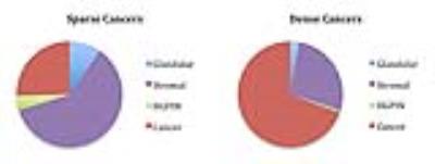 |
Sparse Prostate Cancers on Whole-Mount Histopathology and Multiparametric MRI - permission withheld
Olga Starobinets, Jeffry Simko, Kyle Kuchinsky, Peter Carroll, Kirsten Greene, John Kurhanewicz, Susan Noworolski
The study purpose was to establish incidence and Gleason Score of sparse lesions on whole-mount histopathology in post-prostatectomy samples and to identify imaging characteristics associated with sparse cancers detected on multiparametric MRI (mpMRI). Based on histopathology, sparse lesions were smaller than dense lesions (0.065cc vs 0.916cc), with the majority (56/57 sparse) being low-grade (GS3+3). On imaging, we found statistically significant differences between sparse GS3+3 and benign tissues on apparent diffusion coefficient and peak enhancement maps. This combined with small-size and low-grade, and thus low clinical importance [1] of sparse lesions suggests that current mpMRI capabilities are sufficient to characterize these lesions.
|
08:39
 |
1009.
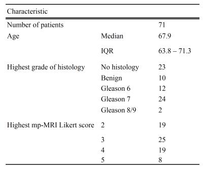 |
Microstructural Diffusion-Weighted (VERDICT) MRI Metrics are Repeatable and Show Potential at Characterising Gleason 7 Prostate Cancer Non-Invasively
Edward Johnston, Elisenda Bonet-Carne, Hayley Pye, Joey Clemente, Ben Yvernault, Dominic Patel, Susan Heavey, Mrishta Appayya, Alexandra Saborowska, Ashwin Sridhar, Greg Shaw, Sebastien Ourselin, David Hawkes, Caroline Moore, Hayley Whitaker, Manuel Rodriguez-Justo, Alexander Freeman, Eleftheria Panagiotaki, Daniel Alexander, Shonit Punwani
In this study we we assess the repeatability of VERDICT (Vascular, Extracellular, and Restricted Diffusion for Cytometry in Tumours) MRI parameters in prostate cancer consider their ability to distinguish between Gleason grades compared with the standard ADC model in 71 patients. Four of the parametric maps derived from the VERDICT technique were found to be satisfactorily repeatable for use as a clinical tool, and are capable of identifying a Gleason 7 component in prostate cancer where ADC failed to do so. VERDICT therefore holds great potential for use in clinical prostate cancer management pathways in the future.
|
08:51
|
1010.
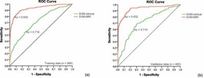 |
Machine learning analysis of multi-parametric MRI helps to improve the predictive performance in prostate cancer
Rui Wang, Xiaoying Wang
We first investigated the systemic outcome of a crowd underwent PSA-based screening and pre-biopsy mp-MRI, and demonstrated the predictive role of pre-biopsy mp-MRI for PCa by using an advanced machine learning-based approach. Here we answer one important question at the beginning of the paper: (1) mp-MRI coupled with PSA screening program can be used to detect PCa. By the constructed nomogram, the outcome of most patients could be accurately predicted in the first 1-yr follow-up period if they received a pre-biopsy mp-MRI examination, even without invasive TRUS biopsy.
|
09:03
|
1011.
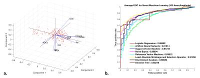 |
A comprehensive study of machine-assisted classifiers for predicting prostate cancer Gleason grade
Jing Wang, Yang Fan, Yudong Zhang
We performed comprehensive study of eight popularized classifiers for predicting prostate cancer (PCa) Gleason score (GS). The multi-parametric MRI data was obtained from 205 histopathology-confirmed PCa. The MR features were modeled using eight classifiers to predict high-GS (4+4) PCa, including Logistic Regression (LR), Artificial Neural Network (ANN), Support Vector Machine (SVM), Naive Bayes (NB), Relevance Vector Machine (RVM), Least Absolute Shrinkage and Selection operator (LASSO), Discriminant Analysis (DA) and Decision Tree (DT) analysis. Results showed that LASSO and DA had significantly higher area under curve than other classifiers, thus could be valuable for automatic prediction of PCa grade.
|
09:15
|
1012.
 |
The value of multiparametric MR Imaging in predicting the volume of clinically significant prostate cancer: a whole-mount step-section analysis
Jian Jiang, Huihui Wang, Xiaoying Wang
This study is to assess the factors influencing multiparametric (MP) MR Imaging occuracy in estimating the volume of clinically significant prostate cancer (Vh) by using whole-mount step-section slides as standard of reference.
|
09:27
|
1013.
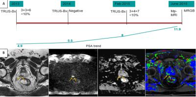 |
MRI guided biopsy in Patients with positive TRUS-biopsy: Necessity or Overkill?
Kareem Elfatairy, Christopher Filson, Adeboye Osunkoya, Rachel Geller, Sherif Nour
TRUS biopsy (TRUS-Bx) is the standard of care for prostate cancer diagnosis but has several limitations. Multiparameteric MRI and MRI guided biopsy (MRGB) show considerable added benefits with better disease detection and stratification. The purpose of this study is to evaluate the role of in-bore MRI guided biopsy in patients with positive TRUS-Bx. 40 patients with 71 TRUS-Bx positive lesions were analyzed for cancer detection rate, false negative rate, and Gleason score upgrade by MRGB. MRGB showed 65% cancer detection rate, 35 % false negative rate, and upgraded disease status in 40% of the whole sample
|
09:39
|
1014.
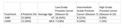 |
Effects of 5 alpha-Reductase Inhibitors on Detection of Prostate Cancer on MRI
Lori Mankowski Gettle, Shivashankar Damodaran , David Jarrard, Frederick Kelcz
5 alpha reductase inhibitors have shown promise in increasing the detection of prostate cancer in prior ultrasound studies. No studies have been performed looking at the effect of 5 alpha reductase inhibitors on increasing the conspicuity of prostate cancer in MRI. The purpose of this study was to determine if treatment with a 5 alpha reductace inhibitor could increase sensitivity for the detection of prostate cancer in the background setting of benign prostatic hypertrophy.
|
09:51
|
1015.
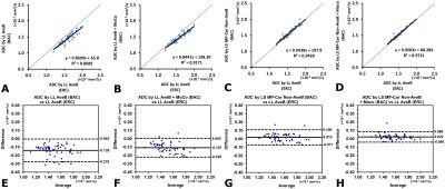 |
Improved Accuracy of Apparent Diffusion Coefficient (ADC) Quantification: Evaluation in Prostate Diffusion Imaging without Using Endorectal Coils
Xiaodong Zhong, Marcel Nickel, Stephan Kannengiesser, Alto Stemmer, Brian Dale, Berthold Kiefer, Mustafa Bashir
In prostate DWI, low SNR often causes inaccuracy in ADC quantification if not compensated, especially when using surface array coils. Endorectal coils can be used, although associated with substantial setup time, patient discomfort and complications. In this work, a noise bias correction framework was developed and validated in a Monte Carlo simulation, a diffusion phantom, and 14 prostate imaging subjects. Using data acquired with an endorectal coil as a reference, this framework showed improved accuracy of ADC quantification in the prostate when only non-endorectal coils were used. This framework may allow quantitative prostate diffusion imaging without requiring endorectal coils.
|
10:03
|
1016.
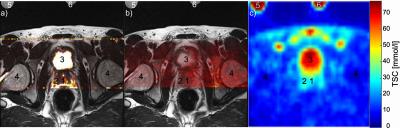 |
Multi-Parametric/-Nuclear 1H/23Na Clinical Protocol of the Prostate at 3T using a Double Resonant Coil
Nadia Paschke, Daniel Hausmann, Lothar Schad, Stefan Schönberg, Frank Zöllner
Prostate cancer is the most common malignancy in men. T2-weighted magnetic resonance imaging (MRI) has been shown to provide detailed information about anatomical structures but suffers from low specificity of morphological abnormalities. To enhance identification of malignant lesions, we set up a multi-parametric and multi-nuclear clinical protocol, in which sodium (23Na-) MRI and diffusion weighted imaging with standard and high b-values are added to the morphological T2-weighted sequences. Using a double resonant 23Na/1H abdominal coil allows acquiring sodium images directly after high resolution clinical proton sequences. Coil changes become unnecessary, which improves clinical feasibility.
|
|