Oral
Body: Breast, Chest, Abdomen, Pelvis
Wednesday, 26 April 2017
| Room 314 |
13:45 - 15:45 |
Moderators: Siarhei Kharuzhyk, Diego Martin |
Slack Channel: #s_body
Session Number: O34
13:45
|
0816.
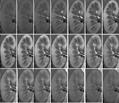 |
An MR compatible kidney perfusion system to assess kidney function and organ preservation. 
Abhishek Pandey, Catherine Min, Zhitao Li, Kevin Johnson, Leah Steyn, William Purvis, Robert C. Harland, Klearchos K. Papas, Puneet Sharma, Diego R Martin, Manojkumar Saranathan, Jean-Philippe Galons
A MR-compatible kidney perfusion system is presented to assess the quality of kidney preservation schemes for transplantation. DCE-MRI is used to estimate and compare the glomerular filtration rate (GRF) in kidneys following a 24 hrs cold gaseous oxygen perfusion (persufflation) Vs a standard 24 hrs cold ischemic storage.
|
13:57
|
0817.
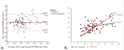 |
The comparative study of MR renography versus 99mTc-DTPA renal scintigraphy for determining split renal function of the transplanted kidney
Jing Wang, Yang Fan, Yudong Zhang
The evaluation of split kidney function is of clinical significance for patients after renal transplantation. This study explored two available approaches, dynamic MR renography vs. 99mTc-DTPA clearance, for determining split renal GFR in 57 transplanted patients. MR-based GFR was measured using an established Baumann-Rudin model, modified two-compartment and three-compartment model. MR results were compared with 99mTc-DTPA, using 24-h creatinine clearance rate as a reference. MRR using a 2C model had significantly higher accuracy (r = 0.925) than 99mTc-DTPA clearance (r = 0.317), thus could be a more promising way for evaluation of transplanted kidney function.
|
14:09
|
0818.
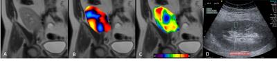 |
Initial experience with magnetic resonance elastography and acoustic radiation force impulse elastography in renal transplant patients
Paul Kennedy, Octavia Bane, Sonja Gordic, Cecilia Besa, Stefanie Hectors, Mathilde Wagner, Rafael Khaim, Madhav Menon, Vinay Nair, Sara Lewis, Bachir Taouli
In this prospective study we compared renal transplant stiffness measured with MRE and ARFI ultrasound in 9 patients. Repeatability of both modalities was determined through test-retest imaging in 5 patients. MRE stiffness was significantly lower than ARFI stiffness as expected. MRE test-retest repeatability was excellent with mean coefficient of variation (CV) of 6%, while ARFI had CV of 30%. In addition, ARFI measurements exhibited a high inter-quartile range in the majority of cases suggesting inconsistency in the measurements. Our results suggest MRE is a more robust choice for renal transplant measurement compared to ARFI.
|
14:21
|
0819.
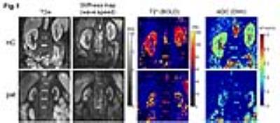 |
In vivo multifrequency MR elastography for the assessment of renal stiffness in patients with IgA nephropathy
Jing Guo, Stephan Marticorena Garcia , Michael Dürr, Florian Dittmann, Sebastian Hirsch, Jürgen Braun, Ingolf Sack
Renal stiffness was investigated using multifrequency MRE in healthy controls and patients with IgA nephropathy. Patients show a reduced renal stiffness as compared to healthy controls. DWI and BOLD imaging were also applied to the subjects. Both ADC and T2* were linearly correlated with wave speed obtained from MRE. By mechanical vascular-solid tissue interactions, wave speed measurements by multifrequency MRE offer a quantitative measure for the noninvasive assessment renal function.
|
14:33
|
0820.
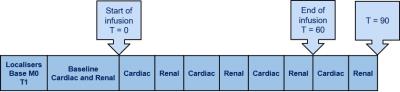 |
Using Cardiorenal MRI to assess fluid balance
Chris Bradley, Damian Bragg, Eleanor Cox, Ahmed El-Sharkawy, Abeed Chowdhury, Dileep Lobo, Susan Francis
We used quantitative MRI measurements to perform a randomised, double-blind crossover study on the effects of isovolumetric and isoeffective infusions of colloid versus crystalloid on structural and haemodynamic (ASL, ADC, flow, volume) changes in the kidney and cardiac output. No significant differences were observed between the blood expanding properties of the infusions, despite using a colloid infusion one-third the volume of the reference crystalloid infusion. We observed a trend of less oedema using an isoeffective volume of colloid. Repeated MRI measurements demonstrated low CoVs, allowing MRI to assess subtle changes in structure and haemodynamics for determining optimal perioperative infusions.
|
14:45
|
0821.
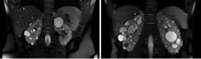 |
Inter-rater Reliability and Translational Implications of MR-based Polycystic Kidney Volume Measurements by Stereology at Early and Late Stage Disease
Rebecca Lepping, Rainer Karcher, Paul Keselman, Darren Wallace, Alan Yu, Laura Martin, William Brooks
Autosomal dominant polycystic kidney disease (ADPKD) is characterized by the presence of fluid-filled cysts that grow over time. Total kidney volume (TKV) is one of the main biomarkers of disease progression, and is estimated through the technique of stereology. We tested whether patients’ kidney size impacted inter-rater reliability using the same stereology protocol. Stereology yielded excellent inter-rater reliability at both early and late stage disease, however, some pathology would still benefit from expert guidance in determining kidney tissue. This technique can easily be translated to animal models of ADPKD.
|
14:57
|
0822.
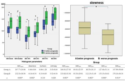 |
Quantitative Volumetric Histogram Analysis of Diffusion-weighted Magnetic Resonance Imaging: An Initial Experience of Solid Renal Cell Carcinoma with Different Prognosis
Anqin Li, Zhen Li, Haojie Li, Daoyu Hu
To evaluate the value of quantitative volumetric ADC histogram analysis for differentiation of clear cell RCC (ccRCC) from papillary RCC (pRCC) and chromophobe RCC (chRCC) which having different prognosis. Differences of ADC histogram parameters between better prognosis group and worse prognosis group were compared. There were significant differences on ADCmean, ADCmedian, ADC10%, ADC25%, ADC75%, ADC90% and skewness between these two different prognosis groups and the ADC10% showed the best diagnostic value. Therefore, quantitative volumetric ADC histogram analysis can be considered a useful and noninvasive method to help distinguish three subtypes renal cell carcinomas which having different prognosis.
|
15:09
 |
0823.
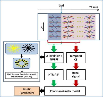 |
Glomerular filtration rate estimation in vivo using 3D radial MRI and a novel multiresolution reconstruction technique
Abhishek Pandey, Jean-Philippe Galons, Kevin Johnson, Diego R Martin, Maria I Altbach, Ali Bilgin, Manojkumar Saranathan
Dynamic contrast enhanced MRI can be used to obtain single-kidney GFR estimates but tradeoffs between spatial and temporal resolution lead to errors. We propose a novel reconstruction technique where AIFs are generated from images reconstructed with high temporal resolution while renal parenchyma signal is extracted from images reconstructed over a larger temporal window to generate images with high spatial resolution and improved image quality. We demonstrate the clinical validity of the method by estimating the GFR using our proposed method and compare it with serum creatinine based eGFR estimate.
|
15:21
|
0824.
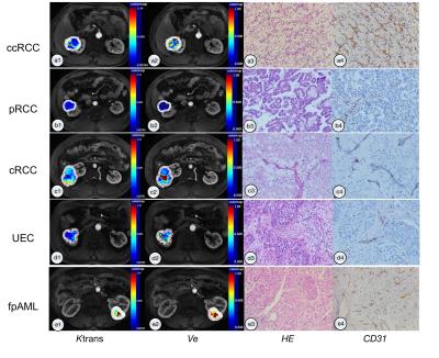 |
Dynamic Contrast-enhanced MRI in Renal Tumors: Common Subtype Differentiation using Pharmacokinetics
Haiyi Wang, Lu Ma, Zihua Su, Ning Huang, Xiao Xu, Zhipeng Sun, Aitao Guo, Huiyi Ye
In this prospective study on pharmacokinetic parameters (Ktrans & Ve) of renal tumors, we enrolled the patients with five common subtypes of renal tumor - clear cell renal cell carcinoma (ccRCC), papillary renal cell carcinoma (pRCC), chromophobic renal cell carcinoma (cRCC), uroepithelial carcinoma (UEC), and fat poor angiomyolipoma (fpAML) to undergo DCE-MRI pharmacokinetic studies. Our results demonstrated that ccRCC, pRCC, cRCC, UEC and fpAML are pharmacokinetically different (Ktrans & Ve). Ktrans could distinguish ccRCC from non-ccRCC (pRCC & cRCC) and differentiate fpAML with non-ccRCC with high specificity and sensitivity, which probably can facilitate the precise treatment of renal tumors in the future clinical practice.
|
15:33
|
0825.
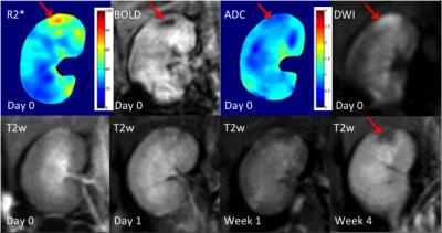 |
Comparison of BOLD-contrast imaging and DW imaging for early prediction of renal damage after microemboli-induced acute kidney injury in animal model
Chengyan Wang, Hanjing Kong, Fei Gao, Wenjian Huang, Lian Ding, Rui Wang, Li Jiang, Yan Jia, Hui Xu, He Wang, Xiaodong Zhang, Li Yang, Jue Zhang, Xiaoying Wang, Jing Fang
Atheroembolic renal disease (AERD) is an important yet under-diagnosed kidney diseases associated with atherosclerosis. This study investigates the utility of BOLD and DWI in the assessment of microemboli-induced AKI, and compares the performances of both techniques for early prediction of renal damage. AKI was induced by injection of microspheres into the right kidney. The results indicate that both techniques can serve as reliable indicators to focal severe regional ischemia before renal damage occurs, while BOLD imaging seems to be more sensitive in early detection of moderate ischemia.
|
|