Monday, 24 April 2017
| Room 316A |
08:15 - 10:15 |
Moderators: Steven Baete, David Bluemke |
Slack Channel: #s_msk
Session Number: O38
08:15
|
0087.
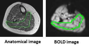 |
Increased muscle BOLD following exercise training in older adults 
Jill Slade, Anne Tonson, David Hurley, Mitchell Rozman, George Abela, Ronald Meyer
Functional MRI (BOLD) of skeletal muscle was used to evaluate changes in microvascular function before and after aerobic exercise training in older adults. Peak BOLD responses increased by ~30% after exercise training, supporting the use and sensitivity of BOLD MRI to assess changes in microvascular function.
|
08:27
|
0088.
 |
Improved Muscle Microstructure Analysis with Diffusion Weighted Imaging and Advanced Tissue Modeling
Nagesh Adluru, Richard Kijowski , Fang Liu
Studies on musculoskeletal systems can benefit by quantitative mapping of the tissue microstructure. Parameters from traditional diffusion tensor imaging (DTI) may serve as bio-markers for assessing muscle fiber health. While these parameters are sensitive to changes of muscle fiber orientation, length and tension, they are non-specific to the changes of microstructure and microcomposition of muscle fibers. In this study, we proposed to use multi-shell diffusion weighted imaging acquisition with advanced diffusion and micro tissue modeling to improve in-vivo muscle fiber analysis and demonstrated the feasibility of applying these methods on in-vivo human thigh muscle imaging.
|
08:39
|
0089.
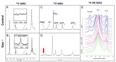 |
13C/31P MRS biomarkers of disease progression and response to gene therapy in a mouse model of Pompe disease
Celine Baligand, Gary Todd, Brittany Lee-McMullen, Ravneet Vohra, Barry Byrne, Darin Falk, Glenn Walter
With the emergence of rAAV-based gene therapy clinical trials in patients with glycogen storage disorders such as Pompe disease, there is a pressing need for early and non-invasive markers to assess treatment efficacy. While 13C-MRS has been used for detection of glycogen in muscle, its clinical implementation remains limited, due to its low natural abundance and inherent low sensitivity. 31P-MRS has higher sensitivity and can probe intermediates of glucose/glycogen metabolism. We sought to identify new biomarkers of Pompe disease progression in muscle using 13C/31P-MRS and 1H-HR-MAS in the mouse model of the disease, and tested their sensitivity to rAAV therapy.
|
08:51
 |
0090.
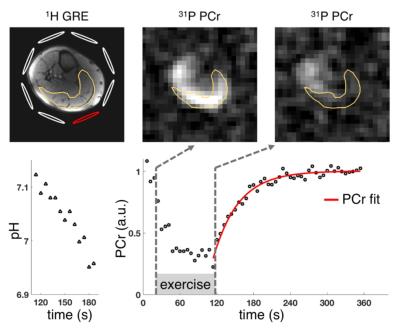 |
Dynamic PCr and pH imaging of the human lower leg muscle during exercise at 3T
Oleksandr Khegai, Guillaume Madelin, Ryan Brown, Prodromos Parasoglou
Dynamic phosphorous MRSI is an established non-invasive method for studying muscle metabolism. It allows quantification of the post-exercise phosphocreatine resynthesis rate, which provides insights into various physiological and pathological conditions. Due to low SNR, 31P imaging experiments are typically limited by long acquisition times relative to the metabolic recovery. We developed an imaging method to measure localized phosphocreatine resynthesis and pH changes in muscles of the lower leg following exercise at 3T with a high temporal resolution of 6 s required for an accurate estimation of quantitative phosphocreatine recovery rates.
|
09:03
|
0091.
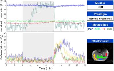 |
Dynamic interleaved NMR measurements of perfusion, deoxymyoglobin and phosphorylated metabolites during ischemic and exercise paradigms in the calf and thigh muscles
Alfredo Lopez Kolkovsky, Benjamin Marty, Bertrand Coppa, Eric Giacomini, Pierre Carlier
NMR allows to investigate multiple aspects of physiological parameters like regional perfusion, blood and tissue oxygenation, intracellular pH or high-energy phosphate metabolism. In the past, interleaved multi-parametric multi-nuclear dynamic NMR imaging and spectroscopy of skeletal muscle was developed on prototype scanners. Here we developed an interleaved pulse sequence combining NMR acquisitions of a perfusion image, 1H deoxy-myoglobin and 31P spectra on a clinical system without any hardware modifications from the customer. We successfully evaluated this sequence in the ischemic calf muscle and exercising quadriceps muscle. Nevertheless, using a surface coil for pulsed-ASL measurements remains a limitation at this time.
|
09:15
|
0092.
 |
Dynamic Diffusion Tensor Imaging in Normal and Compartment Syndrome Calf Muscle with MEDITI
Eric Sigmund, Steven Baete, Karan Patel, Di Wang, Ricardo Otazo, Prodromos Parasoglou, Jenny Bencardino
We describe measurement of skeletal muscle kinematics with a multiple echo diffusion tensor imaging (MEDITI) in clinical scanners. This approach allows characterization of the microstructural dynamics in healthy and diseased muscle. Combining the accelerated MEDITI directional encoding with a radial k-space trajectory and compressed sensing reconstruction allows spatially resolved DTI with a continuous temporal resolution of 16 s. Using an MR-compatible ergometer, post-exercise recovery of DTI metrics in calf muscle were quantified in a pilot cohort of 2 volunteers and 4 subjects with chronic exertional compartment syndrome (CECS). Results indicate anisotropic exercise response and recovery with kinetics differing from relaxation contrast.
|
09:27
|
0093.
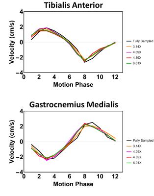 |
Compressed Sensing accelerated time-resolved 3D phase contrast MRI of the lower leg muscles during active dorsi- and plantarflexion
Lukas Gottwald, Valentina Mazzoli, Eva Peper, Qinwei Zhang, Bram Coolen, Pim van Ooij, Gustav Strijkers, Aart Nederveen
Time-resolved 3D phase-contrast MRI can be applied to quantify muscle contraction. 3D coverage with sufficient spatiotemporal resolution (~3x3x5mm3, 160ms) can only be achieved by interleaved acquisitions during many repetitions of a motion task, resulting in long scan times (>10min). In this study we have developed an accelerated protocol, using k-space undersampling and compressed-sensing reconstruction, which was applied on the lower leg of 4 volunteers performing a foot plantar-dorsal flexion motion task. Muscle velocities during the motion cycle of fully-sampled and accelerated protocols were compared. Acceleration was successful up to 6.4X with comparable velocities, which confirmed the benefit of this approach.
|
09:39
 |
0094.
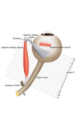 |
Magnetic Resonance Imaging of the Functional Anatomy of the Oblique Muscles in Patients with Primary Oblique Overaction
Qianwen Gong, Longqian Liu, Miroslaw Janowski
The cause of primary eye movement abnormality is unknown. The functional MRI of superior and inferior oblique muscles was instrumental to investigate the cause of overaction. We have shown similar size of superior oblique muscle in patients and controls in the resting state, while the MRI performed during gazes revealed differences in the contractility, what suggests the abnormal innervation as a cause of primary superior oblique muscles. In contrast, the inferior oblique muscle was larger in resting state, without a difference in contractility what indicates the hypertrophy as a basis for primary inferior oblique muscle overaction.
|
09:51
|
0095.
 |
An MRI-based assessment of the correlation between cerebral white matter changes, muscle structure, and muscle function in myotonic dystrophy
Daniel Thedens, Cheryl Smith, Peg Nopoulos, Richard Shields, Laurie Gutmann
The purpose of this work was to study subjects with myotonic dystrophy (DM1) utilizing MRI to assess correlations between global cerebral white matter abnormalities, muscle structure, and muscle function. MRI-based measures of white matter (fractional anisotropy), muscle structure (volume, fat fraction, T2 mapping) along with muscle function testing demonstrated several significant correlations. The combination of neuroimaging and muscle structure assessment with MRI holds considerable promise towards elucidating the relationships between CNS abnormalities and neuromuscular dysfunction.
|
10:03
|
0096.
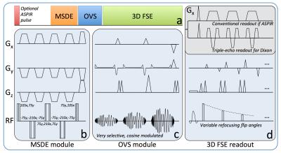 |
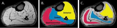
A Flexible Technique for Flow-Sensitive Fat-Suppressed High-Resolution Peripheral Nerve Imaging
Valentina Taviani, Miyoshi Mitsuharu, Kang Wang, Kevin King, Suchandrima Banerjee, Sandip Biswal, Shreyas Vasanawala, Daehyun Yoon, Robert Peters
We developed a flow-sensitive 3D fast spin echo pulse sequence with Dixon-based water-fat separation and compressed sensing for robust and efficient peripheral nerve imaging. Outer volume suppression allows shorter scan times by limiting spatial encoding of the FOV to the anatomy of interest without aliasing concerns. In addition, it improves the performance of spectrally-selective fat suppression methods, that can be advantageous for very high resolution imaging but are typically hampered by B0 inhomogeneity, by allowing shimming over smaller regions. Preliminary data showed good delineation of peripheral nerves in different anatomies, with adequate resolution and clinically feasible acquisiton times.
|
|