Oral
General Cancer Imaging
Tuesday, 25 April 2017
| Room 316BC |
16:15 - 18:15 |
Moderators: Patrick Bolan, Ashley Stokes |
Slack Channel: #s_cancer
Session Number: O41
16:15
|
0620.
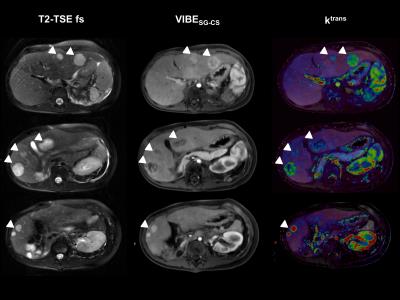 |
Feasibility and evaluation of free-breathing and self-gated cartesian-sampled T1-weighted 4D MRI with compressed sensing resulting in combined diagnostic image quality and perfusion analysis 
Christer Ruff, Jakob Weiss, Ahmed Othman, Petros Martirosian, Marcel Nickel, Robert Grimm, Manuel Kolb, Matthias Kündel, Fabian Bamberg, Konstantin Nikolaou, Mike Notohamiprodjo
In clinical practice, a remaining challenge of dynamic contrast-enhanced (DCE)-MRI is to acquire a high spatio-temporal resolution without motion artifacts and diagnostic quality, despite acquisitions over several minutes. In particular, iterative reconstruction techniques have brought free-breathing acquisitions in clinical range. In this study, we proved feasibility and evaluated a prototype Cartesian-sampled fat-saturated T1-weighted 3D gradient-echo sequence with automated respiratory self-gating and compressed sensing reconstruction (VIBESG-CS) for continuous dynamic contrast-enhanced (DCE) MRI and perfusion analysis including lesion detection of the liver using a Tofts model. VIBESG-CS including perfusion analyisis showed better motion robustness and similar lesion conspicuity compared to a standard morphology sequence (fat-saturated T2-TSE).
|
16:27
 |
0621.
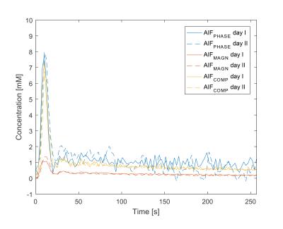 |
Robust arterial input functions by fitting the complex DCE-MRI signal: a test-retest study in prostate cancer
Edzo Klawer, Petra van Houdt, Frank Simonis, Cornelis van den Berg, Floris Pos, Stijn Heijmink, Uulke van der Heide
The arterial input function is still one of the main problems for quantitative DCE analysis. In this study, we show that the repeatability of individual AIFs improve by using the complex signal instead of magnitude or phase signal alone by using test-retest DCE-MRI data of prostate cancer. This will eventually result in an accurate quantitative DCE-MRI analysis by including patient inter and intra-variability.
|
16:39
|
0622.
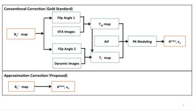 |
Evaluation of Approximation Method for B1+ Correction using Digital Reference Object in Prostate DCE-MRI
Xinran Zhong, Holden Wu, Krishna Nayak, Kyunghyun Sung
A digital reference object (DRO) for prostate dynamic contrast-enhanced MRI application is created by modifying DRO created by the RRSNA Quantitative Imaging Biomarkers Alliance (QIBA). Our previously proposed approximation B1+ correction method for pharmacokinetic modeling was then tested on this DRO assuming B1+ variation of 20%, considering practical considerations such as fitting method, arterial input function selection and noise level. The proposed approximation method introduced error is shown to be negligible compared to B1+ introduced error and noise introduced error.
|
16:51
 |
0623.
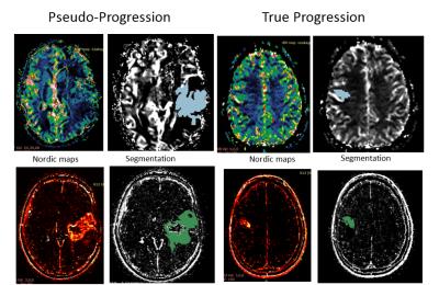 |
Radiomic Texture Features from MR Perfusion images Predicts Pseudoprogression from True Progression in Glioblastoma Patients: A Multi-Institutional Study
Aikaterini Kotrotsou, Nabil Elshafeey, Srishti Abrol , Dunia Giniebra Camejo, Islam Hassan, Ahmed Hassan, Kamel El Salek, Ahmed Shaaban, Samuel Bergamaschi, Fanny Moron, Meng Law, Pascal Zinn, Rivka Colen
Response assessment criteria, such as RANO, struggle to distinguish between true progression and pseudoprogression. In this work we evaluated the performance of radiomic texture features extracted from MR perfusion images (Dynamic contrast enhancement (DCE) and Dynamic susceptibility contrast (DSC)) in discriminating true progression from pseudoprogression. Using a large multi-institutional cohort, we demonstrated that changes in texture features of perfusion maps (DCE and DSC) can be effective predictors of progressive disease. We present a noninvasive, complimentary method that is directly applicable in clinical setting and can assist physicians in diagnosis and therapy planning.
|
17:03
|
0624.
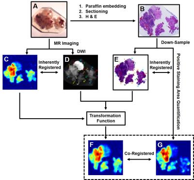 |
Quantitatively Differentiating Multiple Co-Existing Pathologies in High-Grade Glioma
Ze-Zhong Ye, Richard Price, Peng Sun, Ajit George , Chunyu Song, Sonika Dahiya, Albert Kim, Sheng-Kwei Song
We demonstrate diffusion MRI histology (D-Histo) is able to detect, differentiate and quantify various co-existing pathologies and structures including tumor, tumor infiltration, necrosis within human brain tumor specimen while conventional MRI and DTI fails. Quantitative maps of H & E and GFAP were generated and co-registered with D-Histo for voxel-wise correlative analysis. D-Histo-derived restricted-isotropic-diffusion fraction correlated with the area of hematoxylin and GFAP positive stain. D-Histo-derived hindered-isotropic-diffusion fraction successfully predicted the distribution of tumor necrosis corresponding with H & E. D-Histo is promising for brain tumor diagnosis, surgical planning, and treatment response monitoring.
|
17:15
|
0625.
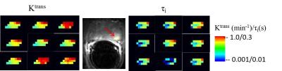 |
Effects of AIF Quantification Variations on Shutter-Speed Pharmacokinetic Modeling of Prostate DCE-MRI Data: A Multicenter Data Analysis Challenge, Part II
Wei Huang, Yiyi Chen, Andriy Fedorov, Xia Li, Guido Jajamovich, Dariya Malyarenko, Madhava Aryal, Peter LaViolette, Matthew Oborski, Finbarr O'Sullivan, Richard Abramson, Kourosh Jafari-Khouzani, Aneela Afzal, Alina Tudorica, Brendan Moloney, Sandeep Gupta, Cecilia Besa, Jayashree Kalpathy-Cramer, James Mountz, Charles Laymon, Mark Muzi, Paul Kinahan, Kathleen Schmainda, Yue Cao, Thomas Chenevert, Bachir Taouli, Thomas Yankeelov, Fiona Fennessy, Xin Li
Prostate tumor DCE-MRI data sets from 11 patients were shared among nine institutions, which determined AIFs using site-specific methods. The managing center performed pharmacokinetic data analysis using the Shutter-Speed model and these AIFs, and their scaled variants obtained with the reference-tissue method. Among the estimated parameters, Ktrans has the highest whereas τi has the lowest variability due to AIF uncertainty. The use of reference-tissue-adjusted AIFs reduces parameter variations. kep and τi are nearly insensitive to AIF scaling, suggesting that they may be robust imaging biomarkers in multicenter DCE-MRI trials where accurate and consistent AIF determination may be unattainable across sites.
|
17:27
|
0626.
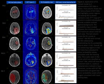 |
DCE time-series characterization with supervised deep learning: Alternative to PK model approaches - permission withheld
Dattesh Shanbhag, Vivek Vaidya, Uday Patil, Sandeep Gupta, Rakesh Mullick
We demonstrate feasibility of using a supervised deep learning method with DCE time-series data to obtain consistent numerical cutoff for tumor regions. DL based characterization is robust to fluctuations in DCE data due to protocol and patient physiology differences, which typically hinders such a classification with PK maps in clinical practice.
|
17:39
|
0627.
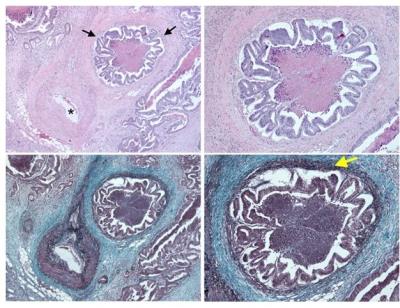 |
MRI Detection of Extramural Venous Invasion in Rectal Cancer: Correlation with Histopathology Utilizing Elastin Stain
Kartik Jhaveri, Hooman Hosseini-Nik, Seng Thipphavong, Naziheh Assarzadegan, Ravi Menezes, Erin Kennedy, Richard Kirsch
Extramural venous invasion (EMVI) in Rectal cancer is an independent predictor of local and distant recurrence, nodal disease and overall survival. Accurate preoperative EMVI detection has implications for treatment decisions. Previously, MRI has been evaluated for this purpose previously against histopathology with Hematoxylin-Eosin(HE) staining. Recently histopathology with Elastin stain has been recommended for EMVI detection given its 2-3-fold increase in accuracy and improved prognostication compared to HE stain. The diagnostic performance of MRI for EMVI detection compared to elastin stain based histopathology is unknown and in this study, we present our results to address this issue.
|
17:51
|
0628.
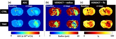 |
Quantifying microstructure changes in a mouse brain tumour model following Temozolomide therapy with VERDICT MRI
Thomas Roberts, Giulia Agliardi, Ben Hipwell, Angela D'Esposito, Andrada Ianus, James Breen-Norris, Valerie Taylor, Mark Lythgoe, Bernard Siow, Eleftheria Panagiotaki, Daniel Alexander, Simon Walker-Samuel
In this study, we use VERDICT (Vascular, Extracellular and Restricted Diffusion for Cytometry in Tumours) MRI to assess longitudinal changes in the microstructure of a mouse model of glioma (GL261), following treatment with Temozolomide, an alkylating chemotherapeutic agent. VERDICT estimates of cell radius and intracellular volume fraction decreased significantly at day-6 of therapy, relative to control animals, and in advance of changes in tumour volume and ADC. These changes were consistent with histological changes measured in the same tumours, and are likely to correspond to microstructural changes induced by apoptosis.
|
18:03
|
0629.
 |
Optimization of mpMRI protocol in differentiating muscle-invasive from non-muscle invasive Bladder Cancer: is there room for Diffusion Tensor Imaging (DTI)?
Giovanni Barchetti, Marcello Grompone, Maurizio Del Monte, Davide Carano, Carlo Catalano, Valeria Panebianco
Staging of BCa is critical, especially in differentiating non-invasive from muscle infiltrative lesions, as patient management strongly differs according to stage. Currently, patients with bladder lesions undergo two invasive procedures to diagnose and eventually treat the tumor if it is superficial. In this study we showed that mp-MRI has high capability for differentiating superficial from deep lesions, thanks also to DTI ability to directly visualize detrusor muscle layers. Mp-MRI could therefore be included systematically in the diagnostic evaluation of patients with suspicious bladder lesions in order to possibly avoid the first diagnostic cystoscopy once detrusor muscle invasion has been excluded.
|
|