Tuesday, 25 April 2017
| Room 313A |
13:45 - 15:45 |
Moderators: Paul Bottomley, Prodromos Parasoglou |
Slack Channel: #s_mrs_molecular
Session Number: O45
13:45
 |
0486.
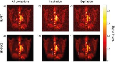 |
Self-gated 23Na-MRI of human lung with separate reconstruction of two respiratory states at 7T 
Tanja Platt, Reiner Umathum, Armin Nagel, Peter Bachert, Mark Ladd, Mark Wielpütz, Hans-Ulrich Kauczor, Nicolas Behl
23Na-MRI at 7T can give valuable insights into lung physiology because 23Na signals are linked to sodium-potassium-ATPase and to fluid balance. However, low NMR sensitivity and low in-vivo concentrations of Na+ ions in-vivo limit the achievable spatial resolution and prolong acquisition times. In this study a retrospective self-gated reconstruction was used to reduce motion artifacts in free-breathing 23Na-MRI of human lung. The presented method allows for the reconstruction of 23Na images which correspond to two respiratory motion states for free-breathing volunteers and patients. 3D Dictionary-Learning Compressed-Sensing reconstruction was shown to markedly reduce image noise.
|
13:57
|
0487.
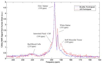 |
Using Hyperpolarized 129Xe MRI to Detect Impaired Cerebral Perfusion in Human Brain with Alzheimer’s Disease
Tao Li, Francis Hane, Jane Lawrence-Dewar, Ayman Hassan, Karl Granberg, Raiili Pellizzari, Jennifer Plata, Mitchell Albert
In this work, we present the first hyperpolarized 129Xe human brain MR spectra and human brain MR images from participants with Alzheimer’s Disease. We found a marked difference in the wash-out of dissolved phase xenon from the brain between the two groups, likely resulting from impaired cerebral blood flow in the Alzheimer’s Disease participants. By exploring this difference, we demonstrate the feasibility and sensitivity of hyperpolarized 129Xe MRI in detecting changes in cerebral perfusion and evaluating this important physiological characteristic during an early stage of mild to moderate Alzheimer’s Disease.
|
14:09
 |
0488.
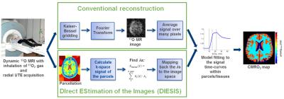 |
DIrect EStimation of 17O MR ImageS (DIESIS) for CMRO2 Quantification in the Human Brain with Partial Volume Correction - permission withheld
Dmitry Kurzhunov, Robert Borowiak, Marco Reisert, Axel Krafft, Michael Bock
Direct 17O-MRI enables quantification of cerebral metabolic rate of oxygen consumption (CMRO2). The low MR sensitivity of the 17O nucleus prevents pixel-wise CMRO2 quantification and the fast T2*≈2ms decay leads to partial volume artifacts. In this work the DIESIS method is proposed, which performs a direct least squares estimation of the 17O-MR signal in image regions (parcels) obtained from coregistered 1H data. CMRO2 values of 1.38-1.87µmol/gtissue/min and 0.51-0.77µmol/gtissue/min were found with DIESIS in gray and white matter consistent with rates from 15O-PET studies. With DIESIS, CMRO2 maps of high resolution can be reconstructed and partial volume artifacts can be reduced.
|
14:21
|
0489.
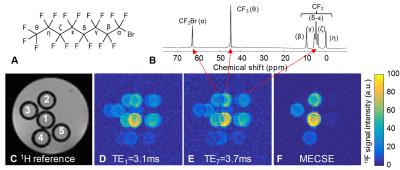 |
Multi-echo chemical shift encoding (MECSE) for sensitive fluorine-19 MRI of complex spectra
Ruud van Heeswijk, Roberto Colotti, Emeline Darçot, Jean Delacoste, Maxime Pellegrin, Davide Piccini, Diego Hernando
Now that several clinical trials are underway, sensitive fluorine-19 MRI of biocompatible perfluorocarbons with complex MR spectra, such as perfluorooctyl bromide (PFOB), is seeing increased interest. We here therefore propose a novel multi-echo chemical shift encoding (MECSE) technique that accounts for all resonances in k-space and reconstructs a high-sensitivity image free of chemical-shift-displacement artifacts. MECSE was developed and characterized in a phantom study, compared to established techniques for imaging compounds with complex spectra, and preliminarily demonstrated in vivo.
|
14:33
|
0490.
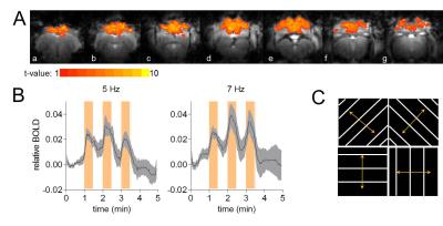 |
Coupling of the glutamate-glutamine cycle rate with both glial and neuronal oxidative metabolism in the visual cortex of the Tupaia belangeri - permission withheld
Sarah Sonnay, Jordan Poirot, Nathalie Just, Anne-Catherine Clerc, Rolf Gruetter, Gregor Rainer, João M.N. Duarte
Cerebral function relies on cooperative interaction between neuronal and glial cells. While neuronal oxidative metabolism has been shown to be coupled to the glutamate-glutamine cycle that represents glutamatergic neurotransmission, it remains unclear whether similar coupling occurs for glial oxidative metabolism. We investigated cortical metabolism in vivo using 13C magnetic resonance spectroscopy (MRS) along with infusion of [1,6-13C]glucose during continuous stimulation of the tree shrew visual cortex (V1). Data indicate that both neuronal and glial oxidative metabolism scale with the glutamate-glutamine cycle.
|
14:45
|
0491.
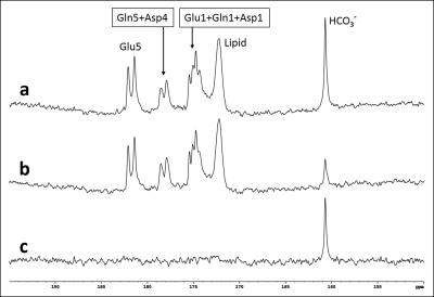 |
Detection of Carbonic Anhydrase Activity in Human Brain in Vivo
Shizhe Li, Li An, Christopher Johnson, Maria Ferraris Araneta, Jun Shen
This study demonstrates the feasibility of detecting carbonic anhydrase activity in the human brain. A very large magnetization transfer effect on the bicarbonate signal was measured in healthy human subjects using 13C MRS upon saturation of carbon dioxide. Despite the large variations in blood glucose response to oral administration of 13C-labeled glucose the magnetization transfer effect was measured with high precision.
|
14:57
|
0492.
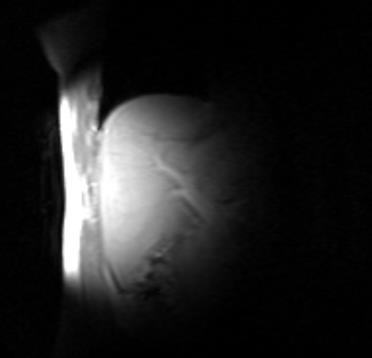 |
MRS measurements of [2-13C] glycine conversion to glutathione in the liver: A new method of measuring hepatic oxidative stress defences in vivo - permission withheld
Stephen Bawden, Bernard Lanz, Mehri Kaviani, Peter Morris, Penny Gowland, Peter Thelwall, Guruprasad Aithal
With rising incidents of fatty liver disease and metabolic disorder there is a need for biomarkers that can assess progression to steatohepatitis and other forms of liver damage. Oxidative stress in the mitochondria may play a central role in disease progression, with glutathione acting as the main antioxidant. In this study we developed a method previously suggested to monitor hepatic glutathione production in vivo by administering oral [2-13C] labelled glycine and using 13C MRS to measure conversion to glutathione. Following optimization, we tested variability in 8 healthy volunteers over two visits.
|
15:09
|
0493.
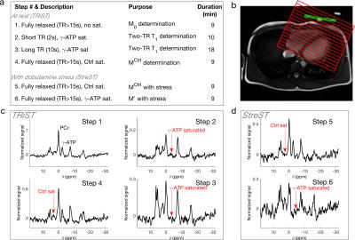 |
StreST: Myocardial Creatine Kinase Rate in Both Rest and Stress.
William Clarke, Jennifer Rayner, Betty Raman, Stefan Neubauer, Oliver Rider, Christopher Rodgers
A method for reduced time and stress myocardial creatine kinase measurements using 31P-MRS at 3T is presented. The new method is validated in healthy controls and demonstrated in 27 obese and heart-failure patients. In addition, the existing TRiST method is applied in 63 subjects for the first time on a Siemens platform.
|
15:21
|
0494.
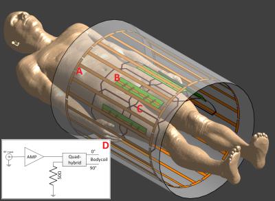 |
Multi-transmit proton MRI combined with high power body transmit and receive array for 7 Tesla phosphorus imaging
Mark Gosselink, Dimitri Welting, Tim Linnartz, Ingmar Voogt, Bart Steensma, Lucian Purvis, Christopher Rodgers, Tijl Velden, Wybe Kemp, Dennis Klomp
We have integrated a body RF coil tuned at the 31P frequency outside the patient tube in a 7T MR system for homogeneous B1 transmit and combined it with a 8 channel 31P receive array for optimal sensitivity. Imaging and B0 shimming is done with radiative antennas integrated in the receiver setup. With this setup, large field of view imaging and broadband 31P MRSI with high SNR can be obtained.
|
15:33
|
0495.
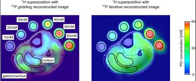 |
Fast quantitative 31P MRI using prior information from 1H MRI
Kristian Rink, Nicolas Behl, Christine Gnahm, Peter Bachert, Armin Nagel
Phosphorus-containing biomolecules play a crucial role in the energy metabolism of the human body. Compared to hydrogen, the in vivo phosphorus MR signal is four orders of magnitude lower. In this study, a conventional gridding reconstruction applied on the phosphocreatine signal of the human calf was compared to a constraint iterative approach, which uses prior knowledge from hydrogen MRI data. For both reconstructions, different acquisition times were tested and phosphocreatine concentrations in the gastrocnemius and soleus muscles were estimated.
|
|