Tuesday, 25 April 2017
| Room 311 |
13:45 - 15:45 |
Moderators: Guillaume Duhamel, Samuel Hurley |
Slack Channel: #s_contrastmechanisms
Session Number: O55
13:45
|
0466.
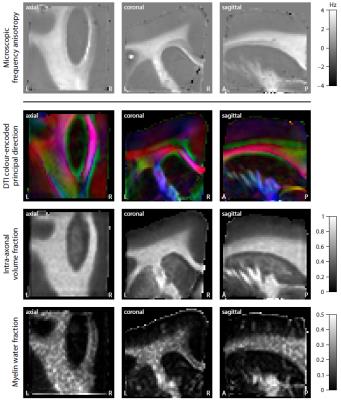 |
Microscopic susceptibility anisotropy mapping 
Enrico Kaden, Irina Barskaya, Nathaniel Kelm, Kathryn West, Mark Does, Daniel Alexander
The gradient-echo signal in brain white matter does not only depend on microscopic tissue features such as myelination, but also on the axon orientation distribution with respect to the external magnetic field. Therefore, to map microscopic susceptibility, we need to factor out the confounding effects of fibre crossings and orientation dispersion. This work adapts the Spherical Mean Technique (SMT), recently introduced in diffusion MRI, to estimate the microscopic susceptibility anisotropy in directionally heterogeneous tissue. Moreover, we show that, in combination with diffusion measurements, the proposed technique may be viable in the clinic.
|
13:57
 |
0467.
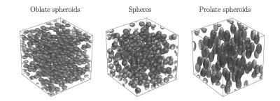 |
Larmor frequency shift in anisotropic heterogeneous media
Alexander Ruh, Valerij Kiselev
The question about the precise value of the proton Larmor frequency in neuronal tissues is a major topic in current research. In this study we present a comprehensive answer for spins moving in magnetically heterogeneous media formed by impermeable susceptibility inclusions. We obtain an analytic result for the mean Larmor frequency in the limit of fast diffusing spins in the space external to the inclusions. This mean frequency explicitly depends on the correlation function of the inclusion. For anisotropic media this results in a nonzero frequency shift, which is confirmed by Monte Carlo simulations.
|
14:09
|
0468.
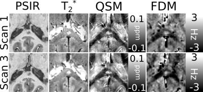 |
Frequency difference mapping as a marker of microstructural integrity of white matter in multiple sclerosis at 7T
Matthew Cronin, Nicholas Geades, Olivier Mougin, Kingkarn Aphiwatthanasumet, Nikos Evangelou, Penny Gowland, Richard Bowtell
Frequency difference maps derived from GRE phase data have been shown to generate orientation-dependent contrast in white matter tracts in the brain due to signal compartmentalization in myelinated nerve fibers. Here, we investigate the use of frequency difference mapping (FDM) as a marker of white matter integrity; comparing FDM with PSIR; T2*-weighted magnitude; and quantitative susceptibility mapping (QSM) images of focal white matter lesions in patients with multiple sclerosis. FDM shows clear contrast between these lesions and the surrounding white matter, suggesting that it has potential as a means of quantitatively identifying changes in white matter integrity in vivo.
|
14:21
|
0469.
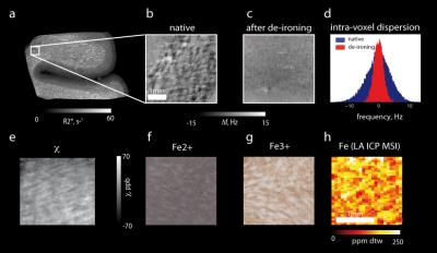 |
More Than Simply Iron: How the Mesoscopic and Cellular Distribution of Iron Impacts the MR Contrast
Evgeniya Kirilina, Katja Reimann, Isabel Weigelt, Thomas Arendt, Andreas Deistung, Jürgen Reichenbach, Steffen Jankuhn, Larissa Müller, Norbert Jakubowski, Markus Morawski, Nikolaus Weiskopf
Iron is an important source of MRI contrast in the brain. Herein, we investigated the influence of the cellular and subcellular iron distribution on the iron-induced MR contrast. Quantitative MRI on post mortem brain samples was combined with quantitative iron mapping and numerical simulations of local field distributions. We show that iron is heterogeneously distributed in both grey and white matter as well as in subcortical nuclei and different scales of heterogeneity play a role for MR contrast in these regions. Our results provide an important step towards quantitative understanding of iron induced MR-contrast and its microstructural underpinnings.
|
14:33
|
0470.
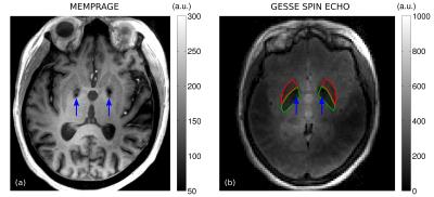 |
Calcification and iron deposition in basal ganglia structures: reversible and irreversible transverse relaxation rates at 7T
Mukund Balasubramanian, Robert Mulkern, Jonathan Polimeni
Gradient-Echo Sampling of the Spin Echo (GESSE) data were acquired at 7T in 16 volunteers (ages: 23-87 years). In globus pallidus and putamen, the reversible and irreversible transverse relaxation rates derived from this data varied with age in a manner largely consistent with prior postmortem studies of iron concentration. The exception to this was when calcifications appeared to be present, leading to outliers in the reversible (but not irreversible) relaxation rates. Our results suggest that consideration of both reversible and irreversible transverse relaxation rates may reveal valuable information about tissue microstructure and may complement measurements based primarily on phase contrast.
|
14:45
|
0471.
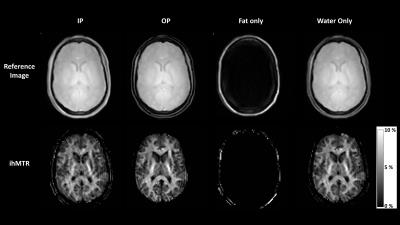 |
Combining inhomogenous Magnetization Transfer (ihMT) with multi-point Dixon (mDixon) for myelin imaging with efficient fat suppression
Ece Ercan, Ivan Dimitrov, Gopal Varma, Xinzeng Wang, Marco Pinho, Ananth Madhuranthakam, Robert Lenkinski, Elena Vinogradov
Inhomogeneous magnetization transfer (ihMT) imaging is a novel enhanced magnetization transfer technique which has recently been applied in human brain and spinal cord. Spinal cord applications of ihMT can especially benefit from a robust fat suppression to help with reducing of the strong fat signal from the large voxel size used by this method. Here we introduce a pulsed ihMT-prepared 3D SPGR sequence with multi-echo Dixon acquisition for a robust fat suppression of the ihMT images. The ihMT multi-echo Dixon method is shown to provide an excellent fat and water separation without a compromise of the observable ihMT effect.
|
14:57
|
0472.
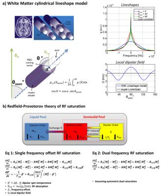 |
Anisotropy of inhomogeneous Magnetization Transfer (ihMT) in White Matter
Olivier Girard, Valentin Prevost, Samira Mchinda, Gopal Varma, David Alsop, Guillaume Duhamel
Inhomogeneous magnetization transfer (ihMT) is a new endogenous contrast mechanism that has been proposed for imaging myelinated tissues. The dipolar interaction underlying the ihMT effect is intrinsically anisotropic, exhibiting the well-known (3cos²θ -1) angle dependency. Here we report experimental evidence of the anisotropy of ihMT in white matter and we derive a realistic theoretical model combining the angular dependency of the myelin lineshape and dipolar-order RF saturation theory.
|
15:09
|
0473.
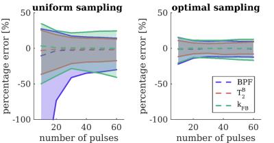 |
Optimal framework for quantitative magnetization transfer imaging of small structures
Marco Battiston, Francesco Grussu, Andrada Ianus, Torben Schneider, Ferran Prados, James Fairney, Sebastien Ourselin, Daniel Alexander, Mara Cercignani, Claudia Gandini Wheeler-Kingshott, Rebecca Samson
Quantitative Magnetization Transfer (qMT) Imaging allows quantification of parameters describing the macromolecular component of tissues, potentially specific for myelin in the central nervous system. To date, applications of qMT in small structures (e.g. the spinal cord) have been hampered by prohibitively long acquisition. We present a framework for robust qMT examinations in small structures. It consists of: a dedicated MT-weighted sequence for reduced field-of-view imaging, explicit modelling of the non-steady state signal, and optimal definition of the sampling scheme. Superiority of the framework compared to a conventional qMT protocol is demonstrated in the healthy spinal cord and in the brainstem.
|
15:21
|
0474.
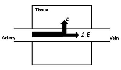 |
Towards a non-contrast assessment of blood-brain-barrier permeability
Zixuan Lin, Yang Li, Pan Su, Jay Pillai, Matthias Osch, Hanzhang Lu
Blood-brain-barrier (BBB) permeability has been shown to be disrupted in many diseases. Current study proposes a non-invasive method to measure the extraction fraction and BBB permeability of water. Studies were performed to provide proof-of-principle and a new sequence was developed to improve efficiency. These methods provided consistent results with previous literature. CO2 effect on permeability was explored and increased measurement sensitivity was demonstrated.
|
15:33
|
0475.
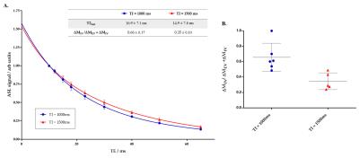 |
Non-invasive Assessment of Vascular Water Permeability in the Mouse Brain using multi-TE ASL
Yolanda Ohene , Ian Harrison , Payam Nahavandi , Ozama Ismail , David Thomas , Mark Lythgoe , Jack Wells
We apply a multi-TE ASL technique in the mouse brain, to separate intravascular and extravascular components of the ASL signal, as a non-invasive assessment of vascular permeability. Methodological development enabled the technique to reliably capture ASL signal compartmentation in the mouse brain, despite inherently low SNR. We report a significant decrease in intravascular fraction of the ASL signal from 0.66 (± 0.17) to 0.35 (± 0.10) as inflow time increases from 1000ms to 1500ms. This technique can be applied to transgenic mouse models of neurodegeneration, and a kinetic model can extract vascular permeability parameters from the ASL signal distribution.
|
|