Oral
Acquisition, Reconstruction & Analysis
Thursday, 27 April 2017
| Room 313BC |
08:15 - 10:15 |
Moderators: Pablo Irarrazaval, Craig Meyer |
Slack Channel: #s_acq_recon_analysis
Session Number: O59
08:15
 |
1037.
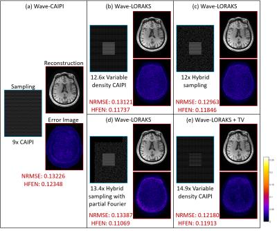 |
Wave-LORAKS for faster Wave-CAIPI MRI 
Tae Hyung Kim, Berkin Bilgic, Daniel Polak, Kawin Setsompop, Justin Haldar
Wave-CAIPI is a novel technique that enables accelerated acquisition with negligible g-factor penalty by using corkscrew readout trajectories, while LORAKS (LOw-RAnk modeling of local K-Space neighborhoods) is a powerful approach to constrained reconstruction that integrates sparse support, phase, and parallel imaging constraints into a unified linear prediction framework. In this work, we propose a new fast imaging technique called Wave-LORAKS, which combines Wave-CAIPI acquisition with LORAKS-based reconstruction. Retrospective undersampling experiments with 3D T1-weighted data show that Wave-LORAKS enables higher acceleration and more flexible sampling compared to traditional Wave-CAIPI, allowing up to 15-fold acceleration with similar quality as 9-fold accelerated Wave-CAIPI.
|
08:27
|
1038.
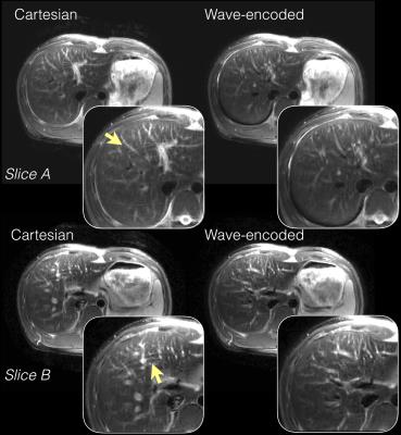 |
Variable Density Single-Shot Fast Spin Echo with Auto-Calibrated Wave Encoding
Feiyu Chen, Valentina Taviani, Joseph Cheng, Tao Zhang, Brian Hargreaves, John Pauly, Shreyas Vasanawala
Wave encoding was implemented in a variable-density single-shot fast spin echo (VD-SSFSE) pulse sequence. Auto-calibrated estimation of the wave-encoding point-spread function (PSF) and coil sensitivity maps was used. Images were reconstructed with parallel imaging and compressed sensing reconstruction. Compared to non-wave-encoded Cartesian imaging, wave-encoded VD-SSFSE achieves improved image quality with reduced aliasing artifacts at higher acceleration factors and with full k-space coverage, providing fast acquisitions and clinically relevant echo times.
|
08:39
|
1039.
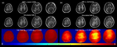 |
Wave-CAIPI ViSTa: Accelerated mapping for direct visualization of myelin water
Zhe Wu, Berkin Bilgic, Hongjian He, Yi Sun, Kawin Setsompop, Jianhui Zhong
Direct Visualization of Short Transverse relaxation time component (ViSTa) is a new myelin water imaging method, which directly extracts myelin water signal through double inversion RF pulses (DIR) to preserve signal from short T1 components. This method is robust and sensitive to demyelinated lesions, but suffers from extremely long scan time. Herein, we propose accelerated ViSTa acquisition through Simultaneous Multi-Slice (SMS) wave-CAIPI while keeping the high fidelity of ViSTa and MWF maps. As the average/maximum g-factor noise amplification is only 3/10% at MultiBand-4, further acceleration is likely to shorten whole brain MWF acquisition to within 5 minutes.
|
08:51
|
1040.
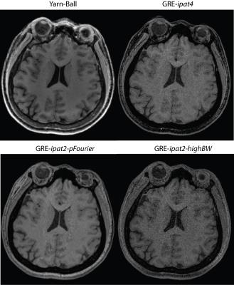 |
Novel 3D Yarn-Ball k-space Acquisition for Fast High-Resolution T1-Weighted Brain Imaging
Robert Stobbe, Peter Seres, Christian Beaulieu
Novel 3D Yarn-Ball k-space acquisition is applied for the first time in vivo with application to T1-weighted human brain imaging and a goal of 0.9x0.9x0.9 mm3 voxels in ~2 minutes at 3T. This efficient, large-looping trajectory allows much more of k-space to be sampled following each excitation than the 1 line of 3D-Gradient-Echo (3D-GRE), facilitating longer repetition times (TR), greater acquisition duty cycle, and full sampling. We demonstrate that as a result images created with Yarn-Ball yield better resolution phantom element distinction, and considerably higher SNR (2x on average) in vivo than the three different 3D-GRE methods tested.
|
09:03
|
1041.
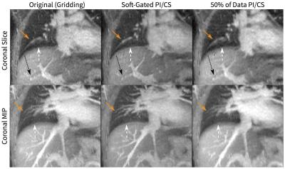 |
Flexibility and Robustness of Cones UTE for Pediatric Abdominal and Chest Imaging
Joseph Cheng, Wenwen Jiang, Michael Carl, Michael Lustig, Shreyas Vasanawala
Three-dimensional cones k-space sampling trajectory in UTE imaging is ideal for pediatric abdominal and chest MRI which are often hindered by patient motion and lengthy scan durations. The scan-time efficiency of the trajectory reduces scan durations with little to no subsampling. With the repeated sampling and oversampling of the k-space center, the acquisition is robust to motion and can flexibly retrospectively tradeoff spatial and temporal resolutions. Lastly, the method enables retrospective delineation of physiological dynamics. These features of cones UTE enable flexibility and robustness that is ideal for pediatric MRI.
|
09:15
|
1042.
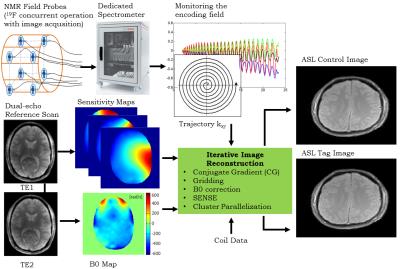 |
Single-Shot Spiral Arterial Spin Labeling MRI Enabled by Concurrent Field Monitoring
Mustafa Cavusoglu, Lars Kasper, Klaas Pruessmann
Spiral k-space sampling holds great potential for many MRI applications particularly for arterial spin labeling which has inherently very low SNR. Besides providing significant increase in SNR by reducing the echo time and readout durations, spiral trajectories with single shot acquisitions are highly robust against motion artifacts. However their sensitivity to encoding deficiencies such as B0 off-resonance, field drifts, eddy currents, gradient coupling, gradient delays and concomitant fields prevents their utilization effectively in practice. In this work, we provide ASL with single-shot spiral readouts that are robust against all those deficiencies by using dynamic field monitoring concurrent with image acquisition.
|
09:27
|
1043.
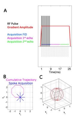 |
Looping Star
Florian Wiesinger, Anne Menini, Ana Beatriz Solana
This abstract describes a novel imaging method called Looping Star. The unique imaging characteristics are 1) continuous 3D radial imaging with close to 100% sampling efficiency, 2) inaudible/silent scanning, and produces 3) multi-gradient echo images at equidistant echo times including an FID image at TE=0. Looping Star is demonstrated in phantom, and in-vivo experiments for T2* weighted imaging and T2* BOLD fMRI.
|
09:39
|
1044.
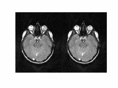 |
Dynamic imaging of eye and optic nerve with golden angle radial MRI.
Saikat Sengupta, David Smith, Alex Smith, E. Brian Welch, Seth Smith
In this abstract we present dynamic imaging of the eyes and the optic nerves in humans using golden angle radial MRI. Continuous 15 s radial scans with azimuthal profile steps of 111.246 degrees are acquired under various eye motion states. Qualitative analyses of the images reveal features of basic eye and nerve mechanics. Image-based characterization of eye mechanics can improve understanding eye physiology and disease.
|
09:51
|
1045.
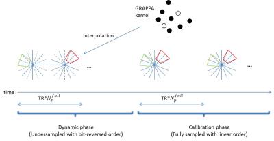 |
A scalable composite through-time radial GRAPPA method
Seng-Wei Chieh, Steen Moeller, Mehmet Akcakaya, Mostafa Kaveh
Through-time radial GRAPPA showed promising reconstruction for cardiac imaging. However, it's challenging to extend 3D Kooshball trajectory because of long calibration scans. We propose a novel and flexible data-driven calibration method. The MRXCAT numerical phantom image results show image similarity with through-time radial GRAPPA.
|
10:03
|
1046.
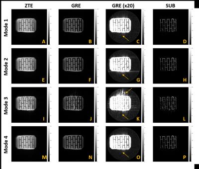 |
Exploring various radial trajectories for optimal relative phase correction in dual-echo subtraction ZTE MRI
Hyo Lee, Markus Weiger, Klaas Pruessmann
In dual-echo subtraction ZTE MRI, it is crucial to correct for gradient delays and phase offsets induced by eddy currents for high image quality. Relative phase correction is a data-driven approach to correct for the gradient hardware imperfection, but it requires that phase offsets induced by eddy current in positive and negative projections to be exact opposite of each other. This work has explored various radial trajectories to satisfy this condition in order to achieve optimal correction for gradient hardware imperfection in dual-echo subtraction ZTE MRI.
|
|