Oral
Acquisition, Reconstruction & Analysis
Thursday, 27 April 2017
| Room 311 |
08:15 - 10:15 |
Moderators: Martina Callaghan, Daniel Gallichan |
Slack Channel: #s_acq_recon_analysis
Session Number: O62
08:15
|
1017.
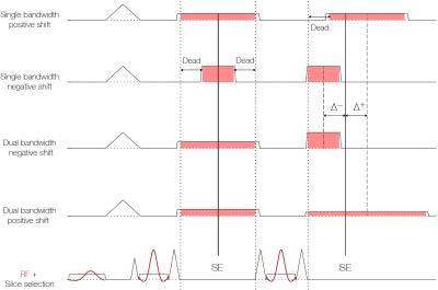 |
T1-weighted two-point fat/water separated PROPELLER acquired with dual bandwidths 
Henric Rydén, Johan Berglund, Enrico Avventi, Ola Norbeck, Stefan Skare
A PROPELLER sequence with dual bandwidths was developed for T1-weighted fat/water separated imaging. The bandwidth of the second readout was adjusted to remove dead time related to shifted readouts in order to improve SNR efficiency. Before PROPELLER reconstruction, bladewise fat/water separation was performed in k-space to remove chemical shift displacement artifacts. This enabled low bandwidth acquisitions without smearing of the fat signal, which further improved SNR efficiency. Strong fat suppression insensitive to B0 inhomogeneity was demonstrated in imaging of the neck and orbits.
|
08:27
|
1018.
 |
Fast, non-local temporally regularized elastic group-wise motion correction for improved fidelity of liver DCE-MRI
Ks Shriram, Dattesh Shanbhag, Jeong Hee Yoon, Uday Patil, Moon Jung Hwang, Jeong Min Lee , Rakesh Mullick, Sheshadri Thiruvenkadam
A novel group-wise registration methodology using non-local regularization is presented for liver DCE-MRI data that can provide robust motion correction even in cases of large deformations, restores DCE data characteristics, generates physiologically relevant PK maps and is feasible for clinical practice due to short computation time.
|
08:39
|
1019.
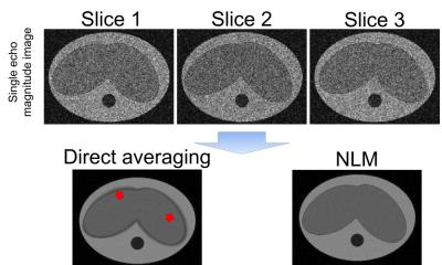 |
A novel fat and iron quantification technique with non-rigid motion-corrected averaging based on non-local means
Huiwen Luo, Curtis Wiens, Ann Shimakawa, Scott Reeder, Kevin Johnson, Diego Hernando
In this study, we developed and validated a free-breathing fat and R2* quantification technique using a multi-average 2D sequential acquisition with non-rigid motion-corrected averaging based on a non-local means (NLM) approach. The proposed technique was applied to simulated data as well as free-breathing liver acquisitions in volunteers. Both direct averaging and the proposed NLM technique were applied to the data to improve SNR. Compared to direct averaging, the proposed NLM technique resulted in improved image quality without motion artifacts, as well as accurate fat and R2* measurements in both simulations and in vivo acquisitions.
|
08:51
|
1020.
 |
Investigation of Simultaneous Multi-slice Imaging Utilized for Respiratory Gating: A Golden Angle Radial CAIPI Free-Breathing Cine Method - permission withheld
Dana Peters, Chenxi Hu, Yuqing Wan, Maolin Qiu
We investigated simultaneous multi-slice as a method for performing respiratory-gating. One slice is used to monitor respiration, while another slice is targeted for imaging. Radial CAIPI data was acquired using a golden angle ordering for free-breathing 2D cine. At each cardiac phase and at each heart-beat, images of the heart and the diaphragm were obtained. Finally, a composite image of the heart was reconstructed using data from all frames acquired at end-expiration, with more projections and higher quality than a single heart-beat image.
|
09:03
|
1021.
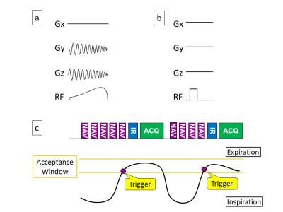 |
Silent Navigator-Triggered Silent-MRI in Abdomen
Yuji Iwadate, Atsushi Nozaki, Yoshinobu Nunokawa, Shigeo Okuda, Masahiro Jinzaki, Hiroyuki Kabasawa
We incorporated a silent navigator (sNav) with the whole volume excitation into zero-TE pulse sequence for respiratory motion corrected silent abdominal imaging. The sNav signals showed an excellent correlation between the bellows signals, and resultant zero-TE images had better contrasts than those acquired without respiratory triggering. The sNav-triggered zero-TE technique is expected to be used in abdominal MRI where acoustic noise is problematic (e.g. pediatric patient imaging with anesthesia).
|
09:15
|
1022.
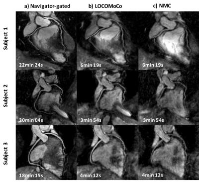 |
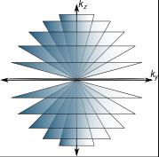
Accelerated motion compensated 3D isotropic coronary MRA using variable density Cartesian sampling
Teresa Correia, Gastao Cruz, Camila Munoz, Rene Botnar, Claudia Prieto
Accelerated whole-heart 3D isotropic coronary MR angiography (CMRA) is achieved by undersampling the acquisition using a variable-density radial Cartesian (VDRC) trajectory and performing non-rigid respiratory motion correction directly in the low-dimensional-structure self-learning and thresholding (LOST) reconstruction. The proposed approach corrects for 2D beat-to-beat translational and 3D bin-to-bin non-rigid motion. The former is estimated from interleaved image navigators and the latter directly from the CMRA data. The VDRC trajectory provides improved respiratory bin reconstructions and initial image estimate for LOST. The proposed approach produces good quality images, comparable to those of a two-fold accelerated navigator-gated acquisition with ~4x longer scan time.
|
09:27
 |
1023.
 |
Evaluation of Self-Navigated Golden-Angle Ordered Conical Ultrashort Echo Time (UTE) MRI of the Abdomen on Pediatric Patients in Multiple Sedation States
Anshul Haldipur, Evan Zucker, Joseph Cheng, Michael Carl, Shreyas Vasanawala
To assess the feasibility of conical k-space trajectory free-breathing UTE abdominal MRI and the effects on image quality of 50% data subsampling (thereby potentially shortening scans) and soft-gated motion correction reconstruction techniques, 42 consecutive pediatric patients were recruited and scanned. The images were scored in blinded fashion by two readers. Adequate delineation was obtained for all evaluated abdominal structures except the hepatic artery. 50% subsampling decreased image quality only slightly, favoring the implementation of a shorter scan time with negligible diagnostic compromise. Overall, motion correction mildly degraded image quality, possibly due to greater noise from data subsampling.
|
09:39
|
1024.
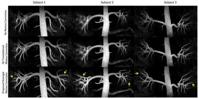 |
Quantification, Analysis, and Correction of Nonrigid Motion in Free-Breathing, Non-Contrast-Enhanced Renal Angiography using 3D Image-Based Navigators
Srivathsan Koundinyan, Corey Baron, Mario Malavé, Nii Addy, Jieying Luo, R. Reeve Ingle, Dwight Nishimura
We present a method that leverages 3D image-based navigators (iNAVs) for nonrigid motion correction in free-breathing, non-contrast-enhanced renal angiography scans. We begin by performing an ROI-based analysis of 3D iNAV motion, with ROI selection based on published biomechanical simulations of the renal arteries during respiration. Then, we demonstrate that localized motion estimates derived from different ROIs agree with the findings of the simulations. Finally, we combine the extracted motion information with an autofocusing technique for respiratory motion compensation. Across all patient studies, the proposed method significantly improves the depiction of the renal arteries as compared to 3D translational motion correction.
|
09:51
|
1025.
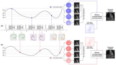 |
Highly efficient respiratory motion corrected dual-phase coronary MR angiography in a 3T PET-MR system
Camila Munoz, Radhouene Neji, Rene Botnar, Claudia Prieto
Respiratory motion remains a major challenge for dual-phase coronary MR angiography (CMRA). Here we propose an efficient acquisition and reconstruction scheme, that allows for inline 2D translational and offline 3D non-rigid motion-corrected systolic and diastolic CMRA using image-based navigators. Results from healthy volunteers show that motion correction improves visualization of the right and left coronary arteries in both cardiac phases. The proposed scheme potentially allows for comprehensive diagnosis of coronary artery disease using simultaneous PET-MR by acquiring coronary anatomy and left-ventricular function with the dual-phase CMRA and myocardial perfusion/viability with PET in an efficient cardiac and respiratory motion-compensated framework.
|
10:03
|
1026.
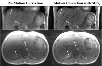 |
Respiratory Motion Compensation in the Liver using Fat-Only Self Gated Signal
Thomas Martin, Tess Armstrong, Ely Felker, James Sayre, Steve Raman, Holden Wu, Kyunghyun Sung
A multi-echo 3D golden angle radial gradient echo (GRE) sequence for fat-only respiratory motion extraction is a promising technique for liver dynamic contrast enhancement MRI (DCE-MRI), because it has inherent motion robustness, while providing other advantages, such as water-only images and R2* mapping. In this work we demonstrate that respiratory motion correction in the liver can be achieved using a fat-only self-navigated signal with minimal error in the fat-water separation (< 5%). Using this technique has implications of a more robust motion correction for liver DCE-MRI due to its inherent separation between respiratory motion signal and contrast uptake.
|
|