Oral
Acquisition, Reconstruction & Analysis
Thursday, 27 April 2017
| Room 312 |
13:00 - 15:00 |
Moderators: Arvin Arani, Lynne Bilston |
Slack Channel: #s_acq_recon_analysis
Session Number: O67
13:00
 |
1132.
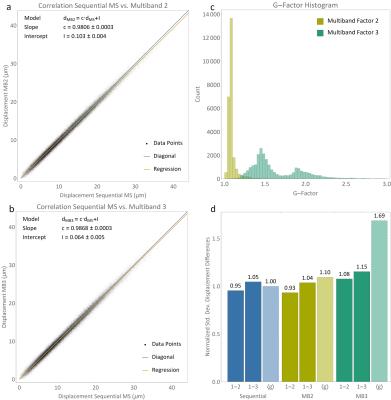 |
Simultaneous Multislice Acquisition for Magnetic Resonance Elastography 
Christian Guenthner, Jurgen Runge, Ralph Sinkus, Sebastian Kozerke
We propose the use of simultaneous multislice acquisition for Magnetic Resonance Elastography (MRE) of the full displacement vector field. To this end, multiband composite RF pulses are used for slice excitation in a fast, “eXpresso” type gradient echo based MRE acquisition. Slice and k-line dependent RF-phases are used to shift simultaneously acquired slices leading to improved unfolding performance (CAIPIRINHA). In this abstract, we demonstrate that multiband MRE with CAIPIRINHA can be used to acquire up to three slices simultaneously with only little SNR penalty in a gel phantom and show the feasibility to acquire full-brain images in-vivo.
|
13:12
|
1133.
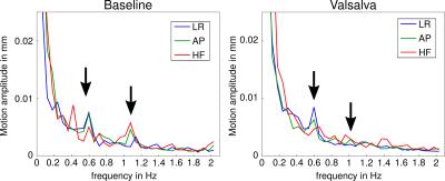 |
Towards single-shot 3D MR elastography: Application to intrinsically activated motion fields in the human brain.
Sebastian Hirsch, Rüdiger Stirnberg, Tony Stöcker, Florian Dittmann, Jing Guo, Eric Barnhill, Jürgen Braun, Ingolf Sack
This study introduces a single-shot 3D EPI sequence for rapid motion field acquisition. The method can be applied for MRE under pulsatile motion or for directly analyzing intrinsic motion fields in pulsatile organs such as the brain. Through 3D k-space sampling, inter-slice phase offset artifacts, which essentially result from 2D k-space MRE, are most effectively avoided. In a first application we used the new method for the measurement of intrinsic brain pulsation in the human brain and analyzed the intensity of deflection field components by stochastic sampling at rest and during Valsalva maneuver.
|
13:24
|
1134.
 |
Preoperative assessment of tumor stiffness and tumor-brain adhesion in patients with vestibular schwannoma using MR elastography-based method
Prateek Kalra, Arunark Kolipaka, PhD, Brian Raterman, Oliver Adunka, MD, Michael Harris, MD, Daniel Prevedello, MD
Microsurgery in vestibular schwannoma patients aims to complete tumor resection without compromising any neurological functionality. Inadequate preoperative knowledge of tumor may prolong surgical time and increase risk of postoperative complications. Previous studies have used magnetic resonance elastography (MRE) based method to determine tumor stiffness and tumor-brain adhesion, separately. But none of the studies have investigated tumor stiffness and tumor-brain adhesion together. The aim of this study is to bring together the two MRE-based outcomes – tumor stiffness and tumor-brain adhesion – and correlate with surgical findings. Preliminary results show a good correlation between preoperative assessment of tumor and surgical findings.
|
13:36
|
1135.
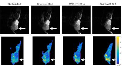 |
Assessing tumor mechanical nonlinearity by MR elastography at different strain levels
Marion Tardieu, Laurent Besret, Lydia Blot, Joaquim Lopes, Ralph Sinkus, Bernard Van Beers, Philippe Garteiser
In this study we attempt to access nonlinear mechanical properties of tumors by applying external loads. Experiments were performed on subcutaneous tumors implanted in mice. MR elastography acquisitions were realized at 600 Hz while deformations were applied, in order to obtain apparent elasticity (G’) values for each strain application. Results showed nonlinear G’ values increasing with loads, when deformations were clearly observed. Some results showed a decrease of G' potentially due to wave displacement polarization. We showed the feasibility of assessing the nonlinear regime of tumor mechanical properties that may potentially be an indicator of internal tumor forces.
|
13:48
|
1136.
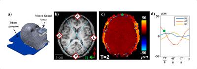 |
Measurement of Human Brain, Scalp, and Skull Motion in vivo using Magnetic Resonance Elastography and Triaxial Accelerometers
Andrew Badachhape, Ruth Okamoto, Curtis Johnson, Philip Bayly
Characterizing motion transmission from the skull to the brain and from skull to external soft tissue would provide valuable insight into traumatic brain injury mechanics and injury assessment by external sensors. In this study, we estimated rigid-body displacement components of brain and scalp using magnetic resonance elastography for comparison with skull motion estimated from three triaxial accelerometers. Comparison of the relative amplitudes and phases of harmonic motion in the skull, scalp, and brain of five human subjects indicated differences between each region. These measured amplitude and phase relationships can improve both simulations and experimental characterization of head biomechanics.
|
14:00
|
1137.
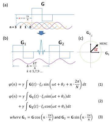 |
Quadrature Motion Encoding (QuME): A Novel Motion Encoding Scheme for MR Elastography
Yi Sui, Kevin Glaser, Ziying Yin, Joshua Trzasko, Jun Chen, Richard Ehman, John Huston III
We propose a novel approach to motion encoding for spin-echo-based MR elastography called quadrature motion encoding (QuME) in which the second motion-encoding gradient (MEG) after the refocusing pulse is used to measure the quadrature component of the in vivo harmonic motion induced by an external mechanical vibration. Unlike the conventional encoding method that shifts the temporal relation between the motion and MEGs, QuME alters the amplitudes of MEGs from one time step to the next. This concept was implemented on a 3T GE scanner and demonstrated the ability to shorten the echo time of SLIM-MRE for human brain MRE.
|
14:12
|
1138.
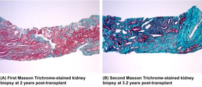 |
Fibrosis in a Renal Allograft: The Role of Magnetic Resonance Elastography as a Non- Invasive Measurement Tool
Jin Kyu Kim, Darren Yuen, General Leung, Serge Jothy, Jeffrey Zaltzman, Ramesh Prasad, Anish Kirpalani
A major reason for poor long-term kidney transplant outcomes is interstitial fibrosis. Currently, percutaneous biopsy, an invasive procedure that samples <1% of the allograft, is the gold standard for detecting renal allograft fibrosis. As the allograft scars, it stiffens due to the deposition of stiff extracellular matrix. This stiffening can be imaged non-invasively using magnetic resonance elastography (MRE). We used serial renal stiffness MRE imaging and serial percutaneous biopsies to measure whole allograft fibrosis preogression in a kidney transplant recipient. We show that renal allograft MRE can detect changes in overall fibrotic burden, as confirmed by biopsy.
|
14:24
|
1139.
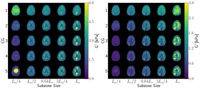 |
Inversion Parameters based on Convergence and Error Metrics for Nonlinear Inversion MR Elastography
Aaron Anderson, Curtis Johnson, Matthew McGarry, Keith Paulsen, Bradley Sutton, Elijah Van Houten, John Georgiadis
Nonlinear inversion (NLI) of brain MRE data has shown the promise in sensitive detection of complex neurodegenerative disease by showing repeatable and accurate assessments of both white matter and gray matter regions in healthy subjects. This study looks to further improve the accuracy of the NLI-MRE framework by characterizing two major inversion parameters: subzone size and conjugate gradient iterations. Additionally, two convergence criteria are proposed as means to quantify the confidence in final reported statistics while fully capturing the distribution of heterogeneity within white matter regions.
|
14:36
|
1140.
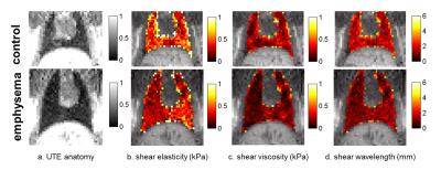 |
Magnetic Resonance Elastography of Emphysematous Rat Lung in vivo
Hongchen Wang, Catherine Sebrié, Georges Willoquet, Jinlong Yue, Felicia Julea, Rose-Marie Dubuisson, Sébastien Judé, Anne Maurin, Xavier Maître
Emphysema is a chronic respiratory disease, which has become the fifth most common cause of death worldwide. Emphysema is characterized by alveolar wall destruction, leading to distal airspace enlargement and decreased elastic recoil. The altered structure and function of the lung is related to modified mechanical properties of pulmonary tissues, which are difficult to probe in vivo by standard techniques. Magnetic Resonance Elastography (MRE) allows to characterize the mechanical properties correlated with lung physiopathologies. Here, we implemented MRE lung imaging in vivo on emphysematous rat models.
|
14:48
|
1141.
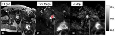 |
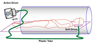
Multifrequency MR elastography of the human prostate by multiple surface-based compressed-air drivers: reproducibility and first patient results
Florian Dittmann, Jing Guo, Heiko Tzschätzsch, Sebastian Hirsch, Rolf Reiter, Patrick Asbach, Andreas Maxeiner, Jürgen Braun, Ingolf Sack
A new setup for prostate MR Elastography is proposed that uses surface-based wave excitation by compressed-air drivers. The post-processing with tomoelastography combining wave fields at drive frequencies of 60, 70, and 80 Hz leads to highly resolved elasticity maps. A study in healthy volunteers demonstrates the good reproducibility of the method. Furthermore, the first patient results show excellent agreement with contrast-enhanced reference images, which motivates future patient studies.
|
|