Wednesday, 26 April 2017
| Room 313BC |
08:15 - 10:15 |
Moderators: Andrada Ianus, Michael Kleinnijenhuis |
Slack Channel: #s_diffusion
Session Number: O68
08:15
 |
0715.
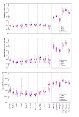 |
Characterisation of cerebellar microstructure with two-compartment Spherical Mean Technique 
Giovanni Savini, Fulvia Palesi, Gloria Castellazzi, Letizia Casiraghi, Francesco Grussu, Alessandro Lascialfari, Egidio D'Angelo, Claudia Gandini Wheeler-Kingshott
Cerebrum microstructure has been extensively investigated with diffusion-weighted MRI, but little attention has been dedicated to the microstructural characterisation of the cerebellum. We considered an anatomical parcellation of the cerebellum and fitted a multi-compartment model to diffusion data exploiting the spherical mean technique, which provides parametric maps unconfounded by the underlying fibre orientation distribution. For each region we report average values for multi-compartment parameters (e.g. intra-neurite volume fraction and intrinsic diffusivity) and diffusion tensor metrics.
Multi-compartment metrics more specific to microstructure provide information complementary to diffusion tensor metrics in the cerebellum, thus giving new insights about microstructural correlations between regions.
|
08:27
 |
0716.
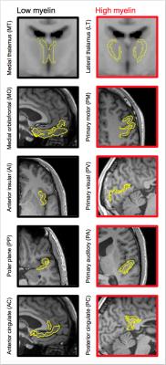 |
Microscopic anisotropy in gray matter is evidence of myelinated axons but not dendrites? An in vivo study using diffusion MRI with variable shape of the b-tensor.
Björn Lampinen, Filip Szczepankiewicz, Mikael Novén, Carl-Fredrik Westin, Elisabet Englund, Johan Mårtensson, Markus Nilsson
Microscopic anisotropy was measured in vivo using a novel tensor-valued diffusion encoding approach. In gray matter, the microscopic anisotropy was generally low, but its variation corresponded well to known differences in myelination. We hypothesize that myelinated axons cause microscopic diffusion anisotropy but that the contribution from dendrites is negligible. This hypothesis is supported by comparisons with independent myelin assessments using T1W/T2W-ratios, T2-mapping, and myelin stains from histology. We also demonstrate that the “neurite density index” detected by NODDI is less sensitive to these changes, and why NODDI cannot map the neurite density accurately.
|
08:39
|
0717.
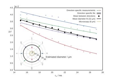 |
Pore size estimation using the mixing time dependence of a double diffusion encoding experiment: experimental validation on a clinical MR system
Vincent Methot, Patricia Ulloa, Martin A. Koch
Diffusion MRI provides information about microstructure, but is limited in complex situations. Double diffusion encoding makes assessment of shape and size possible in these contexts. The time delay (mixing time) between diffusion encodings has not been studied closely outside of the short and long regimes. We present here an experimental study of the mixing time dependence. In spherical pores, the parallel-antiparallel signal difference can be approximately described as an exponential decay with a rate related to diameter. This decay was obtained on a clinical scanner in a water-in-oil emulsion.
|
08:51
|
0718.
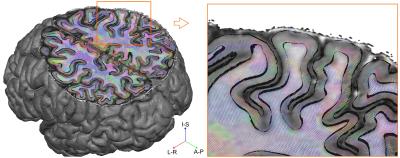 |
Layer-specific analysis of cortical microstructure using in-vivo 7T diffusion MRI
Omer Faruk Gülban, Federico De Martino, An Vu, Kamil Ugurbil, Essa Yacoub, Christophe Lenglet
We present unique in-vivo human 7T diffusion MRI data and a dedicated layer-specific analysis pipeline. We leverage the high spatial and angular resolution of this dataset to improve cortical fiber orientation mapping (i.e. limit gyral bias and identify fiber crossings), and study axonal trajectories within the cortex across depths.
|
09:03
|
0719.
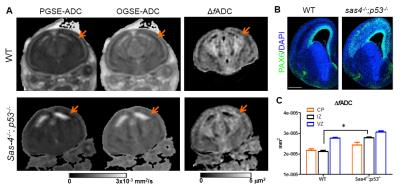 |
HARDI and oscillating gradient diffusion MRI reveal disrupted embryonic cortical microstructure
Dan Wu, Wei Shao, Songhai Shi, Jiangyang Zhang
We investigated the capability of advanced diffusion MRI, including high-angular resolution diffusion MRI (HARDI) and oscillating gradient diffusion MRI, to characterize cortical microstructural organization in the embryonic mouse brains. HARDI-based tractography revealed reduced axons in the intermediate zone of the embryonic cortex in the Sas-4-/-;p53-/- mice compared to the wildtypes. The oscillating gradient diffusion MRI delineated a three-lamina structure in the cortex of the normal embryonic brain, reflecting the neuronal cell distributions during embryonic brain development, which was altered by mislocalized RGPs in extra-ventricular zone, resulting in diminished contrast in the mutant cortex.
|
09:15
|
0720.
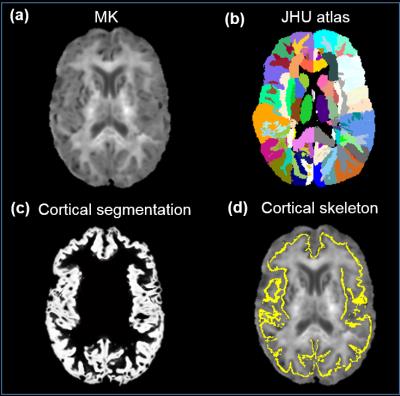 |
Mapping of cerebral cortical microstructure characterized by non-Gaussian water diffusion in aging
Qinmu Peng, King Kevin, Minhui Ouyang, Hanzhang Lu, Hao Huang
The cellular and molecular processes inside the cerebral cortex play a critical role in aging and neurodegenerative disorders. The microstructural changes associated to these cortical processes can be assessed with multi-shell diffusion MRI. Here, we aimed to accurately quantify the regional cortical microstructural changes of the aging brains. Multi-shell diffusion MRI was acquired to measure mean kurtosis (MK) derived from DKI at the center of the cerebral cortical layer for specific cortical regions. Significant MK decreases were found in the primary somatosensory regions, but not in prefrontal and visual regions, suggesting differentiated aging processes in different cortical regions.
|
09:27
|
0721.
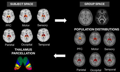 |
Improved tractography-based segmentation of the human thalamus
Carla Semedo, M. Jorge Cardoso, Sjoerd B. Vos, Alex F. Mendelson, Annemie Ribbens, Dirk Smeets, Jonathan D. Rohrer, Sebastien Ourselin
Accurate segmentation of the thalamus and its nuclei is a prerequisite for studying anatomical connectivity and its correlation to neurological diseases. The probabilistic tractography pipeline in FSL is commonly used for thalamus connectivity-based parcellation. However, dMRI data analysis and tractography are done in a mix of standard and subject spaces which can bias anatomical connectivity findings. Here, we presented a framework that improves thalamus parcellation by performing DW data processing and probabilistic tractography in the subject’s native space, as well by generating population- connectivity priors. Higher segmentation accuracy was achieved with it when compared to FSL’s available pipeline.
|
09:39
|
0722.
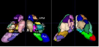 |
Validation of DTI-based parcellation of the thalamus in the squirrel monkey
Yurui Gao, Kurt Schilling, Iwona Stepniewska, Landman Bennett, Adam Anderson
DTI has been used to noninvasively resolve major thalamic nuclei using an unsupervised clustering algorithm. However, rigorous validation of the method has not been studied. Here, we evaluated the method by comparing the parcellation results with histology in the same non-human primate. We found that some nuclei were clustered with larger differences from histology. That probably was because the diffusion properties of these nuclei were not coherent. In addition, the pipeline constructed in this study is also a framework to validate other approaches for thalamic nuclei parcellation.
|
09:51
|
0723.
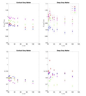 |
In vivo observation and interpretation of time dependent diffusion in human gray matter
Antonios Papaioannou, Dmitry Novikov, Els Fieremans
The temporal scaling of the diffusion coefficient and diffusional kurtosis may reveal the underlying microstructural features of the human brain. Here we demonstrate time-dependent diffusion coefficient and kurtosis in gray matter areas of the human brain in vivo. Our results suggest that the major contribution of time-dependence in gray matter originates from intra-neurite water with the main source of restrictions, e.g. dentrites and beads, being short-range disordered in their placement.
|
10:03
|
0724.
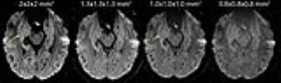 |
High Resolution Diffusion Imaging of the Human Hippocampus
Sarah Treit, Trevor Steve, Donald Gross, Christian Beaulieu
Diffusion imaging of the human hippocampus is typically limited to low spatial resolution due to challenges with low signal-to-noise ratio. Here we demonstrate error in fractional anisotropy/mean diffusivity in high resolution diffusion imaging acquired with multiple gradient directions and 1 average, which is mitigated by acquiring fewer directions and multiple signal averages. Using this approach, 1x1x1 mm3 diffusion data at 3T is shown to produce mean diffusion weighted images with excellent contrast within the hippocampus, acquired in a clinically feasible scan time of 6 minutes. High resolution diffusion imaging will impact the study of numerous disorders affecting the hippocampus.
|
|