Wednesday, 26 April 2017
| Room 316BC |
13:45 - 15:45 |
Moderators: Els Fieremans, Greg Stanisz |
Slack Channel: #s_diffusion
Session Number: O74
13:45
 |
0836.
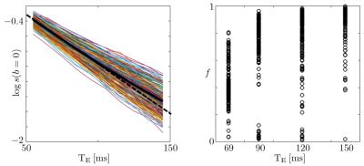 |
Quantifying neuronal microstructure integrity with TE dependent Diffusion Imaging (TEdDI) 
Jelle Veraart, Els Fieremans, Dmitry Novikov
We resolve degeneracies in the estimation of parameters of white matter integrity by exploiting the observed echo time dependency of diffusion MRI signal in human brain white matter. Empirical results and statistical analyses reveal that adding compartment-specific T2 relaxation times to biophysical models of diffusion MRI improves the parameter estimation in terms of precision and accuracy.
|
13:57
 |
0837.
 |
A Novel Method for Assessing Myelination with TE dependence of DTI-derived Parameters
Mu Lin, Qiuping Ding, Xu Yan, Thorsten Feiweier, Hongjian He, Jianhui Zhong
Myelin water is abundant in white matter but myelin signal is often ignored in diffusion models due to its short T2. There is however substantial water exchange between myelin and non-myelin water within typical diffusion times. Using Monte Carlo simulation and in-vivo measurement, we demonstrate that this water exchange might result in an echo-time (TE) dependence of DTI-derived parameters. As myelin water exchange increases with the thickness of myelin sheath, the TE dependence can be used to assess the degree of myelination.
|
14:09
|
0838.
 |
Disentangling in two dimensions in the living human brain: Feasbilty of relaxometry-diffusometry using ultra-strong gradients
Chantal Tax, Umesh Rudrapatna, Thomas Witzel, Derek Jones
Combining multiple, complementary contrasts into one analysis will yield deeper understanding of white matter physiology than using diffusion MRI (dMRI) alone. Varying TE in a PGSE sequence would allow for the exploration of D-T2 spectra in tissue. However, typical hardware and time constraints render the acquisition of such diffusion/relaxation spectra in the living human impractical. In this work, we explore how 300 mT/m gradients of a Connectom scanner could help in further investigating 1) the reported TE dependency of DTI parameters and 2) D-T2 spectra in the living human brain.
|
14:21
|
0839.
 |
T1-induced apparent time dependence of diffusion coefficient measured with stimulated echo due to exchange with myelin water
Hong-Hsi Lee, Dmitry Novikov, Els Fieremans
For diffusion measurements, stimulated-echo acquisition mode (STEAM) has been widely used. To enhance sensitivity to microstructure, previous studies used STEAM to vary the diffusion time by changing the mixing time tM. Here we show that varying tM results in an “apparent” STEAM-measured diffusivity dependence on tM, irrespective of genuine microstructure-specific time dependence. This effect is caused by T1-relaxation and water exchange between myelin water and "free" water (intra- and extra-axonal water). We propose a modified Kärger model considering diffusion+T1-relaxation+exchange, and demonstrate that exchange-induced tM-dependence explains ~20-50% of the total diffusion time dependence, and should be considered while using STEAM.
|
14:33
|
0840.
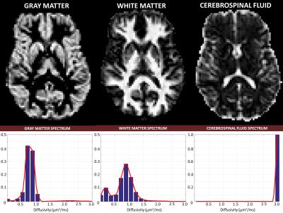 |
Isotropic Diffusion Relaxometry Imaging (IDRI)
Alexandru Avram, Joelle Sarlls, Elizabeth Hutchinson, Peter Basser
We describe a model-free method to quantify the spectrum of water mobilities in fixed and live brain tissues. We eliminate confounds caused by anisotropic diffusion in brain tissues by measuring orientationally-averaged diffusion weighted images over a large range of b-values. Spectra of orientationally-averaged water diffusivities show clear distinctions between white matter, gray matter and cerebrospinal fluid, and could provide new biologically-specific clinical markers for studying and diagnosing ischemic stroke, tumors, and neurodegenerative disorders and diseases, including inflammation.
|
14:45
|
0841.
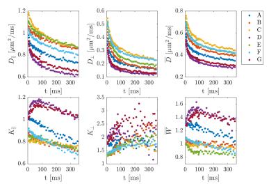 |
Time Dependence Of White Matter Biomarkers From Axially Symmetric Diffusion Kurtosis Imaging
Jonas Olsen, Brian Hansen, Noam Shemesh, Sune Jespersen
Using a recently developed PFG-based framework for fast diffusion kurtosis imaging we observe a strong time dependence of diffusion and kurtosis metrics in fixed spinal cord white matter from 6-350 ms. DKI metrics can be expressed in terms of intra- and extra axonal properties using white matter tract integrity (biexponential modelling), but a sign ambiguity results in two solutions of the inverse problem. The time dependence of the two solutions observed here help identify the correct solution, and allows comparing time-dependent compartment diffusivities with theory.
|
14:57
|
0842.
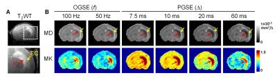 |
Diffusion time dependence of kurtosis reveals microstructural changes after neonatal hypoxia-ischemia
Dan Wu, Frances Northington, Els Fieremans, Dmitry Novikov, Jiangyang Zhang
Apparent diffusion coefficient (ADC) and diffusion kurtosis are both sensitive markers to ischemic brain injury. We investigated the diffusion time (td)-dependency of ADC and kurtosis at nine td’s ranging from 2.5 to 60 ms in a mouse model of neonatal hypoxic-ischemic injury. In the hippocampus, ADCs showed a monotonous decrease with increasing td, whereas kurtosis reached its maximum at td of 5-10 ms and decreased for longer td’s . At the shortest td in this study, we found significant increased kurtosis in the edema region but no significant reduction in diffusivity, suggesting their different sensitivities to microstructural changes after ischemic injury.
|
15:09
|
0843.
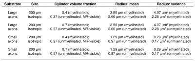 |
Origin of the time dependence of the diffusion-weighted signal in spinal cord white matter
Francesco Grussu, Andrada Ianus, Carmen Tur, Ferran Prados, Torben Schneider, Sébastien Ourselin, Ivana Drobnjak, Hui Zhang, Daniel Alexander, Claudia Gandini Wheeler-Kingshott
Time dependence of the brain white matter diffusion-weighted signal originates from both intra-axonal and extra-axonal spaces. Here, we investigate which of these contributions dominates in spinal cord white matter for clinically feasible acquisitions, to inform accurate model-based microstructural imaging. We analyse data from Monte Carlo simulations and from in vivo scans, and find that for diffusion times of 20-70 ms time dependence has mostly intra-axonal origin. Such a time dependence influences the estimation of axonal volume fraction and extra-axonal diffusivity, and highlights the importance of using long diffusion times to support stick-like models for axons in the spinal cord.
|
15:21
|
0844.
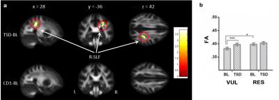 |
Sleep deprivation affects white matter integrity in cognitively vulnerable individuals
Hengyi Rao, Sihua Xu, Zhuo Fang, Fan Yang, Andrea Spaeth, Namni Goel, Mathias Basner, Sumei Wang, David Dinges, John Detre
Using DTI, we examined the effects of one night of acute total sleep deprivation on fractional anisotropy (FA), an index reflecting the degree of anisotropic water diffusion in brain white matter. Sleep deprivation significantly increases FA in the right superior longitudinal fasciculus (SLF) in individuals who were cognitively vulnerable to sleep loss, while no FA changes were observed in cognitively resistant individuals. Vulnerable subjects also showed lower FA in the right SLF than resistant subjects at baseline before sleep loss, suggesting both trait- and state-dependent interactions between SLF microstructure and cognitive vulnerability to sleep deprivation.
|
15:33
|
0845.
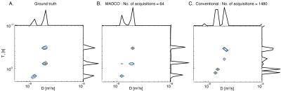 |
Clinically feasible relaxation-diffusion correlation MRI using MADCO
Dan Benjamini, Peter Basser
Even though the brain is microscopically heterogeneous, the majority of currently used quantitative MRI methods in brain research employ idealized models to describe specific structures. Multidimensional relaxation-diffusion correlation (REDCO) is an assumption-free method that measures how water is distributed within the tissue. REDCO had never been used in clinical applications because of the large amount of data it requires. Here we apply the concept of marginal distributions constrained optimization (MADCO) to REDCO-MRI experiments. Using this approach data requirements are vastly reduced, making REDCO-MRI a clinically feasible imaging technique to infer the underlying microstructure, number of compartments, and possibly their function.
|
|