Monday, 24 April 2017
| Room 311 |
13:45 - 15:45 |
Moderators: Peter Koopmans, Andrew Stenger |
Slack Channel: #s_fmri
Session Number: O80
13:45
|
0152.
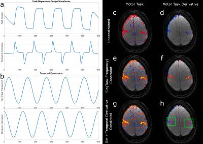 |
Accelerated rank-constrained FMRI data reconstruction informed by external temporal measures 
Mark Chiew, Nadine Graedel, Jostein Holmgren, Dean Fido, Catherine Warnaby, Karla Miller
Reconstruction of highly under-sampled FMRI data using low-rank constraints can suffer from loss of fidelity at high acceleration factors, or when signals are relatively weak. We introduce a method for improving reconstruction fidelity using external constraints, i.e., informative signals that are not data-derived. We show that this improves FMRI reconstruction quality in a number of conditions, including detecting subtle latency shifts between brain regions, and improving resting state network characterization using simultaneously acquired EEG information. We further show that this approach works with noisy or approximate constraints, and the derived benefit is commensurate with the information content they provide.
|
13:57
 |
0153.
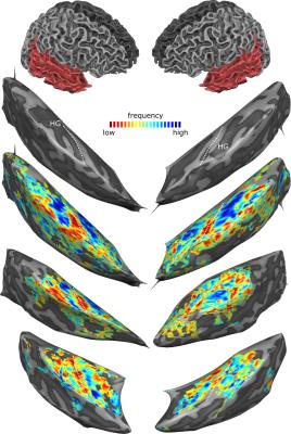 |
Submillimeter 9.4 T fMRI of the human auditory cortex with tones, ripples, and real life sounds
Valentin Kemper, Elia Formisano, Sudhir Ramanna, Essa Yacoub, Federico De Martino
This study demonstrates auditory human fMRI conducted at 9.4T field strength and submillimeter resolution for the first time. Tonotopic maps were measured robustly and reliably. Further, cortical regions with preference for natural sound categories were delineated. We generated ripple control sounds that closely match low level acoustical properties of natural sounds in four natural sound categories, such that the original category is not recognizable. We show that, in areas preferring speech sounds over other natural sounds, ripple control sounds of speech elicit stronger responses than ripple control sounds of non-speech. This indicates tuning to the low-level acoustical properties of speech.
|
14:09
|
0154.
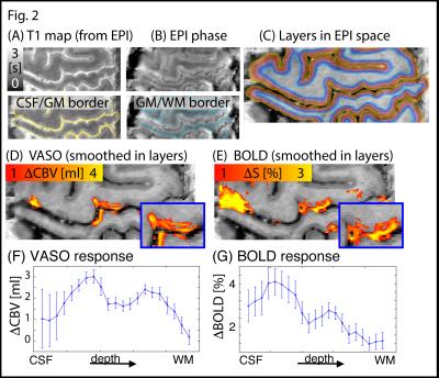 |
Ultra-high resolution blood volume fMRI and BOLD fMRI in humans at 9.4 T: Capabilities and Challenges
Laurentius Huber , Desmond Tse, Kashyap Sriranga, Christopher Wiggins, Kâmil Uludag, Peter Bandettini, Benedikt Poser, Dimo Ivanov
FMRI at ultra-high field strengths of 9.4 T allows functional imaging with submillimeter spatial resolutions. CBV sensitive VASO-fMRI has been suggested to be weighted towards locally specific microvasculature changes close to neural activity changes. Hence, we sought to combine the high physiological specificity of CBV-fMRI with the high signal-to-noise ratio of 9.4 T imaging. In our experiments, we could identify and discuss numerous technical challenges of CBV-fMRI at 9.4 T regarding constraints of RF fields and VASO contrast generation. With the application of advanced imaging methods, we show promising functional results with clearly visible cortical depth-dependent activity patterns.
|
14:21
 |
0155.
 |
Revealing the high frequency brain networks with multiband multi-echo fMRI data
Wenchao Yang, Burak Akin, Fei Wang, Jürgen Hennig, Pierre LeVan
The high frequency networks are hard to be observed with standard fMRI data. Those networks were submerged in the non-BOLD signal and could not be observed with standard fMRI methods. In this work, Multiband Multi-echo (MBME) sequence is used to sample brain fluctuations in high frequency (TR=0.75).By using acquired 8 echoes, T2* and I0 (initial intensity) values are calculated and fluctuations in the brain were separated into BOLD and non-BOLD. Results showed that there is an improved detection in several high frequency networks like LECN, dDMN, Language and high Visual networks by using the separated BOLD signal.
|
14:33
|
0156.
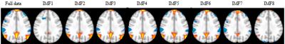 |
Empirical Mode Decomposition and Frequency Characteristics of the Default Mode Network on Group fMRI Resting-State Data
Dietmar Cordes, Muhammad Kaleem, Xiaowei Zhuang, Karthik Sreenivasan, Zhengshi Yang, Virendra Mishra
In this project, high-frequency contributions to functional connectivity of the Default Mode Network (DMN) are studied. Rather than relying on user-defined frequency bands, Empirical Mode Decomposition (EMD) is used to decompose the natural occurring frequency bands of the DMN. The novelty of our approach lies in the data-adaptive and user-independent decomposition of fMRI data using EMD, and identification of a resting-state network based on the frequency characteristics of intrinsic modes in the data, instead of using wavelet- or windowed-Fourier-transform methods. Results are shown for multiband MB8 resting-state data of a group of 22 healthy subjects.
|
14:45
|
0157.
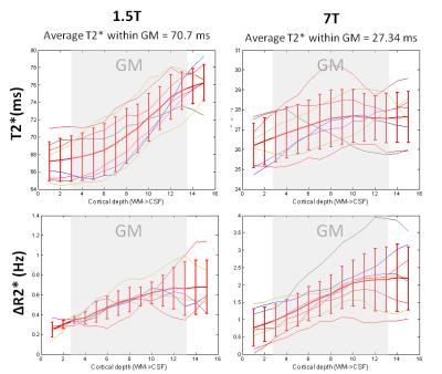 |
An in vivo study of BOLD laminar responses as a function of echo time and magnetic field
Irati Markuerkiaga, Lauren Bains, Jose Marques, David Norris
In this study we evaluate the echo time of laminar BOLD responses of the human primary visual cortex at 1.5 and 7T. It is often assumed that lower magnetic field strengths are increasingly biased towards the signal arising from larger veins located towards the pial surface. In this study (performed with an isotropic resolution of 0.75mm) we found similar shaped laminar profiles at 1.5 and 7T.
|
14:57
|
0158.
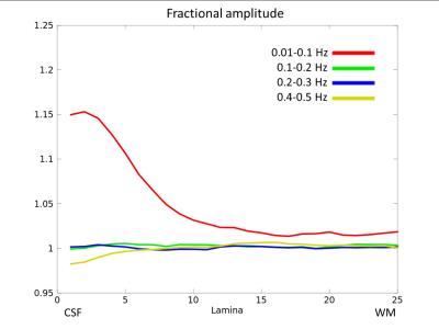 |
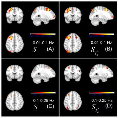
Frequency signature of cortical laminar fMRI
Maria Guidi, Irati Markuerkiaga, Lauren Bains, Laurentius Huber, Harald Möller, David Norris
The nature of spontaneous oscillations in the blood oxygenation level dependent (BOLD) response is in the focus of current research. For resting-state network studies, the low-frequency band (0.01-0.1 Hz) is usually taken to be relevant for neuronal activity. However, this statement is based on low-resolution functional data, where the effect of the draining vasculature cannot always be characterized. This study investigates the distribution of the amplitude of resting-state BOLD fluctuations using a sub-millimeter resolution and shows that the low-frequency band is dominating at all cortical depths, but most of its power is located at the pial surface.
|
15:09
|
0159.
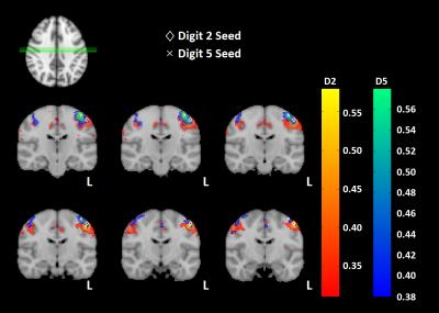 |
Topographic Mapping of Resting State fMRI Data
Eleanor Barratt, Michael Asghar, Matthew Brookes, Susan Francis
Task-based fMRI can provide robust somatotopic mapping of digits of the hand. Resting state fMRI (rs-fMRI) provides the ability to parcellate brain areas based on their connectivity. Here, we use simultaneous multislice to acquire high spatial resolution fMRI resting state data with a short TR to determine whether we can topographically map connectivity within the sensorimotor cortex. Seed based locations of the index finger (Digit 2) and little finger (Digit 5) are defined from somatotopic travelling wave and finger tapping tasks, and used to demonstrate significant topographic mapping in rs-fMRI data.
|
15:21
|
0160.
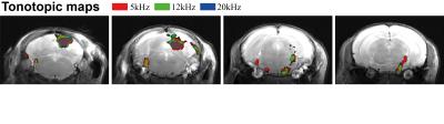 |
Tonotopic mapping in the in vivo mouse via high resolution fMRI
Guilherme Freches, Cristina Chavarrias, Noam Shemesh
The rodent auditory system has been a popular research subject for electrophysiological studies for its complexity, fine tuning and adaptability. More recently, some studies on auditory Functional Magnetic Resonance Imaging (fMRI) in rats have surfaced, aiming to unravel this system’s intricacies by capturing whole brain activity noninvasively. Auditory mapping in the mouse could be highly valuable given its importance vis-à-vis transgenic models and optogenetics. This study provides the first tonotopic mapping in the in vivo mouse via high resolution fMRI. We demonstrate robust activation in the auditory pathway, and specific tonotopy in several prominent regions along the pathway.
|
15:33
|
0161.
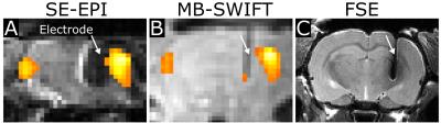 |
MB-SWIFT functional MRI during deep brain stimulation in rats
Lauri Lehto, Djaudat Idiyatullin, Jinjin Zhang, Lynn Utecht, Gregor Adriany, Michael Garwood, Olli Gröhn, Shalom Michaeli, Silvia Mangia
Commercial electrodes used for Deep Brain Stimulation (DBS) cause severe artefacts in conventional echo based MRI. Here we show near artefact free functional MRI during DBS in rats using Multi-Band SWeep Imaging with Fourier Transformation (MB-SWIFT) which allows acquisition at virtually zero-TE. MB-SWIFT showed strong responses in the somatosensory cortex while stimulating the ventromedial. The amplitude and extent of activation recorded with MB-SWIFT were similar with SE-EPI, although activation was flip angle dependent reflecting the possible influence of blood inflow. MB-SWIFT is a promising modality for fMRI in the presence of DBS leads or other severe susceptibility differences.
|
|