Thursday, 27 April 2017
| Room 315 |
13:00 - 15:00 |
Moderators: Clemens Bos, Christiaan Overduin |
Slack Channel: #s_int_eng_safety
Session Number: O81
13:00
 |
1172.
 |
Efficiency improvement in multi-point MR acoustic radiation force impulse imaging 
Henrik Odéen, Joshua de Bever, Lorne Hofstetter, Dennis Parker
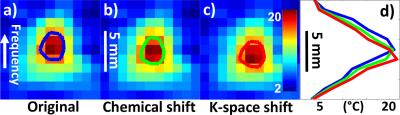
Focused ultrasound (FUS) treatment endpoint can potentially be evaluated using MR acoustic radiation force impulse (MR-ARFI) imaging, which can non-invasively and with very low energies interrogate tissue mechanical properties. In this work we investigate two ways of improving the efficiency of a previously published multi-point (MP)-MR-ARFI pulse sequence which simultaneously measure ARFI displacement and PRFS temperature maps. We investigate view-sharing of higher k-space frequencies from FUS-OFF images to FUS-ON images, and focusing in multiple positions during a single motion encoding gradient lobe as ways to speed up acquisition. Combining these methods full 3D MP-displacement maps could be achieved in a clinically acceptable time of 30-60s.
|
13:12
|
1173.
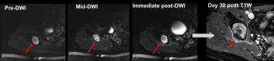 |
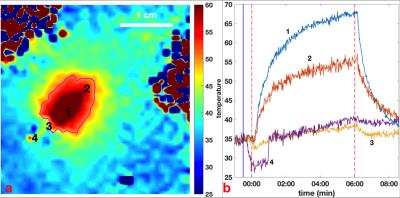
MR guided High Intensity Focused Ultrasound for treating painful bone metastases: relating intra-procedural ADC changes and thermal dose to volume of ablated tissue
Sharon Giles, Matthew Brown, Jessica Winfield, David Collins, Ian Rivens, John Civale, Gail ter Haar, Nandita deSouza
Thermal dose estimates and apparent diffusion coefficient (ADC) changes during MR-guided High Intensity Focused Ultrasound of painful bone metastases (n=11) were correlated with ablated tissue volumes assessed on Gd-T1W imaging acquired immediately, and at 30 days post-treatment. Thermal dose volume and mean maximum temperature were estimated from proton resonance frequency shift thermometry. Ablated tissue volume did not change significantly over 30 days. Mean maximum temperature and thermal dose volume were significant indicators of ablated volume, and mean maximum temperature was significantly higher in those with enduring ADC changes at Day 30. Further work will relate imaging changes to pain outcomes.
|
13:24
|
1174.
 |
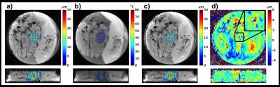
Estimating Acoustic Velocity of Human Skull Bone with CT and MRI
Taylor Webb, Ethan Johnson, Steven Leung, Jeremy Dahl, Norbert Pelc, John Pauly, Kim Butts Pauly
Focusing of transcranial focused ultrasound requires knowledge of the speed of sound in the skull. Clinically, estimates of the speed of sound in the skull are obtained from a CT scan. We measure the acoustic velocity in several human skull fragments and correlate it to the average Hounsfield units, MR-Simulated-CT value (derived from an ultrashort echo time MR sequence), and R2* value of each fragment. Results show that both CT and MR can be used to accurately estimate the acoustic velocity in human skull bone and that replacing CT with MR to plan transcranial FUS treatments is feasible.
|
13:36
|
1175.
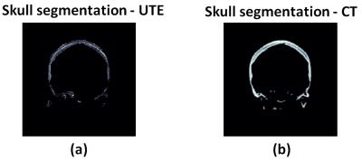 |
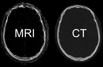
Ultrashort Echo-Time Imaging based Skull Density Ratio Assessment for Transcranial MR guided Focused Ultrasound
Sijia Guo, Jiachen Zhuo, Rao Gullapalli
Transcranial MRI-guided focused ultrasound (tcMRgFUS) applications have been growing steadily on treatment of brain diseases. Typically a CT skull scan is used with the MR scan to perform treatment planning in tcMRgFUS procedures. In this study we examine the use of ultrashort echo time imaging to perform the skull imaging and assess the feasibility of using the information from MRI to perform treatment planning through acoustic and temperature modeling with skull characteristics generated from UTE imaging. We further compared the simulation results with existing data from tcMRgFUS treatment of essential tremor procedure that used CT images of the skull for treatment planning. We demonstrated that UTE based treatment planning is feasible and avoids the use of CT based images thus avoiding unnecessary radiation exposure.
|
13:48
|
1176.
 |
Focus Correction in MR thermography for Precise Targeting in Focused Ultrasound Thalamotomy for Essential Tremor
Chang-Sheng Mei, Bruno Madore, Shenyan Zong, Pei-Hsin Wu, Garth Cosgrove, Nathan McDannold
MRI-guided focused ultrasound thalamotomy has proved an effective method for the treatment of Essential Tremor. Precise alignment of the focus to the target in the thalamus is crucial. Proper targeting is complicated by artifactual shifts in hot spot location, with respect to anatomical images. We identified two sources of errors and proposed solutions to both; the correction scheme does not require any modification to the pulse sequence or imaging protocol. Shifts by 1.0±0.1 mm were detected and corrected, for three separate sonications in one given patient. The size of the shifts agreed with observations from the literature.
|
14:00
|
1177.
 |
Image-registered whole mount histology technique for validation of MRgFUS therapies
Allison Payne, Sara Johnson, Robb Merrill, Nicole Winkler, Rachel Factor, Jill Shea
Clinical pathological review of histological samples is the gold standard for identification of tissue damage. For the development of new magnetic resonance-guided focused ultrasound (MRgFUS) procedures, in vivo post-treatment imaging assessment techniques require correlation to histopathology. Our study presents methodology and preliminary preclinical results of a new MRI-registered whole mount histology technique for efficacy evaluation of MRgFUS therapies.
|
14:12
|
1178.
 |
A Combined Intravascular MRI Endoscope and Intravascular Ultrasound (IVUS) Transducer for High-Resolution Image-Guided Ablation
Xiaoyang Liu, Nicholas Ellens, Emery Williams, Everette Burdette, Parag Karmarkar, Paul Bottomley
An intravascular MRI (IMRI) loopless antenna is combined for the first time with an intravascular water-cooled ultrasound ablation transducer as a possible tool for providing high-resolution MRI-guided ablations of pathological tissue via intravascular access. High resolution anatomical MRI, and real-time MRI thermometry were used to monitor ablation delivery in phantoms and tissue specimens. Results show that IMRI can guide IVUS-mediated directional ablation with minimal image artifacts. This permits the monitoring of thermal dose and therapy titration while minimizing potential thermal damage to the vessel wall.
|
14:24
|
1179.
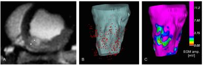 |
MRI-based myocardial ablation lesion extent relates to area of voltage reduction in MR-guided electroanatomical voltage maps
Philippa Krahn, Venkat Ramanan, Labonny Biswas, Nicolas Yak, Kevan Anderson, Jennifer Barry, Mihaela Pop, Graham Wright
In this study we investigated subtle features of acute RF ablation lesions using co-registered MR images and electroanatomic voltage maps (EAVM) acquired under MR guidance. Of particular interest was the relationship between the extent of edema detected in T2 maps and altered electrical activity surrounding the ablation site. The results from this study aim to elucidate mechanisms of high ventricular arrhythmia recurrence rates after successful RF ablation therapy, which may be driven by transient conduction block produced acutely by edema.
|
14:36
|
1180.
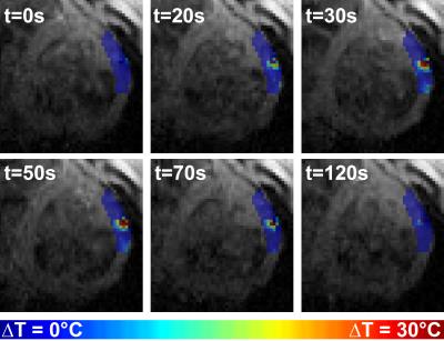 |
In-vivo Imaging of Ablation Lesions during MRI-guided Epicardial Ventricular Ablation in Swine
Sébastien Roujol, Radhouene Neji, Henry Chubb, John Silberbauer, Tom Lloyd, Thomas Pohl, Rainer Schneider, Nick Kampa, James Harrison, Steven Williams, Rahul Mukherjee, Louisa O'Neill, John Whitaker, Matthew Wright, Tobias Schaeffter, Mark O'Neill, Reza Razavi
Cardiac MR (CMR) shows promise for the guidance of ventricular tachycardia (VT) ablation procedures and imaging of ablation lesions. CMR-thermometry is a promising approach for real-time tissue temperature monitoring and tissue characterization for prediction of permanent ablation lesions. In this study, we sought to develop an integrated multi-parametric framework for real-time, hyper-acute and acute imaging of ablation lesions using CMR-thermometry, native T1 mapping and late gadolinium enhancement (LGE), respectively. This framework is evaluated during MR-guided epicardial ventricular ablation in swine.
|
14:48
 |
1181.
 |
Toward hybrid MR thermometry in aqueous and adipose tissue using simultaneous dual contrast weighting with double echo RARE imaging
Hendrik Paysen, Katharina Paul, Michal Pham, Lukas Winter, Thoralf Niendorf
Proton resonance frequency (PRF) shift is the most common MR thermometry (MRTh) technique for water based tissue. For adipose tissue MRTh exploits the temperature dependent relaxation times T1 and T2. Hybrid methods that allow simultaneous acquisition of water and fat based thermometry are of great clinical need. Recognizing this need this work investigates a hybrid temperature mapping technique, designated as 2in1-Thermometry, which combines simultaneous PRF and T2-based temperature mapping using simultaneous dual contrast weighting with double echo RARE imaging.
|
|