Thursday, 27 April 2017
| Room 311 |
13:00 - 15:00 |
Moderators: Özlem Ipek, Benedikt Poser |
Slack Channel: #s_int_eng_safety
Session Number: O84
13:00
|
1122.
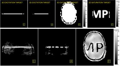 |
Homogeneous high-flip-angle 3D localization by parallel transmission at 9.4T 
Tingting Shao, Yun Zhang, Nikolai Avdievich, Christian Mirkes, Klaus Scheffler, Steffen Glaser, Anke Henning
This work presents in vivo experimental result of high-flip-angle multi-dimensional parallel transmission at a 9.4T human whole-body MRI scanner. A 2D pTx saturation pulse (90°) and a slice selective 3D pTx excitation pulse (60°) were designed by using an algorithm that combines LSQR and optimal control (OC) methods and enables high-flip-angle pTx pulse design with a strict constraint of transmit power. An actual flip angle imaging (AFI) sequence was coded to measure the flip angle map of the pre-saturated slice excitation profile achieved by the sequentially implemented pTx pulses.
|
13:12
 |
1123.
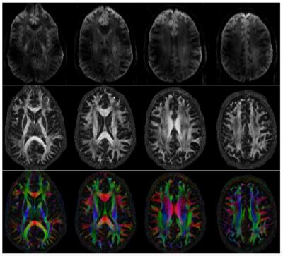 |
High Resolution Multi-shot Diffusion Imaging at 7T without Navigators
Merry Mani, Mathews Jacob, Baolian Yang, Vincent Magnotta
The higher signal-to-noise ratio (SNR) offered by the ultra-high field (UHF) strengths are often exploited to improve the spatial resolution capabilities of several MR imaging modalities. However, SNR advantage of UHF scanners do not often translate to improved diffusion weighted images (DWIs), especially using conventional single-shot echo-planar imaging-based acquisitions. The presence of diffusion gradients limit the lowest echo-time (TE) achievable, while the shortened T2 and T2* values lead to faster decay of the MRI signal, both of which are detrimental to the already signal-starved DWIs. We propose a short-TE acquisition based on multi-shot EPI and partial Fourier acquisition to enable high resolution diffusion imaging at 7T that does not need navigator scans or phase calibration.
|
13:24
 |
1124.
 |
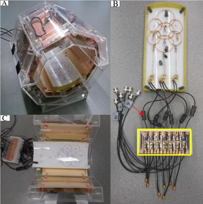
Initial experiences with a fully-removable whole-body birdcage transmit coil and 16-element receive array for cardiac 31P-MRS at 7T
Ladislav Valkovic, Iulius Dragonu, Karsten Wicklow, Ulrich Fontius, Salam Almujayyaz, Alex Batzakis, Liam Young, Lucian Purvis, William Clarke, Tobias Wichmann, Titus Lanz, Stefan Neubauer, Matthew Robson, Dennis Klomp, Christopher Rodgers
This abstract describes our experiences implementing a volume transmit, local receive setup for cardiac 31P-MRS on a Siemens research 7T MRI scanner. Two strands of development work have been performed in tandem: (i) development of a fully removable whole-body transmit RF-coil and testing with the standard 8kW RFPA and SAR monitoring and combined with a 16ch receive array, and (ii) integration of a 35kW RF power amplifier, a new energy chain, and adapted SAR monitoring.
|
13:36
|
1125.
 |
Abdominal Imaging with Heterogeneous Radiofrequency Fields at 7 Tesla
Martijn Cloos, Jan Paška, Zidan Yu, Jakob Assländer, Tiejun Zhao, Riccardo Lattanzi, Graham Wiggins, Daniel Sodickson
Body imaging using ultra-high field systems operating at 7T or more is extremely challenging due to the non-uniformities in the excitation field. Traditionally, these field variations are seen as a nuance that must be calibrated out, leading to a formidable engineering challenge. Recently, a new paradigm was proposed which deliberately interweaves multiple uncalibrated non-uniform RF fields into the scan. In this work we demonstrate these principles in-vivo and show that it is possible to obtain artifact free cross-sectional quantitative maps of the abdomen at 7T across a variety of different subject sizes without the need for any specific calibrations.
|
13:48
|
1126.
 |
An 8/15-Channel Tx/Rx Head Neck RF Coil Combination with Semi-Dynamic B1 Shimming for Improved fMRI of the Cerebellum at 7 T
Viktor Pfaffenrot, Sascha Brunheim, Stefan Rietsch, Thomas Ernst, Oliver Kraff, Stephan Orzada, Harald Quick
Functional MRI of the human cerebellum is challenging at ultrahigh fields, since conventional RF head coils hardly cover the cerebellum with sufficient signal-to-noise ratio and B1+-inhomogeneities introduce challenges. In order to overcome these problems, a coil combination consisting of an 8ch transceiver head coil and a 7ch receive only array are combined to improve imaging of the whole brain with special focus on the cerebellum. A ‘semi-dynamic’ B1+-shimming technique is introduced which provides a tSNR-gain of 29 % and voxels with higher significance in a finger tapping fMRI experiment when comparing the coil combination to a 32ch receive head coil.
|
14:00
|
1127.
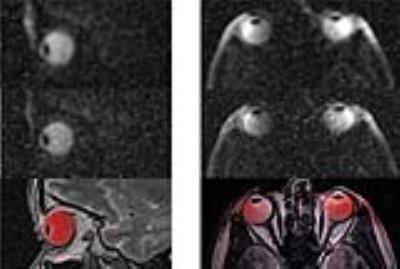 |
Panning for Salt: One Millimeter Resolution In Vivo Sodium MRI of the Human Eye at 7.0 Tesla Using a Six Channel Transceiver Array
Daniel Wenz, Oliver Weinberger, Andre Kuehne, Helmar Waiczies, Armin Nagel, Celal Oezerdem, Till Huelnhagen, Darius Lysiak, Lukas Winter, Oliver Stachs, Soenke Langner, Erdmann Seeliger, Bert Flemming, Thoralf Niendorf
Sodium quantification in the eye may be a valuable aid not only in diagnosis of ocular diseases, but in follow-up after proton therapy of eye tumors. Recognizing this potential, this is the first report on high fidelity in vivo sodium (23Na) MRI of the human eye at 7.0 Tesla. To achieve this goal a six-channel 23Na transceiver array was designed, simulated, built and validated in phantoms. The in vivo studies demonstrated the feasibility of 1 mm isotropic spatial resolution 23Na MRI of the eye and provided encouragement for clinical studies.
|
14:12
|
1128.
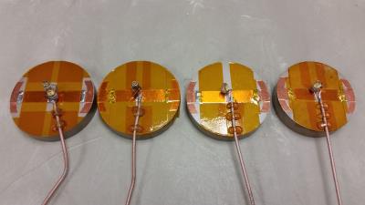 |
High dielectric constant (HDC) disk dipoles for 10.5T imaging
Russell Lagore, Lance DelaBarre, Qing Yang, Michael Lanagan, Yigitcan Eryaman, Sebastian Rupprecht, Wei Luo, Byeong-Yeul Lee, Xiao-Hong Zhu, Kamil Ugurbil, Wei Chen, Gregor Adriany
A small coil element is developed for 10.5T imaging by affixing an end-loaded 82mm long dipole to a high dielectric constant (HDC) flat disk. This coil element produces localized but very high B1 fields similar to a loop of a similar diameter without suffering from diverging B1+ and B1- that loops experience at high field. Furthermore, these elements are inherently highly decoupled in all possible spatial configurations, making them well suited to use in large, densely packed transmit arrays.
|
14:24
|
1129.
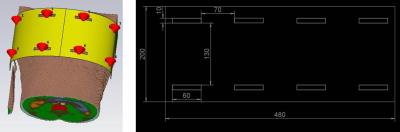 |
Lighter is better: A Flexible Lightweight Eight Channel Slot Antenna Array for Cardiac MRI at 7.0 Tesla
Celal Oezerdem, Till Huelnhagen, Andre Kuehne, Daniel Wenz, Jason Millward, Lukas Winter, Thoralf Niendorf
The wave length in tissue at ultrahigh fields allows for practical realization of RF antenna architectures such as dipole elements, high dielectric resonators and slot antennae. This work presents a novel flexible eight channel slot antenna array customized for cardiac MRI at 7.0 T. The proposed array is lightweight, easy to build, and affords a tight fit for a broad range of upper torso geometries while ensuring good matching and tuning. The in vivo study demonstrated the feasibility of the array for high fidelity, whole heart coverage MRI at 7.0 T and showed rather uniform signal intensity across the heart.
|
14:36
|
1130.
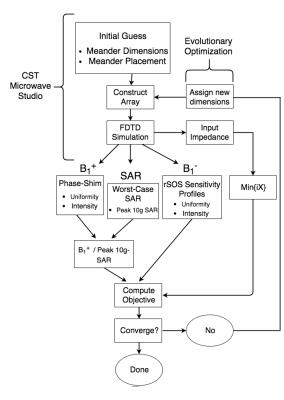 |
Shape-Optimization of Electric Dipoles for Human Head Imaging at 7 Tesla
Ian Connell, Ravi Menon
Dipole antennae have been proposed as an alternative solution for RF transmission at ultra-high field (UHF) strengths. However, adapting dipoles to achieve self-resonance, while minimizing SAR for a given transmission field homogeneity, is challenging given the geometry of the human head. In this study, the design of dipole elements is performed via computer aided shape optimization and is demonstrated to meet several of these design parameters. The final design relies upon meandered-conductor paths to produce electromagnetic fields that minimize local SAR without the use of additional RF shimming or pulse design algorithms.
|
14:48
|
1131.
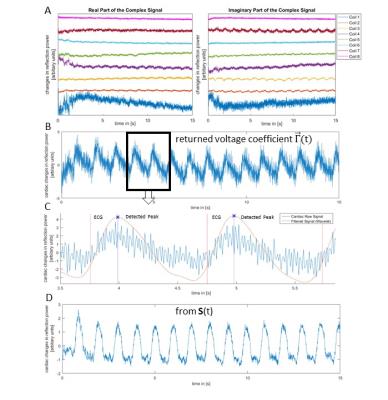 |
Evaluating the Influence of B1-Shimming on Contact-free Cardiac Gating using Scatter of a Parallel Transmit Coil at 7T MRI
Sven Jaeschke, Matthew Robson, Aaron Hess
We propose a contact-free cardiac MRI triggering method based on reflected power measurements that requires no additional hardware other than that provided with a commercial parallel transmit (pTx) MRI scanner and evaluate the influence of B1+ shim on the cardiac information extracted. Time series of scattering matrices of the pTx monitoring system were used with random, uniformly distributed phases to simulate B1+ shims. Preliminary results in 7T MRI are shown with successfully, retrospectively gated 2D-CINE images using the proposed method.
|
|