Poster: Emerging Neuroimaging Techniques
Electronic Power Pitch Poster
Neuro
Thursday, 27 April 2017
| Exhibition Hall |
14:00 - 15:00 |
| |
|
Plasma # |
|
1097.
 |
16 |
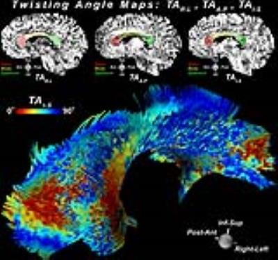
Characterization of White Matter Tortuosity using High-Resolution gSlider-SMS Diffusion Imaging 
Choukri Mekkaoui, Marcel Jackowski, Kawin Setsompop, Qiuyun Fan, Ned Ohringer, William Kostis, Timothy Reese, Alexandra Golby, Susie Huang
Conventional tractography excludes acute directional changes. We define these acute changes as tortuosity and use it to characterize gray-white matter boundaries, as well as quantify deformation and remodeling in pathologic states. Five healthy volunteers and a brain tumor patient were scanned using the gSlider-SMS technique. Diffusion data was processed to create 3D contiguous ribbons that characterize tortuosity. Twisting angles of the 3D ribbons were compared within and between subjects, and were visually and statistically consistent. This highly informative tractographic method could be applied to evaluate tumor infiltration and adjacent brain compression, or traumatic brain injury.
|
|
1098.
 |
17 |
In Vivo Characterization of an Ultrashort-T2 Component in the Brain Reveals a Chemical Shift
Peder Larson, Tanguy Boucneau, Shuyu Tang, Misung Han, Peng Cao, Roland Henry
A new approach for direct measurements of myelin content is to image an sub-millisecond T2 component in the brain, likely associated with myelin membrane protons . This study characterized this ultrashort-T2 component across the whole brain using a novel relaxometry approach at ultrashort echo times (UTEs). This component had an estimated T2* ~ 0.6-1.0 ms at 3T and ~0.2-0.4 ms at 7T, as well as an approximately -3.2 ppm frequency shift from water that has never been measured before in vivo. This verifies that the ultrashort-T2 component primarily arises from methylene protons, as in the myelin phospholipid membranes.
|
 |
1099.
 |
18 |
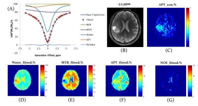
Improved Differentiation of Low- and High-Grade Gliomas by APT Contrast Fitted from Z-Spectrum
Jiaxuan Zhang, Rongwen Tain, Xiaohong Zhou, Wenzhen Zhu, Kejia Cai
Preoperative grading is important for treatment planning in glioma patients. Amide proton transfer (APT) -weighted imaging is helpful in grading since it reflects metabolic changes associated with mobile proteins and peptides. However, the conventional APT based on asymmetrical analysis receives contaminations from semi-solid magnetization transfer asymmetry and nuclear overhauser enhancement effects. Multi-component Z-spectral fitting for the separated quantification of APT can help to remove those contaminations. In this study, we performed such fitting on Z-spectral data from glioma patients. We found that fitted APT provided higher power in differentiating low- and high- grade gliomas compared to the conventional APT quantification.
|
|
1100.
 |
19 |
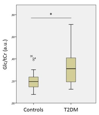
3T 1H PRESS (TE 68 ms) reveals elevated cerebral glucose in patients with diabetes mellitus type 2, which is associated with fasting blood glucose
Frank van Bussel, Tamar van Veenendaal, Miranda Schram, Coen Stehouwer, Walter Backes, Jacobus Jansen
A standard 1H PRESS sequence, with a TE of 68 ms, acquired at 3T is applied to investigate differences in cerebral glucose concentrations in type 2 diabetes mellitus (T2DM ) compared with controls with normal glucose metabolism. Subjects with T2DM (n=38) display an increased cerebral glucose level compared to controls (n=38). These levels are also associated with two blood glucose measures, fasting blood glucose (FBG) and glycated hemoglobin (HbA1c).
|
|
1101.
 |
20 |
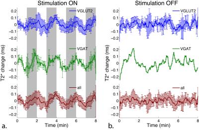
Towards opto-fMRS: Ultra high field MRS measurement of T2* changes due to optogenetic stimulation
Jamie Near, Dan Madularu, Jennifer Robinson, Chathura Kumaragamage, Axel Mathieu, Sylvain Williams, M Rajah, Uzay Emir
Combining optogenetics with fMRI/fMRS (opto-fMRI/opto-fMRS) offers the unique potential to study the whole brain functional or neurochemical effects of stimulating specific neuronal populations within a given brain region. In this study, we investigated the BOLD functional changes and neurochemical changes resulting from optogenetic stimulation of glutamatergic or GABAergic neurons in the medial septum. Stimulation of both glutamatergic and GABAergic neurons in the medial septum resulted in prominent bold activation within the hippocampus, and in other regions. This study represents a unique imaging investigation into the functional response to stimulation of multiple distinct neuronal populations within a single brain region.
|
|
1102.
 |
21 |
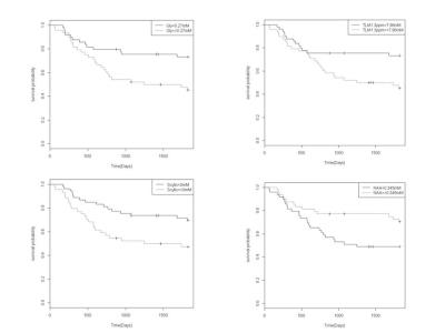
Glycine, a marker of survival in paediatric brain tumours measured with non-invasive Magnetic Resonance Spectroscopy: A five-year survival analysis.
Ben Babourina-Brooks, Sarah Kohe, Simrandip Gill, Martin Wilson, Lesley Macpherson, Nigel Davies, Andrew Peet
Brain tumours have a high mortality rate and are the most common solid tumour of childhood. MRS is a non-invasive imaging technique that measures tumour metabolites which can provide prognostic information to aid clinical management. The metabolite glycine is associated with proliferation and tumorigenesis through the one-carbon metabolic cycle. Glycine concentration has been shown to increase with tumour grade using short echo time MRS at 1.5T, however has not been assessed as a survival marker. This study investigates glycine as a marker of survival in paediatric brain tumours and assesses its added value compared to other established metabolite survival markers.
|
 |
1103.
 |
22 |
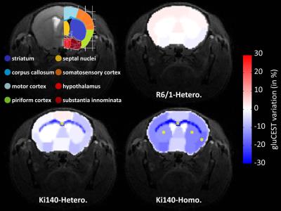
About the complementarity of gluCEST and 1H-MRS for the study of neurodegenerative diseases using animal models
Jérémy Pépin, Clémence Ligneul, Julien Valette, Emmanuel Brouillet, Julien Flament
Animal models of neurodegenerative diseases are useful tools to investigate neurodegenerative diseases. However, there is often a wide variety of described models for a same pathology, each model exhibiting its own characteristics. In this work, we used two mouse models of Huntington’s disease exhibiting very different alterations. Using a protocol combining gluCEST imaging and 1H-MRS, we showed that, while gluCEST may evidence alterations in unexpected brain regions, it may also be blind to disease process in certain situations where glutamate levels are preserved. This highlights the complementarity of both methods to identify relevant biomarkers of the pathology.
|
|
1104.
 |
23 |
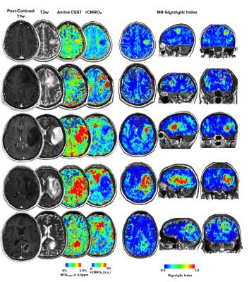
A "Glycolytic Index" for quantifying abnormal metabolism in human gliomas using multi-echo amine chemical exchange saturation transfer spin-and-gradient echo echoplanar imaging (ME-aCEST-SAGE-EPI) at 3T - did not present
Robert Harris, Kevin Leu, Timothy Cloughesy, Whitney Pope, Phioanh Nghiemphu, Albert Lai, Linda Liau, Benjamin Ellingson
Abnormal metabolism is a hallmark of cancer. The current study demonstrates use of a novel imaging technique for fast, simultaneous pH- and hypoxia-weighted images using multi-echo amine chemical exchange saturation transfer spin-and-gradient-echo echoplanar imaging (ME-aCEST-SAGE-EPI). From these data, we demonstrate use of a “glycolytic index”, quantified as the ratio of relative acidity to metabolic rate of oxygen, in estimating metabolically active tumor tissue in 15 patients with gliomas. The glycolytic index showed unique heterogeneous metabolic contrast within the tumor region, and was able to easily stratify tumor from healthy tissue when compared with other imaging techniques.
|
|
1105.
 |
24 |
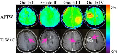
Qualitative and quantitative analysis of amide proton transfer-weighted MR images at 3 Tesla of adult gliomas
Xianlong Wang, Hao Yu, Shanshan Jiang, Yu Wang, Yanyu Wang, Ge Zhang, Chunxiu Jiang, Guodong Song, Yi Zhang, Hye-Young Heo, Jinyuan Zhou, Zhibo Wen
We evaluated the reliability and clinical value of amide proton transfer-weighted (APTW) MR imaging at 3 Tesla in adult gliomas. Fifty-seven patients with primary gliomas were recruited and scanned. Two radiologists evaluated the location and size of the APTW hyperintensity and enhancing areas, and measured the tumor and contralateral normal-appearing white matter (CNAWM) APTW values. The correlation between relative APTW (rAPTW) values and pathologic grades was calculated. Results showed APTW analysis had good reliability. APTW images almost showed the same compared with T1-weighted contrast-enhancing (T1W+C) images. The tumor rAPTW values had a strong positive correlation with pathologic grades.
|
|
1106.
 |
25 |
Cerebral Sodium (23Na) Magnetic Resonance Imaging in Patients with Migraine - permission withheld
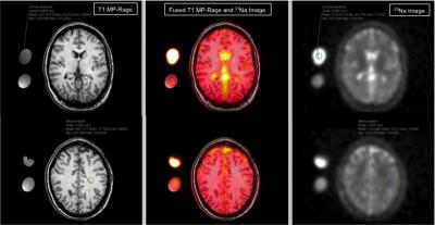
Melissa Ong, Alexander Schmidt, Simon Konstandin, Justus Benrath, Mathias Meyer, Lothar Schad, Stefan Schoenberg, Stefan Haneder
To evaluate sodium concentrations in subgroups of patients with clinically manifest migraine, 12 patients underwent a cerebral 23Na-magnetic resonance imaging examination using a dual-tuned (23Na/1H), dedicated head-coil. 23Na-sequences were reconstructed according to a T1 MP-RAGE, allowing direct cross-referencing of predetermined regions-of-interest (ROI). Significant differences in sodium concentrations could be observed for the white matter and anterior cerebrospinal fluid in patients with and without accompanying aura (p<0.05). These data suggest, that cerebral sodium concentrations may have the potential to differentiate between different subgroups of migraine.
|
|
1107.
 |
26 |
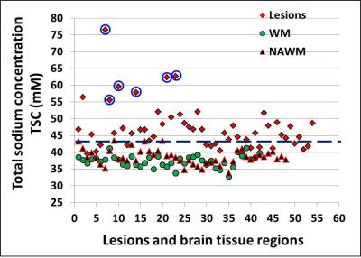
Differentiating subtypes of multiple sclerosis lesions using sodium MR imaging
Yulin Ge, Yongxian Qian, Jean-Christophe Brisset, Fernando Boada
Although conventional 1H MRI is commonly used for diagnosis and monitoring disease progression in multiple sclerosis (MS), it is not specific to pathology and cell vitality, and is limited in differentiating underlying pathology in MS lesions . In this study, using sodium (23Na) MRI, we demonstrated several subtypes of MS lesions with different sodium concentration changes, which may represent various stages of demyelination and axonal injury. Such information that is not available on conventional imaging may have value in characterize early versus chronic inactive lesions in MS.
|
|
1108.
 |
27 |
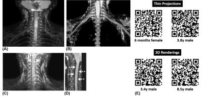
Imaging of the Brachial Plexus using a 3D Dixon-TSE Pulse Sequence with Blood Vessel and CSF Signal Suppression: Preliminary Experience in Children
Barbara Cervantes, Amber Pokorney, Jan Kirschke, Patricia Cornejo, Jeffrey Miller, Dimitrios Karampinos, Houchun Hu
We demonstrate the clinical feasibility and utility of a 3D Dixon TSE sequence in imaging the brachial plexus nerves in children. The employed high spatial resolution MR Neurography (MRN) technique utilizes a refocusing flip angle train to maximize nerve signal and to suppress cerebrospinal fluid signal. Additionally, a T2-preparation with motion-sensitizing gradients was employed to suppress flowing blood signal and Dixon-based chemical-shift water-fat imaging was used to suppress fat signal in the head, neck, and chest. We illustrate this MRN technique in delineating the brachial plexus nerves and associated pathological conditions in 25 pediatric patients (age range: 6 months-21 years).
|
|
1109.
 |
28 |
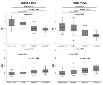
Diffusion-Tensor-Imaging MR-Neurography for the detection of polyneuropathy in Type 1 diabetes
Michael Vaeggemose, Mirko Pham, Steffen Ringgaard, Hatice Tankisi, Niels Ejskjaer, Sabine Heiland, Per L. Poulsen, Henning Andersen
Diabetic peripheral neuropathy (DPN) is an irreversible complication that often remains undiagnosed until advanced stages. Thus, to prevent progression of DPN, early diagnosis is important emphasizing the need for more sensitive diagnostic techniques. Diagnosis is based on a neurological examination, nerve conduction studies, and quantitative sensory testing. Magnetic-resonance-neurography (MRN) enables microstructural nerve fascicle imaging of peripheral neuropathies (1–4). In a previous study on patients with severe DPN we found that diffusion-tensor-imaging (DTI) is a reproducible method in detection of DPN (5). Furthermore, DTI-MRN provided a more accurate group separation than by changes based on T2 or proton-density contrast.
|
|
1110.
 |
29 |
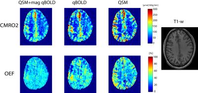
Optimal quantitative mapping of Cerabral Metabolic Rate of Oxygen (CMRO2) by combining quantitative susceptibility mapping (QSM)-based method and quantitative BOLD (qBOLD)
Junghun Cho, Youngwook Kee, Pascal Spincemaille, Thanh Nguyen, Jingwei Zhang, Yi Wang
Current two CMRO2 mapping methods, QSM-based method and qBOLD, suffer from the issues in their own model assumptions, such as blood flow challenge and linear relationship assumption between cerebral blood volume (CBV) and flow (CBF) in QSM-based method and the high sensitivity to noise and recent one-compartment assumption in qBOLD. We combined these two models with regularization based on that vein blood volume fraction and vein oxygenation are shared, which removes the challenge and linear assumption in QSM method and alleviates the high noise sensitivity and one-compartment assumption issue in qBOLD. The proposed model provided more uniform OEF and CMRO2.
|
|
1111.
 |
30 |
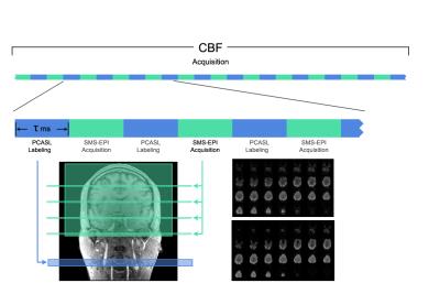
Asynchronous Local Analysis of simultaneous BOLD ASL Multislice Acquisition (ALABAMA): Toward Whole-Brain Noninvasive Estimation of Resting-State Neuronal-Vascular Coupling
Vincent Schmithorst, Vince Lee, Ashok Panigrahy
A method (ALABAMA) for regional estimation of the coupling between R2* fluctuations and log signal intensity (S0) (related to R2*-CBF coupling via a calibration factor) during the resting state is proposed. The method incorporates pseudo-continuous ASL (pCASL) labeling together with simultaneous multi-slice (SMS) and double-echo EPI acquisition for fast temporal resolution. The sequence yields robust positive ΔR2*-Δln S0 correlations in most resting-state networks, which may then be used to estimate resting-state neuronal-vascular coupling (NVC) with an additional calibration scan. However, negative correlations were found in posterior DMN and central executive network (CEN) regions, possibly reflective of altered flow-metabolism relationships.
|
|