Poster: Best of Cardiovascular MR: Hemodynamics & Atherosclerosis
Electronic Power Pitch Poster
Cardiovascular
Tuesday, 25 April 2017
| Exhibition Hall |
09:15 - 10:15 |
| |
|
Plasma # |
|
0332.
 |
1 |
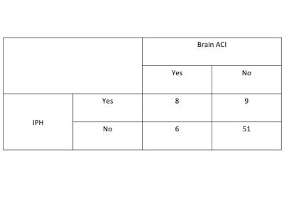
Utilizing Quantitative Measurements of Carotid Intraplaque Hemorrhage can Improve on Presence Alone in Classifying Patients with and without Acute Cerebral Infarcts - permission withheld
Li Dong, Zhaoqi Zhang, Wei Yu, Sheng Wang, Qiang Shen, Chun Yuan
Histological studies have shown that intraplaque hemorrhage (IPH) size might be important in assessing disease severity. We hypotheses that the quantitative measurements of IPH by MRI provide additional value towards classify acute cerebral infarcts (ACI) in the carotid territory by brain MRI. We found that the subjects with ACI had larger max % IPH measurements (AUC=84.7%, p=0.015) and IPH closer to the lumen (AUC=85.4%, p=0.012). Further, using the size and distance measurements simultaneously improved the AUC to 96.9%. Beyond the presence of IPH, quantitative measurements of IPH may improve the predictive value of carotid plaque imaging for future stroke.
|
|
0333.
 |
2 |
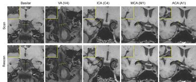
Whole-Brain Vessel Wall MR Imaging Using Inversion-Recovery Prepared SPACE: Reproducibility and Accuracy of Intracranial Artery Morphology 
Na Zhang, Fan Zhang, Zixin Deng, Qi Yang, Xiaoming Bi, Debiao Li, Xin Liu, Zhaoyang Fan
Intracranial atherosclerosis disease is one of the main causes for cerebrovascular events. A T1-weighted whole-brain vessel wall imaging method, inversion-recovery prepared SPACE (IR-SPACE), was developed at 3T demonstrating advantages over conventional SPACE. This study was aimed to determine the reproducibility of scan-rescan, intra-, and inter-observer as well as the accuracy when using the technique for morphology assessment of the intracranial vessel wall. In general, this study demonstrated excellent reproducibility and good agreement between the 3D and 2D techniques. In conclusion, IR-SPACE is a reproducible and accurate MR method for intracranial vessel wall imaging.
|
 |
0334.
 |
3 |
A Preliminary Report on Time-Resolved Coronary Vessel Wall MRI in Heart Transplant Recipients
Giulia Ginami, Jerome Yerly, Jessica Bastiaansen, Ruud van Heeswijk, Nathalie Lauriers, Juan Iglesias, Sophie Degrauwe, Andrea Zuffi, Roger Hullin, Matthias Stuber
In this study, we applied a recently developed and robust coronary vessel wall MRI method that includes a golden angle trajectory and k-t sparse SENSE to a cohort of heart transplant patients. The coronary vessel wall was visualized with high contrast in all patients and the method holds great promise for quantitative and non-invasive characterization of coronary allograft vasculopathy (CAV) in transplanted patients.
|
|
0335.
 |
4 |
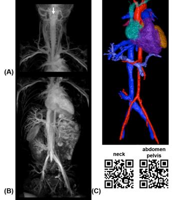
Non-Gadolinium-Contrast Relaxation-Enhanced MR Angiography in Children with an Inversion Recovery and T2-Prepared 3D mDIXON Gradient-Echo Technique: Preliminary Experience
Amber Pokorney, Jonathan Chia, Dianna Bardo, Mittun Patel, Smita Bailey, Scott Jorgensen, Deepa Biyyam, Scott Willard, Jeffrey Miller, Houchun Hu, Masami Yoneyama
We demonstrate the preliminary clinical feasibility and utility of a 3D non-Gadolinium MR angiography sequence called REACT (Relaxation-Enhanced MR Angiography without Contrast and Triggering) in 21 pediatric patients (age range: 1.7 – 16.1 years). REACT, which collectively utilizes T2-preparation, inversion recovery, and water-fat separation to suppress unwanted signals and exploit the long T1 and T2 times of unenhanced blood, is particularly beneficial in children, a population where Gadolinium administration should be minimized given recent concerns with residual intracranial Gadolinium deposition. We illustrate and compare REACT to Gadolinium-based MRA in the neck, upper and lower extremities, and the chest, abdomen, and pelvis.
|
|
0336.
 |
5 |
Ultra-High Spatiotemporal Resolution 4D Flow for Valve and Coronary Arterial Delineation
Shreyas Vasanawala, Furhawn Shah, Marcus Alley, Joseph Cheng
4D flow provides single sequence rapid protocol for assessment of blood flow, ventricular function, and anatomy in congenital heart disease. Though valve function has been well validated for regurgitant flow fraction, assessment of valve leaflet delineation has been limited by relatively low spatiotemporal resolution. Similarly, coronary origin delineation has been limited. Here, we develop an ultra-high spatiotemporal resolution 4D flow acquisition technique and assess its performance for valve and coronary delineation. 4D flow provides superior delineation of coronaries than echo, is highly likely to depict coronary origins, and is highly likely to provide good valve leaflet delineation.
|
 |
0337.
 |
6 |
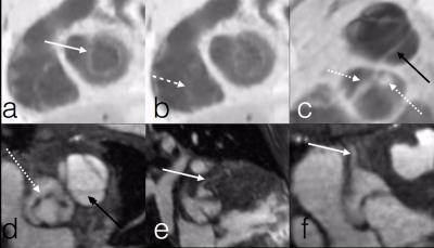
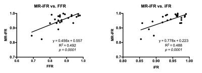
Noninvasive Functional Evaluation of Coronary Stenosis Using MR Instantaneous wave-Free Ratio (MR-iFR): Pilot patient study using invasive fractional flow reserve as a reference
Zixin Deng, Sang-Eun Lee, Zhaoyang Fan, Christopher Nguyen, Yibin Xie, Jianing Pang, Xiaoming Bi, Qi Yang, Byoung-Wook Choi, Jung-Sun Kim, Daniel Berman, Hyuk-Jae Chang, Debiao Li
In patients with suspected coronary artery disease undergoing invasive coronary angiography, approximately half has nonsignificant stenosis (stenotic lesions that may not induce ischemia), leading to frequent and unnecessary invasive procedures. Previously, we proposed a noninvasive technique for functional evaluation of coronary stenosis using PC-MRI and navier-stokes equations. In this study, we evaluated the feasibility of the technique in patients, using invasive fractional flow reserve as a reference. Good correlation was observed between noninvasive and invasive techniques with high specificity and negative-predictive-value, demonstrating the potential of the proposed technique in identifying patients with functionally nonsignificant stenosis and eliminating unnecessary invasive procedures.
|
|
0338.
 |
7 |
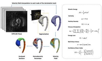
Analysis of 4D flow hemodynamics parameters in BAV patients using a finite element method
Julio Sotelo, Lydia Dux-Santoy, Andrea Guala, Jose Rodríguez-Palomares, Arturo Evangelista , Daniel Hurtado, Sergio Uribe
Bicuspid aortic valve (BAV) is one the most common cardiac defect. The progression of the defect can vary with the time and may lead to ventricular dysfunction, heart failure and death of the patient. In this work we proposed a method to obtain several hemodynamics parameters including WSS, OSI, vorticity, helicity density, viscous dissipation, energy loss and kinetic energy from 4D-flow data sets of BAV patients and healthy volunteers using a finite-element approach. We found that the viscous dissipation, helicity density, vorticity, WSS and energy loss are the most relevant hemodynamics parameters in the ascending aorta of those patients.
|
|
0339.
 |
8 |
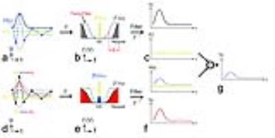
Phase-Contrast MRI with Hybrid One- and Two-sided Flow-Encoding and Velocity SPectrum SepAration (HOTSPA)
Da Wang, Jiaxin Shao, Daniel Ennis, Peng Hu
In this work, we propose a new and more efficient flow encoding and velocity calculation strategy for PC-MRI using a temporal modulation technique to double the temporal resolution or reduce the scan time by 50%. This is the first study to examine a temporal modulation strategy for an under-sampled M1-space (gradient first moment space including FC, FEx, FEy and FEz encoding steps). Our strategy can be combined with conventional acceleration techniques, i.e. parallel imaging and compressed sensing, to further shorten the scan time of PC-MRI.
|
|
0340.
 |
9 |
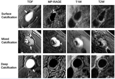
Association between Carotid Atherosclerotic Plaque Calcification and Intraplaque Hemorrhage: A High Resolution Magnetic Resonance Imaging Study
Shuo Chen, Ruolan Lin, Gaifen Liu, Rui Li, Yunjing Xue, Xihai Zhao
Carotid intraplaque hemorrhage (IPH) is associated with cardiovascular events. Calcification, frequently accompanied with IPH, may play a role in occurrence of IPH. In this study we aimed to investigate the associations between calcification characteristics and IPH in carotid plaques. Our results suggest that surface calcification and multiple calcification in carotid atherosclerotic plaques are independently associated with presence IPH. Both quantity and location of calcification may play important roles in occurrence of IPH. These findings may provide novel insights for understanding the mechanism of IPH.
|
|
0341.
 |
10 |
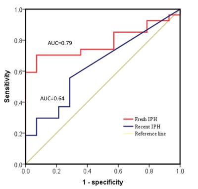
Association between Age of Intraplaque Hemorrhage and Fibrous Cap Rupture in Carotid Artery Atherosclerosis: A High Resolution Magnetic Resonance Imaging Study
Yuanyuan Cui, Xihai Zhao, Huiyu Qiao, Dongxiang Xu, Mingming Lu, Xiaoyi Chen, Lu Ma, Jianming Cai
It has been shown that presence of carotid artery intraplaque hemorrhage (IPH) is associated with fibrous cap rupture (FCR), which is one of the major causes of ischemic cerebrovascular events. However, the role of different age of IPH in occurrence of IPH remains unclear. This study investigated the association between the age of IPH and FCR in carotid arteries using MR imaging. We found that fresh IPH volume was independently associated with FCR (odds ratio: 1.826; 95% CI: 1.130-2.949; P=0.014). Compared with recent IPH, fresh IPH was a stronger predictor for FCR (area under the curve: 0.79 vs. 0.64).
|
|
0342.
 |
11 |
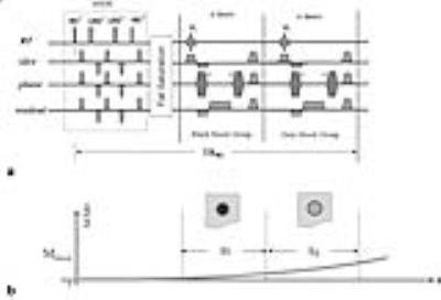
Compressed Sensing based Simultaneous Black- and Gray-blood Carotid Vessel Wall MR Imaging
Bo Li, Hao Li, Guofu Huang, Xia Qian, Wei Wang, Li Dong
We sought to achieve high-quality simultaneous black- and gray-blood MR imaging using the CS based dual contrast imaging technique proposed on 2015. The performances on the blood suppression efficiency, image quality and morphological measurements for the CS based dual-contrast imaging technique were investigated. Seven healthy volunteers and five patients with carotid artery stenosis were recruited in this study. STR, CTR were calculated. LA and WA were measured. The comparisons for the black- and gray-blood image quality and the presence of plaque calcifications were implemented. This technique provides spatially matched black- and gray- images and excellent visualization for vessel wall imaging.
|
|
0343.
 |
12 |
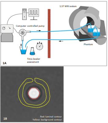
Comparison of acceleration algorithms in whole-heart 4D flow MRI for aortic and mitral valve flow assessment
Jos Westenberg, Pankaj Garg, Pieter van den Boogaard, Sven Plein
Accelerated acquisition is required to make 4D flow MRI clinically feasible. In this study, three commonly used acceleration approaches are compared. Validation of flow volume and velocity assessment is performed in phantoms and comparison against conventional 2D phase-contrast is done across the aortic and mitral valve in 25 healthy volunteers. 4D flow MRI with echo-planar-imaging shows largest in vitro error in velocity assessment, however, the bias is within clinically acceptable margins. In volunteers, 4D flow MRI with echo-planar-imaging produces most reliable quantitative results in flow volume and velocity and presents the shortest acquisition time with satisfactory image quality.
|
|
0344.
 |
13 |
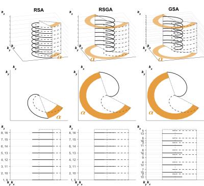
Golden Step, Golden Angle, Spiral-Cartesian Imaging for Flexible Gated Three-dimensional Angiography
Grzegorz Kowalik, Jennifer Steeden, David Atkinson, Kristian Mortensen, Vivek Muthurangu
The purpose of this study was to develop and validate a novel 3D hybrid spiral-Cartesian MR angiographic sequence that utilized a golden step, golden angle acquisition strategy to enable flexible reconstruction. We demonstrate that it is possible to acquire GSA-MRA and reconstruct it in a flexible manner (Real-time and cardiar and/or respiratory gated reconstructions). This may enable better interrogation of anatomy.
The new flexible 3D MRA provide novel ways of assessing cardiac disease.
|
|
0345.
 |
14 |
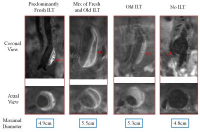
Distribution of Intraluminal Thrombus Composition in Abdominal Aortic Aneurysms by Diameter: a High Resolution MRI study
Chengcheng Zhu, Bing Tian, Joseph Leach, Qi Liu, Jianping Lu, David Saloner, Michael Hope
Intraluminal thrombus (ILT) composition as identified by MRI has been suggested to be a marker of likely abdominal aortic aneurysm (AAA) progression. However, little is known about the distribution of ILT composition across different sizes of AAAs. This study investigated the ILT composition by AAA diameter in 62 patients from two centers. We found ILT distributed differentially by size, with an increasing mix of fresh and old ILT in larger AAAs. ILT composition is a potential indicator of AAA progression. Larger, prospective studies are needed to clarify its prognostic value in managing AAA patients.
|
|
0346.
 |
15 |
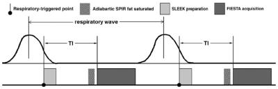
Evaluation of Portal Vein System in patients after liver transplantation by Unenhanced MR Angiography Using Spatial Labeling with Multiple Inversion Pulses Sequence and by CT portography - did not present
hao tang, daoyu hu, xiaoyan meng, zi wang, zhen li, yanchun wang
Unenhanced MR angiography with spatial labeling with multiple inversion pulses (SLEEK) is a reliable method for depicting portal vein system in patients with liver transplantation compared with computed tomographic portography (CTP) results. In a study of 20 patients who underwent liver transplantation, we found that there was excellent correlation between SLEEK and CTP in presenting the diameter of portal vein. Unenhanced MRA using SLEEK is relatively inexpensive and is not associated with renal complications. It can be as a good choice for screening portal vein system in patients with liver transplantation, especially in patients with renal insufficiency.
|
|