Poster: Body MRI Quantitative
Electronic Power Pitch Poster
Body: Breast, Chest, Abdomen, Pelvis
Monday, 24 April 2017
| Exhibition Hall |
14:45 - 15:45 |
| |
|
Plasma # |
|
0112.
 |
1 |
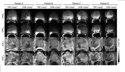
Accelerated Segmented Diffusion-Weighted Prostate Imaging for Higher Resolution, Higher Geometric Fidelity, and Multi-b Perfusion Quantification 
Pelin Ciris, Jr-yuan Chiou, Daniel Glazer, Shelley Zhang, Tzu-Cheng Chao, Bruno Madore, Stephan Maier
An accelerated multi-shot diffusion imaging scheme was developed for prostate imaging. Its robustness to regularization was evaluated. Two-fold improvement in spatial resolution and over three-fold improvement in geometric fidelity were obtained as compared to single-shot EPI, in twenty-five prostate cancer patients. In contrast to the standard protocol, which involves separate scans with high (b=1400 and 0 s/mm2) and intermediate (b=500 and 0 s/mm2) diffusion weighting, the proposed accelerated protocol extended b-factor coverage to yield an additional 8 b-factors and enabled multi-b perfusion quantification in half the scan time (5 min 43 s vs. 11 min 48 s).
|
 |
0113.
 |
2 |
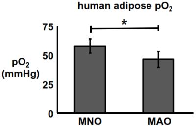
Towards validation and non-invasive interrogation of the hypoxia-driven insulin resistance hypothesis
Scott Beeman, Gordon Smith, Joel Garbow, Joseph Ackerman
Insulin resistance is a defining feature of type 2 diabetes – a disease associated with severe morbidities and mortality. Recent studies have suggested that adipose tissue hypoxia is a major common pathway to systemic insulin resistance. The goals of this work are to: (i) directly observe evidence of the hypoxia-driven insulin resistance hypothesis in human subjects via gold-standard invasive pO2 measures and (ii) establish an R1-based pO2 metric for future non-invasive studies of adipose pO2 in metabolic disease. Herein, we report initial progress towards these goals.
|
|
0114.
 |
3 |
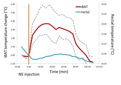
Measuring temperature in brown adipose tissue using the proton chemical shift
Clemens Diwoky, Renate Schreiber, Rudolf Zechner
Within this work an approach based on the temperature dependence of the proton resonance frequency (PRF) of water and methylene bound protons is followed to monitor in-vivo thermogenesis in the interscapular brown adipose tissue (iBAT) of mice. Measuring the change in chemical shift difference of water and fat rather than water alone, known problems of PRF-based temperature measurements such as magnetic field drift and the temperature dependence of magnetic susceptibility are circumvented. The study determines the temperature coefficient in ex-vivo iBAT tissue extracts and presents the application of the technique in measuring norepinephrine stimulated in-vivo iBAT thermogenesis.
|
|
0115.
 |
4 |
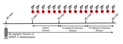
Development of a Noninvasive Beta Cell Functional Assay Using a Novel Zinc-Sensitive MRI Contrast Agent in Non-Human Primates
Catherine Hines, Veronica Clavijo-Jordan, Liza Gantert, Stacey Conarello, Christian Preihs, Sarah Chirayil, Rachel Ortiga, Shu-An Lin, Michael Klimas, A. Dean Sherry, Jeff Evelhoch
Pancreatic beta cells secrete insulin to maintain normal blood glucose levels, and the integrity and function of pancreatic beta cells have been found to be compromised in Type-1 and Type-2 diabetes. Therefore, non-invasive beta cell function measurements may provide valuable information for improving diabetes diagnostics and disease management. Currently available diabetes assays lack functional information and spatial identification of properly functioning beta cells. In this work, we introduce a new assay to assess the function and identify functional beta cells in vivo in the non-human primate pancreas non-invasively with MRI using a Gd-based zinc sensor as contrast agent.
|
|
0116.
 |
5 |
Feasibility of Estimating Placental Oxygen Metabolism in Pregnant Women $$$in$$$ $$$vivo$$$: Initial Experience
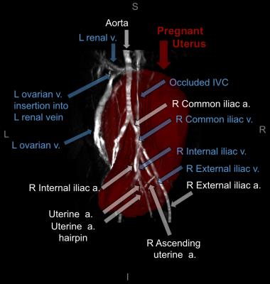
Ana Rodríguez-Soto, Michael Langham, Nadav Schwartz, Felix Wehrli
The development of methods to assess placental oxygen metabolism would allow to clinically evaluate its function. Here, we evaluated the feasibility of estimating oxygen extraction and blood flow in some abdominal and fetal draining veins. Ovarian veins appear to play an important role in draining blood from the uterus in the supine position, where flow increased from the 2nd to 3rd trimester (16.4±8.1 versus 34.3±4.1mL/min/100g). Additionally, elevation of oxygen saturation (61.6±6.6% versus 68.3±5.0%) at the umbilical vein occurred from the 2nd to 3rd trimester, potentially reflecting increased fetal oxygen demand as pregnancy progresses.
|
|
0117.
 |
6 |
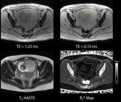
Free-breathing R2* Characterization of the Placenta During Normal Early Gestation Using a Multiecho 3D Stack-of-Radial Technique
Tess Armstrong, Dapeng Liu, Thomas Martin, Alto Stemmer, Yutaka Natsuaki, Sherin Devaskar, Carla Janzen, Teresa Chanlaw, Rinat Masamed, Daniel Margolis, Kyunghyun Sung, Holden Wu
Abnormal placental vascular development leads to ischemic-hypoxia thereby causing fetal growth restriction, preterm labor, and spontaneous abortion. Multiecho Cartesian MRI can characterize placental hypoxia by quantifying R2*, but is susceptible to motion artifacts. We have developed a new free-breathing (FB) multiecho R2* quantification technique using 3D stack-of-radial imaging (Radial). In n=16 subjects as part of an IRB-approved study, we observed an R2* range of 5 – 30s-1 at 3 T using the new FB Radial technique in the placenta during early normal gestation. Our new technique and the measured normative range of R2* may improve management of pregnancies with placental ischemic-hypoxia.
|
|
0118.
 |
7 |
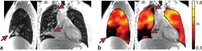
Respiratory $$$\alpha$$$-mapping of cystic fibrosis at 1.5T
Orso Pusterla, Grzegorz Bauman, Sylvia Nyilas, Philipp Madörin, Bernd Jung, Michael Ith, Enno Stranzinger, Urs Frey, Philipp Latzin, Oliver Bieri
Respiratory $$$\alpha$$$-mapping is based on native 1H multi-volumetric ultra-fast balanced steady-state free precession (ufSSFP) breath-hold imaging of the lung and provides whole lung isotropic pulmonary ventilation-related information. In this work, respiratory $$$\alpha$$$-mapping is evaluated in pediatric patients with cystic fibrosis (CF) and compared to functional lung parameters from nitrogen multiple-breath washout (N2-MBW). The percentage of respiratory $$$\alpha$$$-impairments measured with $$$\alpha$$$-mapping is strongly correlated with the lung clearance index (LCI), a parameter for global ventilation inhomogeneity.
|
|
0119.
 |
8 |
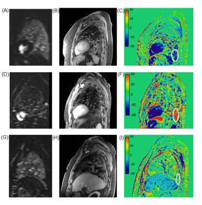
5D MRI for late enhancement dynamics in lung fibrosis
Maria Teodora Buzan, Julien Dinkel, Christopher Rank, Claus Peter Heussel, Marc Kachelrieß , Robert Grimm, Andreas Wetscherek
We analyzed the contrast agent late dynamics in fibrotic lung lesions of different severity, assessed with perfusion MRI, using a 5D reconstruction (respiratory 4D at different time points) of a 3D radial stack-of-stars spoiled gradient echo sequence with golden-angle spacing acquired in free-breathing. Idiopathic pulmonary fibrosis (IPF) shows late peak enhancement regardless of severity level and possibly no wash-out within the scan interval, while non-IPF lesions have faster time to peak and slower accumulation rate with increasing severity. The method might prove useful for a more thorough characterization of lung fibrosis burden in the context of new anti-fibrotic treatments available.
|
|
0120.
 |
9 |
Quantification of short-T2* Signal Components in the Liver using Radial 3D UTE Chemical Shift-Encoded MRI
Ante Zhu, Diego Hernando, Kevin Johnson, Scott Reeder
Recent studies have suggested the presence of a short-T2* signal component in the liver. The origin and MR properties of this signal have not been determined but have been shown to confound the liver fat quantification when using short echo times. In this work, we developed a UTE chemical shift-encoded MRI technique and a multi-component reconstruction to characterize short-T2* liver signals. A short-T2* signal fraction of 11.6±2.4% with an R2* of 2222±281s-1 was measured in seven healthy volunteers. This study demonstrated the presence of the short-T2* signal component in healthy livers and provided an initial estimate to guide future studies.
|
|
0121.
 |
10 |
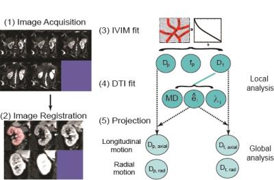
REnal Flow and Microstructure AnisotroPy (REFMAP) MRI in Normal and Peritumoral Renal Tissue
Andrea Liu, Artem Mikheev, Henry Rusinek, William Huang, Hersh Chandarana, Eric Sigmund
Using a recently developed joint intravoxel incoherent motion (IVIM)- diffusion tensor imaging (DTI) protocol for kidney evaluation, we present reproducibility analysis of its metrics in normal volunteers, as well as pilot assessments in several patients with renal masses prior to surgery. Reproducibility analysis indicates a subset of robust parameters, including structural and microcirculation markers in both cortex and medulla, for clinical application. Preliminary results in renal mass patients suggest multifactorial differences from controls, supporting the need for advanced diffusion characterization in assessing renal functional reserve.
|
|
0122.
 |
11 |
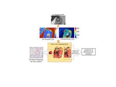
Addressing Metabolic Heterogeneity in Clear Cell Renal Cell Carcinoma with Quantitative Magnetic Resonance Imaging
Yue Zhang, Durga Udayakumar, Ling Cai, Zeping Hu, Payal Kapur, Eun-Young Kho, Andrea Pavía-Jiménez, Michael Fulkerson, Alberto DiazdeLeon, Qing Yuan, Ivan Dimitrov, Takeshi Yokoo, Jin Ye, Matthew Mitsche, Hyeonwoo Kim, Jeffrey McDonald, Yin Xi, Ananth Madhuranthakam, Robert Lenkinski, Jeffrey Cadeddu, Vitaly Margulis, James Brugarolas, Ralph Deberardinis, Ivan Pedrosa
MRI fat fraction (FF, Dixon) and % enhancement (DCE) measurements in vivo in clear cell renal cell carcinoma (ccRCC) were correlated with intracellular fat (Oil Red O), lipidomic profile (mass spectrometry), and cellular metabolomics of tissue samples isolated from anatomically co-registered locations in the same tumor. In vivo FF correlated positively with histologic fat content, spectrometric cholesterol and triglycerides; and negatively with spectrometric free fatty acids and phospholipids. ISUP grade 2 and 3 tumors exhibited marked intra-tumoral heterogeneity in FF whereas grade 4 tumors had reduced lipid accumulation. MRI-derived FF and % enhancement correlated with altered metabolic features of ccRCC.
|
|
0123.
 |
12 |
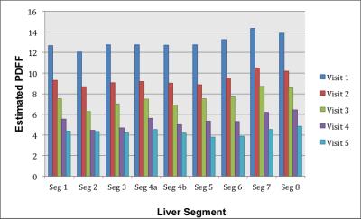
Liver Fat Reduction Following Bariatric Weight Loss Surgery is Greater in the Right Lobe of the Liver
Soudabeh Fazeli Dehkordy, Tanya Wolfson, Cheng Hong, Alexandra Schlein, Yesenia Covarrubias, Jennifer Cui, Ethan Sy, Adrija Mamidipalli, Gavin Hamilton, Scott Reeder, Claude Sirlin
As liver fat is heterogeneously distributed, longitudinal changes in liver fat may vary between liver segments. We used confounder-corrected chemical-shift-encoded MRI to examine longitudinal changes in proton density fat fraction (PDFF) of individual liver segments in obese adults following a weight-loss program comprising a very low calorie diet (VLCD) followed by bariatric weight loss surgery (WLS). We observed that changes in PDFF in the 5-month postoperative period vary across segments, with right-lobe segments having more rapid reduction in liver fat.
|
|
0124.
 |
13 |
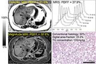
In Vivo Biochemical and Histological Validation of Proton Density Fat Fraction as a Quantitative Biomarker of Hepatic Steatosis - permission withheld
Scott Reeder, Curtis Wiens, Nathan Artz, Jeffrey Schwimmer, Rashmi Agni, Rao Watson, Tanya Wolfson, Anthony Gamst, Guilherme Campos, Santiago Horgan, Luke Funk, Garth Jacobsen, Jacob Greenberg, Alexandra Schlein, Yesenia Covarrubias, Jonathan Hooker, Michael Middleton, Gavin Hamilton, Benjamin Ratliff, Alan McMillan, Diego Hernando, Claude Sirlin
The purpose of this study was to validate complex- and magnitude-based quantitative chemical-shift encoded MRI (CSE-MRI) to quantify PDFF as an accurate biomarker of hepatic steatosis. A biopsy-MRI correlation study was performed in 95 obese subjects undergoing bariatric weight loss surgery. The results of biochemical triglyceride, conventional histological, digital histological, and MR spectroscopy analyses were used as reference standards. Strong correlations were found between MRI-PDFF measured using both complex- and magnitude-based CSE-MRI methods vs. each reference standard. This is the first cross-sectional in-vivo study validating PDFF as a biomarker of hepatic steatosis using biochemical triglyceride concentration as the reference.
|
 |
0125.
 |
14 |
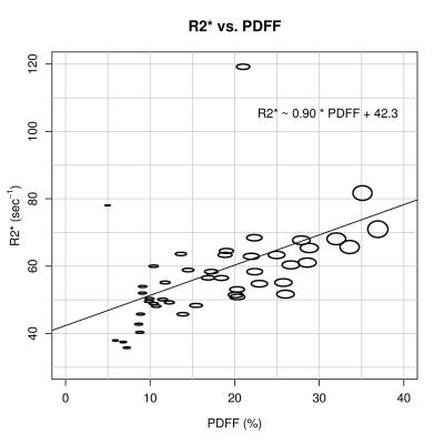
Hepatic MRI-PDFF is positively correlated with R2* across a range of fat spectral models
Cheng Hong, Adrija Mamidipalli, Jonathan Hooker, Gavin Hamilton, Tanya Wolfson, Soudabeh Fazeli Dehkordy, Michael Middleton, Scott Reeder, Rohit Loomba, Claude Sirlin
Confounder-corrected estimation of proton-density fat fraction (PDFF) concurrently estimates R2* (1/T2*), a parameter modeled to account for R2* signal decay. Although they are derived from the same mathematical model, PDFF and R2* are generally considered independent parameters. Emerging evidence, however, suggests that PDFF and R2* are positively correlated. This study confirms that PDFF and R2* are positively correlated, and this association is not a spurious result of the applied fat multipeak spectral model.
|
|
0126.
 |
15 |
Anatomical and functional deficits of the placenta identified by MRI in a rat model of preeclampsia
Emily Waters, Pamela Monahan, Chad Haney, Michael Fritsch, Thomas Meade, Kelly Mayo
Though a large percentage of poor pregnancy outcomes such as stillbirth and preterm birth are related to placental dysfunction, the gold-standard of diagnosing placental pathology remains examination of the placenta by a pathologist after delivery. We demonstrate that anatomical and functional MRI can detect features of placental pathology correlating with ex vivo measures in a rat model of pre-eclampsia.
|
|