Poster: Cancer Imaging in the Body
Electronic Power Pitch Poster
Body: Breast, Chest, Abdomen, Pelvis
Wednesday, 26 April 2017
| Exhibition Hall |
09:15 - 10:15 |
| |
|
Plasma # |
|
0660.
 |
16 |
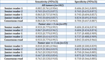
Interobserver Agreement and Diagnostic Performance of LI-RADS v2014 on contrast-enhanced MRI for non-HCC malignancies. 
Natally Horvat, Ines Nikolovski, Niamh Long, Scott Gerst, Jian Zheng, Linda Pak, Junting Zheng, Lorenzo Mannelli, Richard Do
Patients at risk for hepatocellular carcinoma (HCC) are often also at risk for intrahepatic cholangiocarcinoma (ICC) and combined hepatocellular cholangiocarcinoma (cHCC-ICC). The purpose of this study was to evaluate the sensitivity and specificity of MRI in 4 radiologists using LI-RADS v2014 and their inter-reader agreement for diagnosing non-HCC malignancies (ICC and cHCC-ICC). Applying LI-RADS v2014, we found sensitivities between 74% to 88% for the diagnosis of non-HCC malignancies with moderate to substantial interreader agreement for LI-RADS category as LR-M or non LR-M. However, lower sensitivity was seen for smaller tumors, which are known to overlap with HCC in imaging appearance.
|
 |
0661.
 |
17 |
Quantification of hepatocellular carcinoma tumor heterogeneity with multiparametric MRI
Stefanie Hectors, Mathilde Wagner, Octavia Bane, Cecilia Besa, Sara Lewis, Romain Remark, Nelson Chen, M. Isabel Fiel, Hongfa Zhu, Sacha Gnjatic, Miriam Merad, Yujin Hoshida, Bachir Taouli
We assessed tumor heterogeneity in hepatocellular carcinoma using multiparametric MRI (mpMRI) combining DWI, BOLD-MRI, TOLD-MRI and DCE-MRI measurements. Histogram characteristics (central tendency parameters mean and median and heterogeneity parameters standard deviation, kurtosis and skewness) of mpMRI data were quantified in the lesions and correlated between MRI methods and with histopathology and gene expression levels in a subset of patients. We observed that central tendency and heterogeneity parameters were largely complementary in terms of the assessed correlations. The proposed histogram analysis is therefore promising for noninvasive HCC characterization on the functional, immunohistochemical and genomics level.
|
|
0662.
 |
18 |
3D MR Elastograpy in Prediction of Tumor Capsule Formation of Hepatocellular Carcinoma (HCC) in Patients with Hepatitis B Virus Infection
Jin Wang, Hao Yang, Yong Liu, Jingbiao Chen, Tianhui Zhang, Kevin Glaser, Xin Li, Jun Chen, Yao Zhang, Qungang Shan, Bingjun He, Zhuang Kang, Yin Meng, Dzyubak Bogdan, Venkatesh SK, Ronghua Yan, Xi Long, Richard Ehman
Hepatocellular carcinoma (HCC) is one of the leading causes of cancer-related deaths around the world. Tumor capsule formation is a significant and independent predictor of survival and recurrence. We explored the potential value of HCC stiffness using 3D MR elastograpy for the prediction of tumor capsule formation. Results in 50 examinations showed that HCC stiffness has promise for the preoperative prediction of tumor capsule formation, thus providing motivation for further evaluation of HCC characteristics with MRE.
|
|
0663.
 |
19 |
Dynamic Contrast Enhanced MR Imaging of Hepatopancreatobiliary lesions in Combined use of Parallel Imaging and Compressed Sensing
Takayuki Masui, Motoyuki Katayama, Yuji Iwadate, Naoyuki Takei, Kang Wang, Kevin King, Kei Tsukamoto, Mitsuteru Tsuchiya, Yuki Hayashi, Masako Sasaki, Takahiro Yamada, Kenichi Mizuki, Harumi Sakahara, Koji Yoneyama, Yuki Takayanagi
The feasibility of dynamic Gd-contrast study using turbo LAVA with ARC and CS for evaluation of pancreatobiliary lesions was evaluated. Acceptable image quality and good temporal resolutions with selective recognition of vasculatures, and lesion detections in the liver and pancreas could be made. With this technique, dynamic contrast imaging with high temporal and spatial resolutions can cover the wide area. It takes acceptable time for imaging reconstruction with ARC and CS at clinical 3T MR unit.
|
|
0664.
 |
20 |
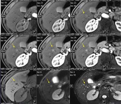
Detection and measurement of neuroendocrine tumors liver metastases using Gd-EOB-DTPA enhanced MRI: comparison between multiple arterial phases, hepatobiliary phase, and DWI
Jia Xu, Xuan Wang, Hua dan Xue, Shi tian Wang, Hui Liu, Zheng yu Jin
The detection and evaluation of liver metastasis (LM) is important in initial staging and follow-up examinations of neuroendocrine tumors patients. Our aim is to compare different sequences including multiple arterial phases (MA), hepatobiliary phase (HA), diffusing-weighted imaging (DWI), and T2WI in Gd-EOB-DTPA-enhanced MRI, to find which sequence is better for detecting LM and which is better for size measurement. MA using CDT-VIBE were superior for the detection of small lesions. HBP imaging shows better repeatability in size measuring and may be a better choice for lesion size measurement during follow-up.
|
|
0665.
 |
21 |
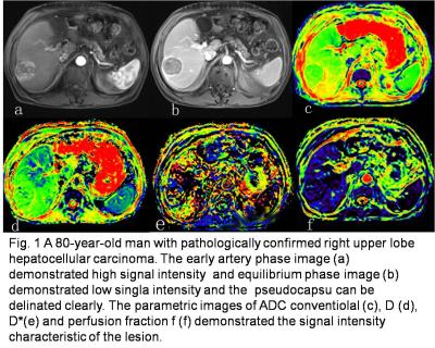
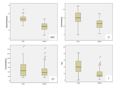
Integrated slice-specific shimming (iShim) intravoxel incoherent motion diffusion-weighted MR imaging in the liver: the value of differential diagnosis between benign and malignant hepatic tumors - did not present
Hongxia Wang, Qingbo Li, Bin Wang, Qinglei Shi, Yan Feng, Xingyue Jiang, Peigong Zhang
This study investigated the value of ADC value, D, Dstar, ADC500~800 acquired with prototype iShim sequence at 3T. All those parameters demonstrated high diagnostic capacity in distinguishing benign and malignant hepatic lesions and in distinguishing different types of malignant hepatic lesions, among which ADC500~800 demonstrated the best diagnostic performance , which may have great value in clinical practice in future.
|
|
0666.
 |
22 |
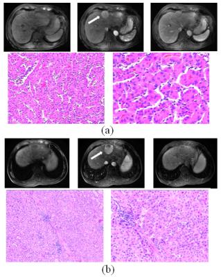
Quantitative texture feature to predict Microscopic portal vein invasion of Hepatocellular carcinoma with contrast-enhanced MR images
Wu Zhou, Qiyao Wang, Su Yao, Guangyi Wang, Zaiyi Liu, Changhong Liang, Lijuan Zhang
Portal vein invasion of Hepatocellular carcinoma (HCC) is a well-known prognostic factor for patients after hepatic resection or liver transplant. Typical risk factors of microscopic venous infiltration (MVI) include large tumor size, multifocality, poor histological grade and non-smooth tumor margins. In this work, we adopt a new techniue for quantitative texture feature Gray-level-run-length nonuniformity (GLRLN) to predict MVI of HCC based on the contrast-enhanced magnetic resonance images (CE-MRI). The present study showed that the proposed texture feature GLRLN in the portal vein phase of CE-MRI yielded best performance as compared with typical markers for the prediction of MVI.
|
|
0667.
 |
23 |
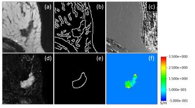
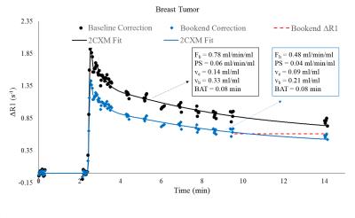
Conductivity of different malignancy grades of invasive ductal carcinomas and fibroadenomas
Ulrich Katscher, Mussa Gagiyev, Naoko Mori, Keiko Tsuchiya, Jochen Keupp, Hiroyuki Abe
In ex vivo studies, breast tumors exhibit a significantly altered electric conductivity, measurable in vivo using “Electric Properties Tomography”. A significant conductivity difference was reported between benign and malignant breast tumors and between invasive and in situ carcinomas. This study tested a correlation between conductivity and WHO grade of invasive ductal carcinomas (IDCs) and benign fibroadenomas. A clear conductivity difference was found between IDC grade 1 and grade 2, as well as between IDC grade 1 and grade 3. No clear difference was found between fibroadenoma and IDC grade 1, as well as between IDC grade 2 and grade 3.
|
|
0668.
 |
24 |
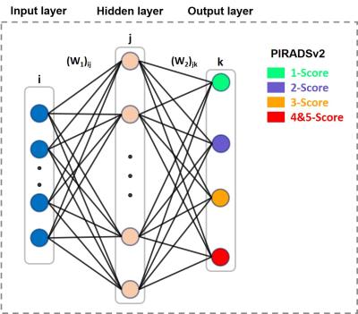
A computer aided diagnosis (CAD) scoring tool: prostate cancer risk evaluation with PI-RADS v2 Guidelines
Lian Ding, Ge Gao, Yajing Zhang, Chengyan Wang, Jue Zhang, Xiaoying Wang, Jing Fang
The second version of the Prostate Imaging Reporting and Data System (PIRADSv2) indicates the likelihood of a clinically significant cancer with a simplified 5-point scale. To assist radiologists in making diagnostic decisions consistent with the PIRADSv2, we proposed a machine learning-based computer aided diagnosis (CAD) scoring tool of prostate cancer risk evaluation by combining apparent diffusion coefficient (ADC) and T2-weighted MRI-based features. The tool could provide a malignancy prediction color map of 5 scores. The statistical results of the total score test for 130 patients between radiologist graded and the CAD tool showed high accuracy and AUC.
|
 |
0669.
 |
25 |
Deep learning to improve prostate cancer diagnosis
Nikolaos Dikaios, Edward Johnston, Harbir Sidhu, Mrishta Appaya, Alex Freeman, Hashim Ahmed, Shonit Punwani
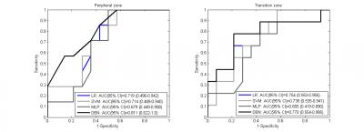
Multiparametric MRI (mp-MRI) can localize tumour within the prostate, guide biopsy, and assess disease burden. Nevertheless, mp-MRI itself remains imperfect. Almost 40% of mp-MRI studies are reported as indeterminate for significant cancer. An indeterminate mp-MRI confers no patient benefit, such patients require either repeat interval mp-MRI and/or subsequent biopsy. There remains a clear unmet need to improve diagnostic imaging over and above standard mp-MRI protocols. Previous work has developed zone specific logistic regression (LR) models for the determination of significant cancer (any cancer-core-length (CCL) with Gleason>3+3 or any grade with CCL≥4 mm) in the peripheral (PZ) and the transition zone (TZ) based on quantitative mp-MRI parameters following MR imaging. This work proposes a state-of-art deep learning method to detect prostate cancer both in the PZ and the TZ. The proposed model is trained on a cohort of patients imaged at a 3T scanner, and validated on independent cohort of patients imaged at a 1.5T scanner. The performance of the model was compared with LR, Support Vector Machines and traditional Neural Networks.
|
|
0670.
 |
26 |
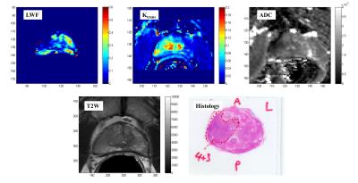
Comparing the Diagnostic Accuracy of Luminal Water Imaging with Diffusion-Weighted and Dynamic Contrast-Enhanced MRI for Evaluation of Prostate Cancer.
Shirin Sabouri, Silvia Chang, Richard Savdie, Edward Jones, S. Larry Goldenberg, Peter Black, Piotr Kozlowski
Luminal water imaging (LWI) is a newly developed MRI technique that has shown promising results for diagnosing and grading of prostate cancer. Incorporating LWI in clinical settings requires a profound investigation on its diagnostic accuracy by comparing it to existing clinical protocols. In this pilot study we have acquired and analyzed LWI, DWI, and DCE-MRI data from 15 patients to compare the diagnostic accuracy of LWI with the other two techniques. Our results show that LWI provides equal or higher accuracy in detection of tumors and better correlation with tumor grade compared to DWI and DCE-MRI.
|
|
0671.
 |
27 |
Novel Informatics Modeling of Magnetic Resonance Imaging Metrics for Characterizing Prostate Lesions with Pathology Correlation. - permission withheld
Katarzyna Macura, Vishwa Parekh, Seyed Saeid , Michael Jacobs
Precision medicine is increasingly being used in radiological applications. Advanced machine learning coupled with informatics modeling of clinical and radiological variables can provide the foundation to relate to precision medicine in patients with prostate cancer. We test our modeling using multiparametric prostate MR imaging (mpMRI) and MR-guided prostate biopsy with magnetic resonance-transrectal ultrasound (MR-TRUS) fusion to correlate the imaging features with histopathological results.
|
|
0672.
 |
28 |
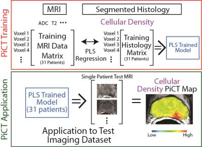
Predictive Cytological Topography (PiCT): a Radiopathomics Approach to Mapping Prostate Cancer Cellularity
Amy Kaczmarowski, Kenneth Iczkowski, Sarah Hurrell, Sean McGarry, Kenneth Jacobsohn, William Hall, Mark Hohenwalter, William See, Peter LaViolette
This study combines radiographic images and pathological microscopy with machine learning to generate predictive maps of pathological features (i.e. new contrasts) based on segmented histological features. Predictive cytological topography (PiCT) maps of cellularity were utilized to detect additional pathologically confirmed high-grade prostate cancer tumors missed by radiologists.
|
|
0673.
 |
29 |
Estimating breast tumor blood flow and blood volume using MRI: DCE vs IVIM
Leonidas Georgiou, Nisha Sharma, Daniel Wilson, David Buckley
There is an increasing interest on whether IVIM parameters can be used as surrogates of perfusion and thus complement information on tumor microstructure. DCE-MRI techniques on the other hand have been widely used to characterise perfusion and hence offer the opportunity to test this hypothesis. In this study, we use high temporal resolution DCE-MRI and IVIM-DWI techniques to monitor patients with advanced breast cancer at various stages of their neoadjuvant chemotherapy, and assess the physiological relationship between perfusion and IVIM parameters.
|
|
0674.
 |
30 |
Discrimination of Malignant versus Benign Mediastinal Lymph Nodes Using Diffusion MRI with An IVIM Analysis
Li-Ping Qi, Wan-Pu Yan, Ke-Neng Chen, Zheng Zhong, Kejia Cai, Xiao-Ting Li, Ying-Shi Sun, Xiaohong Zhou
Noninvasive discrimination of mediastinal lymph nodes (MLN) is crucial for management of cancer patients such as those with non-small cell lung carcinoma. This study aims at investigating the value of IVIM diffusion parameters (D, D*, and f) as well as ADC for discrimination of malignant and benign MLN. A total of 91 MLN from 35 patients with histopathologic proven lesions were studied. D and f of malignant mediastinal lymph nodes were significantly different from those in the benign ones. The combination of f and D produced the highest diagnostic performance as evaluated by a receiver operating characteristic (ROC) analysis.
|
|