Poster: Marching on Musculoskeletal
Electronic Power Pitch Poster
Musculoskeletal
Wednesday, 26 April 2017
| Exhibition Hall |
09:15 - 10:15 |
| |
|
Plasma # |
|
0645.
 |
1 |
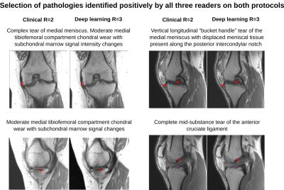
Accelerated knee imaging using a deep learning based reconstruction 
Florian Knoll, Kerstin Hammernik, Elisabeth Garwood, Anna Hirschmann, Leon Rybak, Mary Bruno, Tobias Block, James Babb, Thomas Pock, Daniel Sodickson, Michael Recht
The goal of this study is to determine the diagnostic accuracy and image quality of a recently proposed deep learning based image reconstruction for accelerated MR examination of the knee. 25 prospectively accelerated cases were evaluated by three readers and show excellent concordance to the current clinical gold standard in identification of internal derangement.
|
|
0646.
 |
2 |
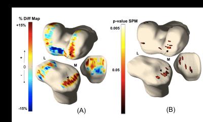
Anterior Tibial Translation Following ACL Reconstruction is Associated with Postsurgical Cartilage Matrix Changes.
Alan Li, Valentina Pedoia , Keiko Amano, Jonathan Ochoa, Qi Li , Benjamin Ma, Xiaojuan Li
Biomechanical abnormalities and accelerated cartilage matrix changes are commonly seen following ACL injury; however, the association between the two remains unclear. The purpose of this study was to analyze the relationship between altered joint kinematics and long term cartilage health. Utilizing voxel based relaxometry with T1p and T2 mapping sequences, in conjuction with MRI kinematics of the tibia and femur bone, allowed for the assessment of local cartilage matrix changes 2 years following ACL reconstruction. Notably, anterior translation of the tibia in the injured knee was associated with greater cartilage degeneration in the medial femoral and tibial cartilage.
|
 |
0647.
 |
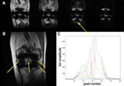
3 |
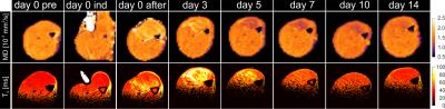
Longitudinal characterization of deformation-induced skeletal muscle damage by T2-mapping, DWI and MRE - permission withheld
Jules L. Nelissen, Willeke A. Traa, Larry de Graaf, Cees W. J. Oomens, Jurgen H. Runge, Ralph Sinkus, Klaas Nicolay, Aart J. Nederveen, Martijn Froeling, Gustav J. Strijkers
Skeletal muscle injury is often accompanied by fibrosis, fatty infiltration, and edema. There is great need for imaging readouts to detect and quantify such compositional changes, which would aid understanding and greatly assist in the development of emerging therapies. The goal of this work was to use a multi-modality approach, combining magnetic resonance elastography (MRE; muscle stiffness, fibrosis) with diffusion-weighted imaging (DWI; myocyte integrity) and T2-mapping (edema, inflammation) to provide a comprehensive assessment of muscle injury development and regeneration. The multi-modality assessment provided differential readouts of the deformation-induced muscle injury development and regeneration process.
|
|
0648.
 |
4 |
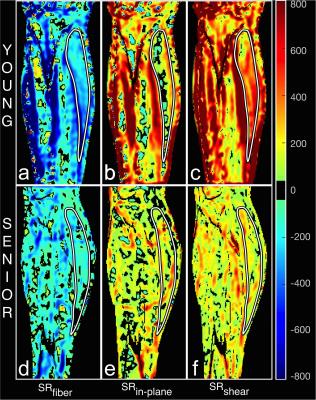
Age Related Differences in Shear Strain in Medial Gastrocnemius: Implications for Lateral Transmission of Force
Vadim Malis, Usha Sinha, Robert Csapo, Shantanu Sinha
The disproportionate loss of muscle force in comparison to loss of muscle mass with age remains unexplained. Recent studies indicate that the remodeling of the extracellular matrix (ECM) may disrupt lateral force transmission pathways mediated by the ECM. Shear strain is the mechanism that supports lateral transmission of force. We quantified shear strain in muscle from the strain rate tensor derived from velocity encoded phase contrast dynamic images of the in-vivo human calf muscle under isometric contractions. The maximal shear strain was significantly lower in the older cohort compared to the younger cohort which potentially identifies that lateral force transmission decreases with age.
|
|
0649.
 |
5 |
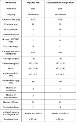
Metal Artifact Reduction MRI for the Assessment of the Rotational Alignment Knee Arthroplasty Implants: Compressed Sensing SEMAC TSE versus High-Bandwidth TSE - permission withheld
Filippo Del Grande, Benjamin Fritz, Satre Stuelke, Steven Stern, Susanne Bensler, Jan Fritz
Malrotation of knee arthroplasty implants is a source of pain following total knee arthroplasty and maybe measured on metal artifact reduction MR images; however, it is unclear which technique is best. Therefore, we compared the reliability, repeatability, and precision of rotational alignment measurements that were obtained on optimized high-bandwidth and compressed sensing SEMAC TSE MR images and their relationships with symptoms. CS-SEMAC TSE MRI afforded the highest reliability, repeatability and precision of rotational alignment measurements, which correlate best with symptoms. CS-SEMAC TSE MRI may be preferred over high-BW TSE for rotational alignment measurements of knee arthroplasty implants.
|
|
0650.
 |
6 |
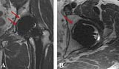
Ability of MAVRIC MRI to Predict Component Loosening in Total Hip Arthroplasty
Alissa Burge, Gabrielle Konin, Jennifer Berkowitz, Matthew Koff, Douglas Padgett, Hollis Potter
Improved metal reduction techniques for conventional MRI pulse sequences and the addition of MAVRIC (multiacquisition variable resonance image combination) have been established as useful in assessing component osteolysis and synovial reactions. The purpose of this retrospective study was to determine the utility of MRI in assessing loosening of total hip arthroplasty in a cohort of patients using surgical confirmation of loosening as the gold standard. Our results show that loosening can be predicted with high sensitivity and specificity; however, intraoperative variability in assessing loosening and the overall low frequency of implant loosening are limiting factors.
|
|
0651.
 |
7 |
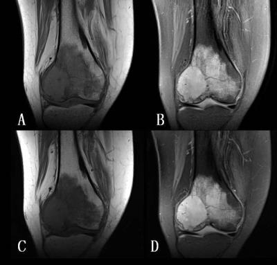
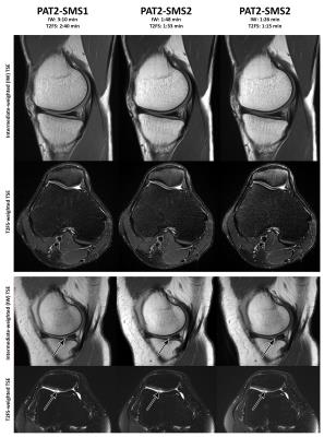
Simultaneous multi-slice TSE for clinical MR Imaging of lesions in the knee
Xiaona Li, Zhigang Peng, Yi Sun, Panli Zuo, Dingxin Wang, Jianling Cui
To introduce simultaneous multiple slices (SMS) turbo spin echo (TSE) and to evaluate its image quality and diagnostic accuracy for lesions in the knee. Participants were examined by SMS and routine TSE sequences. Both sequences were evaluated by three radiologists with subjective and objective scores in T1- and PD-weighted images. The diagnostic value in lesions was evaluated. SMS requires less scan time and offers similar imaging quality and diagnostic rate compared to routine TSE sequence. SMS is a valuable technique for MR examination of the knee.
|
|
0652.
 |
8 |
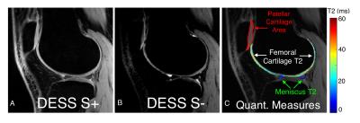
Simultaneous T2 Relaxometry and Morphometry of Cartilage and Meniscus with Double-Echo in Steady-State in Five Minutes
Akshay Chaudhari, Marianne Black, Bragi Sveinsson, Garry Gold, Brian Hargreaves
Quantitative MRI parameters such as T2 relaxation times and morphometry are potential biomarkers for tracking spatial and temporal changes in musculoskeletal diseases and injuries. While cartilage has been widely studied, the short-T2 of meniscus makes it challenging to quantify in short scan times. In this study, we optimized the double-echo steady-state (DESS) pulse sequence to produce high-resolution and high signal-to-noise ratio images in five minutes. We compared the relaxometry and morphometry measures against established methods and found no significant differences. This suggests that a five-minute DESS scan could be used for simultaneous relaxometry and morphometry of the cartilage and meniscus.
|
|
0653.
 |
9 |
Soft Tissue Tumors: Use of Intravoxel Incoherent Motion MR Imaging for Assessment of Diffusion and Perfusion for the Differentiation of Benign from Malignant Tumors
Haijun Wu, Changhong Liang
The apparent diffusion coefficient (ADC) had better diagnostic performance than other Intravoxel incoherent motion (IVIM)-derived parameters did for differentiating malignancies from benign soft-tissue tumors. The f values of intermediate soft-tissue tumors are significantly lower than those of benign and malignant soft-tissue tumors. The combination of ADC and f values is significantly better than other IVIM parameters at differentiating soft-tissue tumors.
|
|
0654.
 |
10 |
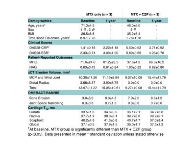
1-year Follow-Up of T1? for Assessing Radiocarpal Cartilage Matrix Changes after Anti-TNF treatment for Rheumatoid Arthritis: Preliminary Results
Eric Ku, Valentina Pedoia, Matthew Tanaka, Hyo Jin Choi, Ursula Heilmeier, Andrew Burghardt, Jonathan Graf, John Imboden, Thomas Link, Xiaojuan Li
MR T1ρ is used to investigate biochemical changes in cartilage composition, but few studies have looked at its application in radiocarpal wrist cartilage. In this study, we assess 1-year changes in T1ρ following rheumatoid arthritis treatment and its relationship with changes in clinical disease activity scores and patient-reported outcomes. Changes between 1-year and baseline T1ρ values correlated significantly with disease activity score changes and approached significance with patient-reported outcome changes during the same period. Changes in T1ρ values at 3-months also correlated significantly with 1-year changes in metacarpophalangeal and wrist bone erosion volume measured with high-resolution peripheral quantitative CT (HR-pQCT).
|
|
0655.
 |
11 |
Simultaneous Multi-Slice Accelerated High Resolution MRI of the Knee: Comparison with In-plane Parallel Imaging Acceleration - permission withheld
Jan Fritz, Benjamin Fritz, Jialu Zhang, Dharmdev Joshi, Gaurav Thawait, Li Pan, Dingxin Wang
Simultaneous multi-slice acceleration techniques excite, acquire, and reconstruct multiple slices simultaneously and provide the potential to create substantially accelerated clinical TSE protocols with similar spatial and contrast resolution to current TSE protocols. We quantified the signal-to-noise and contrast-to-noise ratios of various combinations of parallel and simultaneous multi-slice acceleration and compared two 4-fold accelerated TSE protocols against a clinical 2-fold accelerated TSE standard. Our results demonstrate that 4-fold TSE acceleration enables 43-60% shorter acquisition times with similar image quality, structural visibility and observer satisfaction than standard parallel imaging acceleration.
|
|
0656.
 |
12 |
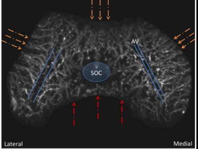
New insights into the predilection sites of Juvenile Osteochondritis Dissecans using Quantitative Susceptibility Mapping
Jutta Ellermann, Casey P Johnson, Luning Wang, Ferenc Toth, Kevin Shea, Cathy Carlson, Mikko J Nissi
The unique utilization of tissue-inherent MRI contrast using QSM for depicting the intraepiphyseal vascular supply provides high resolution and accuracy without using exogenous contrast making it a noninvasive tool for future in vivo studies. A better understanding of the development of the epiphyseal cartilage vascularity during skeletal maturation may shed light on the etiology of many developmental diseases, such as Juvenile Osteochondritis Dissecans that are precursors to osteoarthritis.
|
|
0657.
 |
13 |
Pile up correction for 3D-Multi Spectral Imaging using Gaussian Spectral Modeling and Bin Expansion
S Kaushik, Kevin Koch
The difficulty of imaging around metal implants has been overcome using pulse sequences such as MAVRIC SL and SEMAC. While the images are largely artifact-free, subtle intensity fluctuations, or ‘pile up’ artifacts still remain. The post-processing approach presented here uses information extracted from a spectral-domain model to identify and correct local fluctuations in image intensity. In these regions, the spectral bins are expanded using a moving average filter. When the expanded bins are combined, the pile up is significantly reduced. This pile up correction can improve the aesthetic quality of the images, and significantly improve their diagnostic ability.
|
 |
0658.
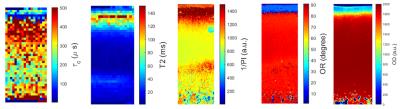 |
14 |
Correlation Time Mapping of Articular Cartilage: correlation with tissue composition and structure
Hassaan Elsayed, Stefan Zbyn, Mikko Nissi, Jari Rautiainen, Matti Hanni, Miika Nieminen
Correlation time ($$$\tau_c$$$) is a physical property of the tissue that describes the dynamics of water molecules in their microenvironment. In this study, $$$\tau_c$$$ and T2 maps were obtained from enzymatically digested bovine samples. To evaluate the relationship between $$$\tau_c$$$and macromolecular components of cartilage, depth-wise $$$\tau_c$$$ profiles were correlated with quantitative histological measurements. The same comparison was performed for T2 data. Our results suggest that $$$\tau_c$$$ is sensitive to the laminar architecture of cartilage and to the proteoglycan content in the radial zone. $$$\tau_c$$$ provides complementary information to conventional T2 mapping.
|
|
0659.
 |
15 |
Correlation of 7T gagCEST MRI with Electromechanical and Biochemical Properties of Femoral Articular Cartilage
Sander Brinkhof, Razmara Nizak, Sotcheadt Sim, Vitaliy Khlebnikov, Dennis Klomp, Daniel Saris
The purpose of this study was to validate the 7T 3D gagCEST measurements obtained in patients in vivo, using correlation of MRI GAG values with electromechanical mapping of the articular cartilage and biochemical analyses. Five patients were scanned before their total knee replacement, after which the extracted cartilage samples were used for electromechanical mapping and biochemical analyses. GAG content as determined by gagCEST MRI shows to be significantly correlated with biochemically measured GAG dry weight and with electromechanical mapping. This work shows that the electromechanical properties of cartilage are correlated with gagCEST MRI values.
|
|