Poster: New Molecular & Metabolic Imaging Approaches
Electronic Power Pitch Poster
Molecular Imaging
Tuesday, 25 April 2017
| Exhibition Hall |
17:15 - 18:15 |
| |
|
Plasma # |
|
0555.
 |
16 |
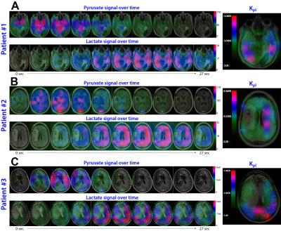
Dynamic Hyperpolarized 13C Metabolic Imaging of Patients with Brain Tumors 
Ilwoo Park, Peder Larson, Jeremy Gordon, Lucas Carvajal, Hsin-Yu Chen, Mark VanCriekinge, Robert Bok, Jason Crane, Adam Elkhaled, Joanna Phillips, James Slater, Marcus Ferrone, John Kurhanewicz, Dan Vigneron, Susan Chang, Sarah Nelson
Dynamic 13C data were acquired following injection of hyperpolarized [1-13C]pyruvate from 3 patients previously diagnosed with glioblastoma. Pyruvate, lactate and bicarbonate signal with high SNR were detected in human brain. Lactate/Pyruvate appeared to be relatively high in the contra-lateral, normal appearing brain. In contrast, tumor regions produced higher Lactate/Bicarbonate than contra-lateral brain. The contrast-enhancing lesion of one patient, who underwent surgical resection shortly after 13C imaging due to suspected recurrence, produced a relatively low level of Lactate/Pyruvate compared to contra-lateral brain and Lactate/Bicarbonate similar to the value in contra-lateral brain. Subsequent biopsy of the contrast-enhancing lesion indicated treatment effect.
|
|
0556.
 |
17 |
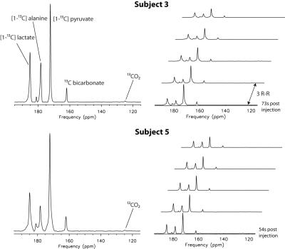
Hyperpolarized 13C MRS of the Human Heart
Albert Chen, Justin Lau, Benjamin Geraghty, William Perks, Idan Roifman, Graham Wright, Kim Connelly, Charles Cunningham
The feasibility of acquiring hyperpolarized 13C images from human hearts following injections of HP [1-13C]pyruvate solution has been recently demonstrated. Following the rapid multi-slice 13C imaging acquisition, 13C spectroscopic data were acquired from the whole heart to detect any residual hyperpolarized magnetization. 13C pyruvate, lactate, alanine and bicarbonate were observed in all 5 subjects. 13CO2 was also detected in two of the datasets. Good inter-subject consistency in the observed metabolite ratios from these spectra demonstrated the potential for changes in cardiac metabolism due to disease to be interrogated with simple whole-heart spectroscopy.
|
|
0557.
 |
18 |
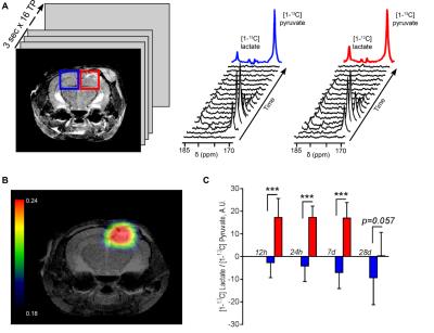
In vivo metabolic imaging of neuroinflammation following traumatic brain injury using hyperpolarized [1-13C] pyruvate
Caroline Guglielmetti, Austin Chou, Karen Krukowski, Maria Serena Paladini, Lara-Kirstie Riparip, Susanna Rosi, Myriam Chaumeil
This study demonstrates that metabolic imaging of hyperpolarized [1-13C] pyruvate can detect increased hyperpolarized lactate production in vivo in a preclinical model of Traumatic Brain Injury. Correlative assays further demonstrate that this increased lactate is linked to the presence of pro-inflammatory macrophages that upregulate pyruvate dehydrogenase kinase 1, subsequently leading to regional pyruvate dehydrogenase inhibition. Metabolic imaging of hyperpolarized [1-13C] pyruvate thus has great potential to provide a novel tool for in vivo detection of neuroinflammation.
|
|
0558.
 |
19 |
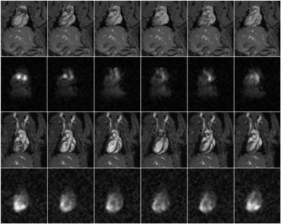
Toward Dynamic 3D Cardiac Perfusion Imaging Using bSSFP and Hyperpolarized tert-Butanol
Timothy Pagliaro, Gopal Varma, Li Zhao, David Alsop, Aaron Grant
Perfusion imaging is a promising application for hyperpolarized tracers, as they provide high signal with no endogenous background. Hyperpolarized 13C labeled tert-butanol is a perfusion agent with long T1 and T2 relaxation times in vivo. Moreover, because it diffuses freely through tissue, bolus injections of tert-butanol are largely extracted from the vasculature on a first pass, and the residence time in tissue is on the order of tens of seconds. This provides a long time frame for dynamic imaging. Here we demonstrate the feasibility of time-resolved 3D cardiac perfusion imaging in rats.
|
|
0559.
 |
20 |
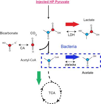
Detection of Bacteria-specific metabolism using hyperpolarized 13C pyruvate
Renuka Sriram, Jinny Sun, Javier Villanueva-Meyer, Justin DesLos Santos, Christopher Mutch, Oren Rosenberg, Mark Van Criekinge, John Kurhanewicz, David Wilson, Michael Ohliger
Bacterial infection is a major health problem, with high morbidity and mortality. Current imaging techniques have limited ability to differentiate infection from either tumor or sterile inflammation, and invasive tissue sampling is frequently required. There is currently no clinically-available non-invasive method to directly detect living bacteria in vivo. We describe a method for detecting bacteria specific metabolism using hyperpolarized 13C pyruvic acid.
|
|
0560.
 |
21 |
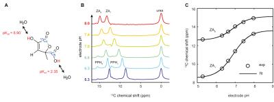
In vivo pH imaging using hyperpolarized 13C-labelled zymonic acid - permission withheld
Stephan Duewel, Christian Hundshammer, Malte Gersch, Benedikt Feuerecker, Axel Haase, Steffen Glaser, Markus Schwaiger, Franz Schilling
Pathologies which overrule natural pH regulatory mechanisms such as ischemia, inflammation or tumors can trigger local pH changes in the human body. Currently, clinical routine measurement of extracellular pH is limited to measuring systemic blood pH. We synthesized, characterized, calibrated and applied [1,5-13C2]zymonic acid (ZA) as a novel hyperpolarized 13C pH biosensor. With its demonstrated features both in vitro and in vivo (bladder, healthy kidney, Mat B III adenocarcinoma), we believe that ZA could help localize and quantify regions with aberrant acid-base balance, allowing for improved understanding, diagnostics and therapy of common diseases.
|
|
0561.
 |
22 |
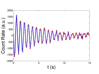
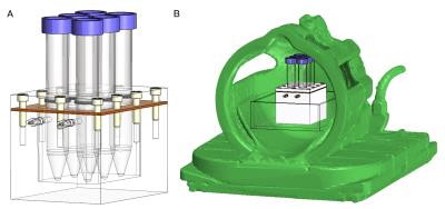
A new method for measuring T$$$_{1\rho}$$$ of hyperpolarized radioactive isotopes using gamma rays
Yuan Zheng, Gordon Cates, William Tobias, G. Miller
We have developed a novel technique that combines MR and gamma detection for measuring T1ρ of radioactive isotopes. Gamma-ray emission is anisotropic with respect to the polarization direction of hyperpolarized radioactive nuclei (spin>1/2). When the spins are flipped into the transverse plane and subjected to a spin-locking field, the emission anisotropy also precesses and gamma detectors in the transverse plane will observe an oscillating count rate. Decay of the oscillation amplitude during SL can be used to fit T1ρ. This technique also vastly reduces the number of spins needed for a meaningful measurement since gamma-ray detection is highly efficient.
|
 |
0562.
 |
23 |
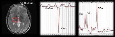
Occupational Manganese Exposure: Reversibility of Increased GABA Levels and Brain Mn Accumulation
David Edmondson, Ruoyun Ma, Chien-Lin Yeh, S. Zauber, Sandy Snyder, Eric Ward, Ulrike Dydak
While manganese (Mn) is known in the pre-clinical community as a T1 contrast agent, it is also notorious as a neurotoxin that can cause irreversible parkinsonian symptoms at high enough exposure levels. In an occupational setting, workers are exposed to Mn through processes such as welding, smelting, and other metalwork. As the workload changes over time, so does the level of exposure. Using MRI and MRS, effects of exposure such as elevated thalamic GABA levels and brain Mn deposition can be detected and show evidence of reversibility. This may help identifying meaningful no-observed-adverse-effect levels (NOAEL) as used in occupational settings.
|
|
0563.
 |
24 |
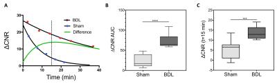
CM101: an optimized MR probe targeting type I collagen for detection of liver fibrosis
Christian Farrar, Richard Kennan, Eric Gale, Ian Ramsay, Ricard Masia, Gunisha Arora, Kailyn Looby, Lan Wei, Michelle Bunzel, Chunlian Zhang, Yonghua Zhu, Taro Akiyama, Michael Klimas, Shirly Pinto, Himashinie Diyabalanage, Valerie Humblet, Bryan Fuchs, Peter Caravan
Recent molecular MR approaches targeting collagen demonstrated the promise of noninvasive detection and staging of liver fibrosis and monitoring treatment response, but the molecular probe used was not suitable for clinical translation due to the low stability of the Gd chelator chosen. CM-101 is a new peptide based probe using the highly stable Gd-DOTA chelate that is rapidly eliminated from plasma intact into the urine and shows no sign of Gd accumulation. CM-101 robustly detected liver fibrosis in a bile duct ligation model in rats and in a CCl4 mouse model.
|
|
0564.
 |
25 |
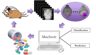
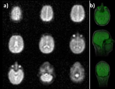
A High Throughput, MEMRI-Based Imaging Pipeline to Study Mouse Models of Sporadic Human Cancer
Harikrishna Rallapalli, I-Li Tan, Alexandre Wojcinski, Alexandra Joyner, Daniel Turnbull
A high-throughput imaging pipeline is presented to quantify the heterogeneity in longitudinal disease progression in mouse models of human brain cancer and to test the efficacy of novel anti-cancer therapeutics in accurate mouse models of sporadic human cancer.
|
|
0565.
 |
26 |
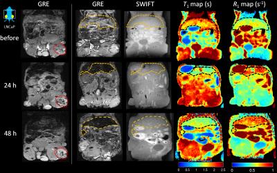
In vivo quantification of IONP-labeled PAR T-cells using positive contrast MRI
Jinjin Zhang, Sidath Kumarapperuma, Qi Shao, Lakmal Kotelawala, John Bischof, Carston Wagner, Michael Garwood
Immunotherapies have received increasing attention as novel therapeutics for the treatment of cancer and autoimmune disease. In this study, IONP labeled PAR T-cells were tracked and quantified in vivo using the SWIFT sequence for positive contrast imaging and T1 mapping. The longitudinal relaxation rate constant (R1) showed a linear dependence on the cell density in vitro and thus was used to quantify T cell density in vivo in liver. These preliminary results demonstrate how positive contrast from an ultra-short T2 sensitive sequence can provide a tool to quantify the bio-distribution of T cells.
|
|
0566.
 |
27 |
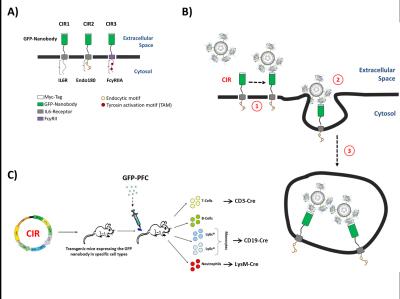
A Unique ´Cargo Internalization Receptor (CIR)´ System for In Vivo Tracking of Individual Cell Populations by 19F MRI
Pascal Bouvain, Paul Baran, Tuba Güden-Silber, Sebastian Temme, Jens Moll, Doreen Floss, Christoph Grapentin, Jürgen Scheller, Ulrich Flögel
Synopsis: We demonstrate the capability of unique ‘cargo internalization receptors’ (CIR) for specific cell tracking. A nanobody for green fluorescent protein (GFP) was used to engineer surface receptors which undergo rapid internalization after GFP binding. For 19F MR visibility, the GFP carrier was equipped with perfluorocarbon (PFC) contrast cargo. PFC cargo uptake after CIR transfection was verified by flow cytometry, confocal microscopy, and 1H/19F MRI. The results show that this approach can be successfully used for targeted contrast agent internalization, which can be extended to an cell-specific CIR expression in transgenic mice for in vivo cell tracking by 19F MRI.
|
|
0567.
 |
28 |
Characterization of Gd-DOTA-APC, a novel cancer-targeting MRI contrast agent
Christina Brunnquell, Ray Zhang, Benjamin Cox, Anatoly Pinchuk, Alan McMillan, Jamey Weichert
In this work we characterize the relaxivity, chemical stability, and tumor-specific uptake of a cancer-targeting T1-shortening contrast agent, Gd-DOTA-APC. We observe striking longitudinal relaxivity of up to 16.5 s-1/mM and 10.6 s-1/mM at 1.5T and 3.0T, respectively. High chemical stability was measured, with less than 0.1% free Gd3+ detected after dissolution of the agent in buffer. Finally, we observe tumor-specific T1-weighted signal enhancement following Gd-DOTA-APC delivery, sustained out to seven days after administration. These observations indicate that Gd-DOTA-APC holds potential as a safe and effective agent for targeted cancer imaging.
|
|
0568.
 |
29 |
CMRO2 Quantification in Human Brain with Direct 17O-MRI: Profile Likelihood Analysis for Optimization of Temporal Resolution - permission withheld
Dmitry Kurzhunov, Robert Borowiak, Ali Özen, Michael Bock
In this study the temporal resolution of 17O-MRI protocols for quantification of the cerebral metabolic rate of oxygen consumption (CMRO2) in human brain were optimized using the profile likelihood (PL) method. For this, retrospective analysis of the influence of temporal resolution on the CMRO2 values and their PL-based confidence intervals (CI) was performed. Dynamic 17O-MRI was implemented with a 3D golden-angle radial acquisition during and after inhalation of 17O gas. The results showed that 17O-MRI data acquired at a temporal resolution 120≤Δt≤165 seconds gave identifiable CMRO2 values of 0.82-1.18/0.92-1.42 µmol/gtissue/min in WM/GM brain regions.
|
|
0569.
 |
30 |
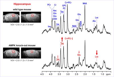
Region-Specific Effects of AMP-Activated Protein Kinase on the Neurochemical Profiles of the Hippocampus and Midbrain in Mice
Ivan Tkac, Biplab Dasgupta, Raghavendra Rao
Adenosine monophosphate-activated protein kinase (AMPK) is an evolutionarily conserved signaling molecule essential for cellular energy balance. AMPK senses metabolic stress and integrates diverse physiological signals to restore energy balance. Its role in the normal brain development is not well understood. The purpose of this study was to assess whether neurochemical profiles of developing mouse brain are affected by knocking out of AMPK enzyme in a region-specific manner. Observed changes in metabolite levels (Lac, Glu) indicate reduced energy metabolism in AMPK knockout mice relative to WT controls. In addition, changes in myo-Ins suggest osmotic stress.
|
|