Poster: Contrast Mechanisms: New Horizons
Electronic Power Pitch Poster
Contrast Mechanisms
Thursday, 27 April 2017
| Exhibition Hall |
09:15 - 10:15 |
| |
|
Plasma # |
 |
0992.
 |
16 |
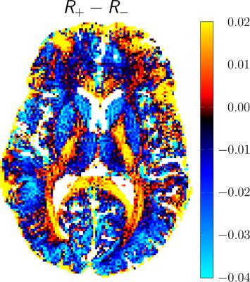
On the decay of SSFP configurations 
Damien Nguyen, Rahel Heule, Carl Ganter, Oliver Bieri
In this work, we explore the decay of negative and positive steady state configurations as a mean to assess the tissue microstructure. Steady state configurations are retrieved up to a high order from an exhaustive sampling of the frequency response profile of low-angle balanced SSFP scans. Subsequently, the decay of configurations (termed DECO) is analyzed using a single-pole matrix pencil analysis yielding positive and negative DECO images. Any asymmetry in the configuration decay is directly linked to asymmetric frequency content within a voxel and is captured in the DECO difference image.
|
 |
0993.
 |
17 |
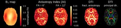
Asymmetries of the balanced SSFP profile allow to probe microstructure anisotropy at 9.4 Tesla
Philipp Ehses, Mario Báez-Yánez, Michael Erb, Klaus Scheffler
The bSSFP signal profile exhibits tissue-dependent asymmetries that can be used as a novel contrast mechanism and have been hypothesized to relate to the tissue microenvironment. In this work, we investigate this effect at ultra high-field using phase-cycled bSSFP at an isotropic resolution of 1.2 mm. As in the original publication, we also observe strong asymmetries in white matter and a comparison to DTI data reveals that the largest asymmetries occur in white matter tracts oriented orthogonal to the main magnetic field.
|
|
0994.
 |
18 |
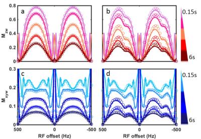
Quantitative modeling of exchange in bSSFPX
Shu Zhang, Robert Lenkinski, Elena Vinogradov
bSSFP was shown to be sensitive to exchange and is explored as an alternative way for CEST/T1ρ experiments (the bSSFPX method). In this abstract, an analytical solution is derived for the magnetization behavior in the bSSFPX. The solution describes the transient signal fluctuations for short saturation times. The solution is verified by comparing it to full, step-wise computation of Bloch-McConnell Equations. Overall, this solution is in good agreement with Bloch-McConnell Equations and follows the transient signal oscillations well. Work is in progress to use this solution to quantify exchange rates experimentally.
|
|
0995.
 |
19 |
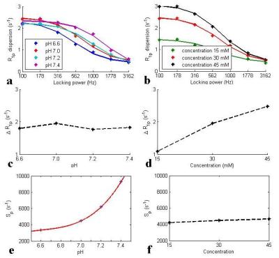
Spin-lock imaging of exogenous exchange-based contrast agents to assess tissue pH
Zhongliang Zu, Hua Li, Xiaoyu Jiang, John Gore
We measured spin-lock relaxation rates as a function of locking field to quantify tissue pH and the concentration of an exogenous X-ray contrast agent, iohexol, based on chemical exchange effects. Results show that spin-lock imaging can be used to detect exchange-based agents and the effects of tissue acidification.
|
|
0996.
 |
20 |
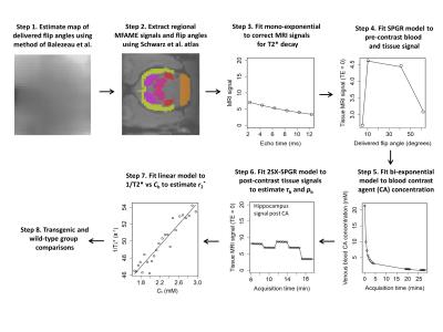
Quantification of trans-endothelial water exchange and vessel geometry using contrast-enhanced MRI and alterations in a transgenic rat model of Alzheimer’s disease
Ben Dickie, Hervé Boutin, Jose Ulloa, Laura Parkes, Geoff Parker
MRI measurements of cerebrovascular function and structure are important for understanding neurodegenerative disease mechanisms. In this study, a novel contrast-enhanced multi-flip angle multi-echo (MFAME) MRI technique capable of simultaneously quantifying vessel permeability surface area product to water (PSw), blood water population fraction (pb), and contrast agent r2* is presented, and applied to a transgenic rat model of Alzheimer’s disease (TgF344-AD). Transgenic rats exhibit higher pb and lower PSw in the hippocampus compared to wild-types, suggesting MFAME MRI may be sensitive to regional pathologic microvascular alterations in this model.
|
|
0997.
 |
21 |
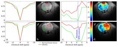
CEST-weighted MRI at 21.1 T: application to glioma and ischemic rat model
Tangi Roussel, Jens Rosenberg, Samuel Grant , Lucio Frydman
This study explores and demonstrates the opportunities opened by ultrahigh fields for in vivo CEST-weighted imaging. CEST-weighted fast spin-echo imaging was performed on two neurological models: hypoxic ischemic and glioblastoma. A remarkably strong CEST contrast (≈15%) was observed for the tumors at 3.32 ppm originating mainly from an APT increase and a strong decrease in NOE and MT. Ischemic lesions were still more robustly detected with standard T2-weighted images than with CEST. Potential explanations and dependence with high magnetic field are discussed.
|
|
0998.
 |
22 |
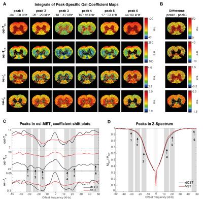
Offset-Saturation-Induced (osi-) Variations in Multiexponential T2 at 16.4T: A New Dimension for Probing White Matter Contrast
Teresa Serradas Duarte, Noam Shemesh
Whether Magnetization Transfer could affect Multiexponential T2 (MET2) Relaxometry remains poorly explored. Here, the effects of irradiation at different offset frequencies on the MET2 components were evaluated as novel contrast mechanisms for white matter in the rat spinal cord at 16.4T. MET2 coefficients were found to shift with off-resonance saturation, showing unique tract-specific signatures across the irradiation frequency. Offset-saturation-induced (osi-) MET2 Relaxometry maps show strong contrasts between microstructurally-distinct rat spinal cord tracts. The potential of exploiting the osi-MET2 shift phenomenon to increase white matter contrast is discussed.
|
|
0999.
 |
23 |
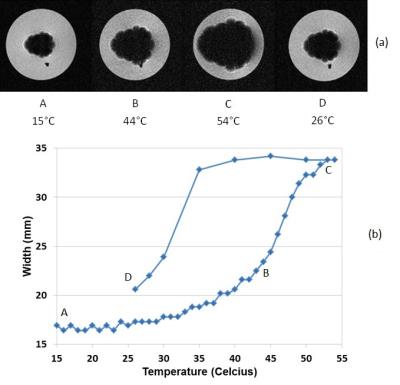
Magneto-Caloric Materials as Tunable and Switchable Labels for MRI
Mladen Barbic, Tim Harris, Stephen Dodd, H Morris, Alan Koretsky, Barbara Marcheschi, Alan Huston, Neil Dilley
We present the case for the use of magneto-caloric materials as tunable and switchable labels for MRI. Sharp magnetic phase transitions these materials have at typical physiological temperatures and in the presence of the large DC magnetic field values associated with MRI machines make them uniquely suitable for the development of novel MRI contrast agents. We present physical and MRI measurements of a prototypical magneto-caloric material Iron-Rhodium (FeRh) that clearly demonstrate the MR image contrast changes due to the temperature tunable magnetic state of the material in the MRI compatible magnetic field range and physiologically relevant temperature range.
|
|
1000.
 |
24 |
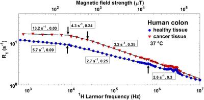
The T1-Dispersion Curve as a Biomarker of Colorectal Cancer
Vasileios Zampetoulas, Lionel Broche, Graeme Murray, David Lurie
A graph of T1 versus applied magnetic field, obtained via Fast Field-Cycling (FFC) NMR relaxometry, can be used as a diagnostic tool thanks to the information it provides about molecular dynamics. In this work, FFC NMR relaxometry, extended to magnetic fields below 17 μT, was used to investigate new biomarkers of colorectal cancer. The acquired results indicated that there were significant differences in the molecular motions with correlation times 0.1-10 ms and 0.5–1.4 μs between the healthy and cancer tissues examined, showing great potential for diagnosis, staging and monitoring response to treatment.
|
|
1001.
 |
25 |
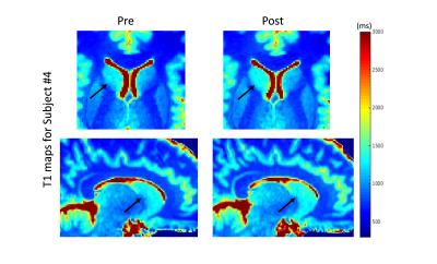
Detecting regional changes in brain tissue quantitative T1 values due to hydration status
Sofia Chavez
Volumetric/morphometric changes in brain structures are often investigated as markers for disease or drug-induced effects. Brain tissue has been shown to shrink during mild dehydration which is not typically controlled for in MRI studies thus potentially confounding the results. Quantitative T1 is expected to change in response to changes in water content of tissue. Here, we show for the first time, that T1 maps, generated as we suggest, can capture regional water shifts that result from changes in hydration status. These can be used to control for water shifts in volumetric/morphometric studies and may aid in the interpretation of results.
|
|
1002.
 |
26 |
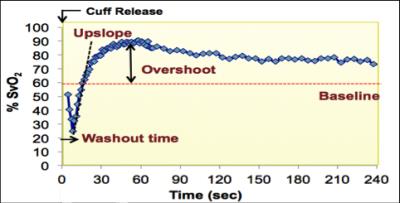
In vivo whole-blood $$$T_2$$$ versus $$$HbO_2$$$ calibration by modulating blood oxygenation level in the femoral vein through intermittent cuff occlusion
Michael Langham, Ana Rodríguez-Soto, Nadav Schwartz, Felix Wehrli
The relationship between whole-blood T2 and blood hemoglobin oxygen saturation (HbO2) can be modeled as $$${1\over T_{2b}}={1\over T_{2o}}+K(1-HbO_2)^2$$$, where K and T2o are determined empirically in vitro. The feasibility of estimating K and T2o in vivo is investigated with T2-prepared bSSFP at 1.5T in the superficial femoral vein (SFV) with intermittent cuff occlusion. In this manner a range of HbO2 were achieved allowing quantification of venous oxygen saturation via MR susceptometry, a method that had been validated rigorously against blood gas analysis. Initial result (K=19.6Hz, T2o=185ms) is lower than the literature value (24.6Hz, 254ms) but not unexpected because transient bSSFP signal is acquired while disturbing the T2-prepared magnetization.
|
|
1003.
 |
27 |
Visualizing local mechanical properties of agar phantoms and meningioma patients using magnetic resonance rheology
Sebastian Theilenberg, Jakob Bindl, Anna-Lisa Kofahl, Carsten Urbach, Karl Maier
Magnetic resonance rheology is a novel method to create an imaging contrast based on the mechanical properties of brain tissue. It is based on a short fall of the head that creates a broadband excitation of the tissue. The resulting deflections of the tissue elements are depicted using motion sensitive phase imaging. This contribution presents measurements on agar phantoms as well as four meningioma patients to show the feasibility of the method to depict local alterations of the mechanical properties of the investigated material.
|
|
1004.
 |
28 |
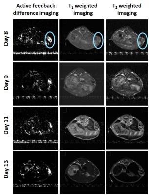
Early Cancer Detection Using Paramagnetic Liposome by a Novel Contrast Mechanism with Active-feedback Magnetic Resonance Imaging
Sayoni Ray, Chao-Hsiung Hsu, Zhao Li, Fang-Chu Lin, Ying-Chih Lin, Yung-Ya Lin
The detection of early tumors requires creating the contrast between healthy and tumor tissues that share a common morphology, making it difficult to distinguish them by relaxation-based MRI technique. Here we have exploited the small magnetic differences between the healthy and tumor tissues to develop a detection technique of early tumors using theranostic nanoparticle paramagnetic (Gd) liposome along with continuous wave (CW) irradiation in the presence of the feedback magnetic field from an active-feedback electronic device for in vivo subcutaneous glioblastoma multiforme mouse models and obtained significantly superior contrast compared to the conventional MRI technique, in agreement with spin-dynamics simulations.
|
|
1005.
 |
29 |
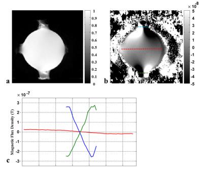
Analysis of magnetohydrodynamic effects in current injection induced magnetic flux density images at very high magnetic fields
Atul Minhas, Munish Chauhan, Rosalind Sadleir
Magnetic resonance imaging based electrical conductivity imaging of biological tissues could be highly challenging at magnetic fields of the order of 18.8T. We could see significant magnetohydrodynamic (MHD) effects in low viscosity materials at such fields. In this study, we investigated these effects using saline and agarose phantoms and explained the MHD mechanism using a fluid flow model coupled with electromagnetic field equations. Experimental and simulation results showed a correlation between the higher magnetic field and velocity. This relation changed from exponential to linear as the viscosity of material increased. The velocity was negligible for highly viscous materials such as agarose.
|
|
1006.
 |
30 |
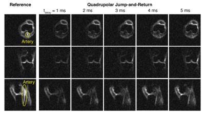
Quadrupolar jump-and-return sequence for sodium knee MRI at 7 tesla
Jae Seung Lee, Ding Xia, Ravinder Regatte
Glycosaminoglycans (GAGs) is an important biomarker for the diseases related to the degradation of cartilage tissues. The GAG content in cartilage tissue is known to be well correlated with the sodium concentration. In addition, the collagenous extracellular matrices of cartilage tissues provide sodium ions with ordered environments. Recently, the so-called quadrupolar jump-and-return (QJR) sequence has been developed, which can selectively detect sodium ions in ordered environments. In this work, we demonstrate the feasibility of the QJR sequence for in vivo knee MRI by assessing its performance on the contrast modification to cartilage tissues and fluid.
|
|