Poster: Reconstruction Highlights
Electronic Power Pitch Poster
Acquisition, Reconstruction & Analysis
Tuesday, 25 April 2017
| Exhibition Hall |
14:45 - 15:45 |
| |
|
Plasma # |
|
0441.
 |
16 |
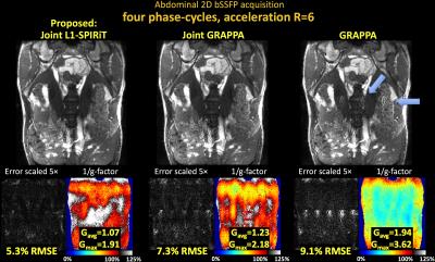
Joint Reconstruction of Phase-Cycled Balanced SSFP with Constrained Parallel Imaging 
Berkin Bilgic, Thomas Witzel, Himanshu Bhat, Lawrence Wald, Kawin Setsompop
Balanced SSFP is an SNR-efficient sequence with unique T2/T1 contrast, but suffers from banding artifacts due to B0 sensitivity. These artifacts can be mitigated through RF phase-cycling at a cost of significant increase in scan time. Here, we propose Joint L1-SPIRiT to jointly reconstruct highly accelerated/undersampled phase-cycled images and convert the different banding artifacts of these images into additional spatial encoding. This enables highly accelerated 2D and Simultaneous Multi-Slice imaging, thus mitigating scan time burden of phase-cycled bSSFP while producing banding-free images. In particular, Joint L1-SPIRiT provides around 2-fold decrease in both the average g-factor and RMSE relative to GRAPPA.
|
 |
0442.
 |
17 |
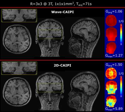
Wave-CAIPI for Highly Accelerated MP-RAGE Imaging
Daniel Polak, Kawin Setsompop, Stephen Cauley, Borjan Gagoski, Himanshu Batt, Florian Maier, Lawrence Wald, Berkin Bilgic
We introduce a highly accelerated T1-weighted MP-RAGE acquisition that utilizes a novel reordering scheme and Wave-CAIPI encoding to retain high image quality. R=9-fold accelerated in vivo MP-RAGE scans were performed in 71 sec, with maximum and average g-factor of gmax=1.27 and gavg=1.06. Compared to the state-of-the-art 2D-CAIPIRINHA method, this is a factor of 4.6/1.4 improvement in gmax/gavg. In addition, we demonstrate a 57 sec acquisition at 7T with R=12-fold acceleration. This acquisition had a g-factor performance of gmax=1.15 and gavg=1.04. Wave encoding overcomes the g-factor noise amplification penalty and allows for an order of magnitude acceleration of MP-RAGE acquisitions.
|
 |
0443.
 |
18 |
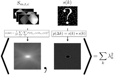
Differential Domain Analysis for 3D Cartesian Sampling
Evan Levine, Brian Hargreaves
Selection of arbitrary 3D Cartesian sampling patterns for support-contrained MRI, parallel MRI, and dynamic MRI can be heuristical, and g-factor calculations require a computationally expensive simulation. To provide theoretical guidance and a method to optimize 3D Cartesian sampling, a novel concept of a differential distribution is introduced to represent a distribution of pairwise differences between sample locations, and is related to point-spread-functions. Its relationship to noise amplification in a generalized sensitivity encoding model and linear reconstruction is then used to efficiently optimize multidimensional k-space sampling. Examples in support-constrained MRI, parallel MRI, and dynamic MRI demonstrate reduced noise amplification
|
|
0444.
 |
19 |
Comparison of 2D and 3D MR Liver Elastography in 600 Patients
Bogdan Dzyubak, Kevin Glaser, Sudhakar Venkatesh, Richard Ehman
2D Magnetic Resonance Elastography (MRE) is a validated method for staging liver fibrosis via liver stiffness measurements produced by introducing and imaging acoustic waves. Newer 3D MRE additionally images waves propagating out-of-plane. This retrospective study compared 2D and 3D liver MRE stiffness measurements across 600 patient exams. The two methods had excellent agreement, with 3D MRE yielding 6% lower stiffness and having lower failure rate. 3D MRE, which has the same acquisition time, is a good alternative for liver imaging as it does not suffer from out-of-plane propagation bias and has the added potential to characterize smaller abdominal organs.
|
|
0445.
 |
20 |
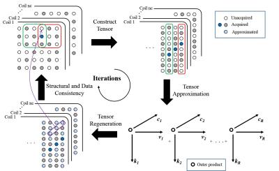
Calibrationless Parallel Imaging Reconstruction Using Hankel Tensor Completion (HTC) - permission withheld
Yilong Liu, Jun Cao, Mengye Lyu, Ed Wu
Autocalibrating parallel imaging requires sufficient autocalibration signals (ACS) for reliable estimation of coil sensitivity. However, this is not feasible in some applications, for example, spectroscopic imaging where matrix size is relatively small. Recent publications (SAKE, P-LORAKS, and ALOHA, etc.) proposed to construct k-space data into block-wise Hankel matrix, and perform parallel imaging reconstruction with low rank matrix completion. In this study, we proposed to construct a block-wise Hankel tensor, and use tensor completion techniques to synthesize the unacquired samples. This method can also be extended to reconstruct multiple slices simultaneously and provide more accurate reconstruction.
|
|
0446.
 |
21 |
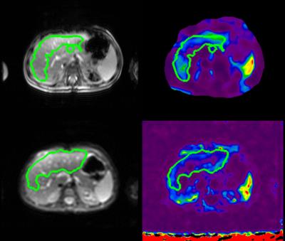
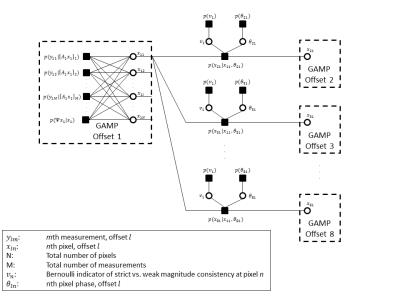
Highly Accelerated Magnetic Resonance Elastography via Bayesian Modeling
Christopher Ebersole, Rizwan Ahmad, Adam Rich, Lee Potter, Arunark Kolipaka
While magnetic resonance elastography (MRE) provides a non-invasive method of estimating tissue stiffness, which is indicative of a variety of diseases, MRE scans typically require lengthy breath-holds which are prohibitive for many patients. We have extended a recently proposed Bayesian imaging method, called ReVEAL, for MRE. This method is capable of reconstructing images from highly undersampled data by leveraging both sparsity and the near equal magnitude across multiple offsets, inherent to MRE acquisition, as reconstruction constraints. This reconstruction method is validated against SENSE using a retrospectively downsampled phantom dataset and three retrospectively downsampled in vivo liver datasets.
|
|
0447.
 |
22 |
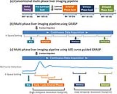
RACE-GRASP: Respiratory-weighted and Aortic Contrast Enhancement-guided GRASP MRI
Li Feng, Krishna Shanbhogue, Daniel Sodickson, Hersh Chandarana, Ricardo Otazo
This work proposes a technique named RACE-GRASP (Respiratory-weighted and Aortic Contrast Enhancement guided Golden-angle RAdial Sparse Parallel imaging) for robust free-breathing DCE-MRI of the liver. First, the aortic contrast enhancement curve is automatically detected from continuously acquired data and is used to guide k-space sorting to ensure precise capture of desired contrast-enhancement phases. Second, k-space data of each contrast phase are binned into different respiratory motion states, and each motion bin is weighted differently during image reconstruction to reduce motion blurring. The performance of RACE-GRASP was demonstrated in 5 healthy volunteers and was compared with conventional GRASP.
|
|
0448.
 |
23 |
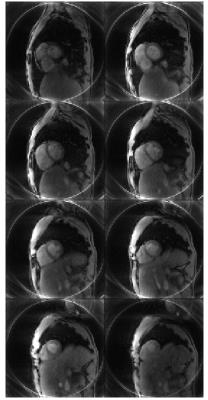
Real-time 3D cardiac MRI using through-time radial GRAPPA and GPU-enabled reconstruction pipelines in the Gadgetron framework
Dominique Franson, James Ahad, Jesse Hamilton, Wei-Ching Lo, Yun Jiang, Yong Chen, Nicole Seiberlich
An accelerated 3D cardiac dataset (8 partitions; in-plane spatial resolution: 2.34 x 2.34 mm2; partition thickness = 8 mm) can be reconstructed using through-time radial GRAPPA and GPU-enabled pipelines in the Gadgetron framework in approximately 515.5 ms. This work may enable real-time, 3D imaging in the heart with on-line reconstruction.
|
|
0449.
 |
24 |
Navigator-free EPI ghost correction using low-rank matrix modeling: Theoretical insights and practical improvements
Rodrigo Lobos, Tae Hyung Kim, W. Hoge, Justin Haldar
While the formation of ghost-free images from EPI data can be a difficult problem, recent low-rank matrix modeling methods have demonstrated promising results. In this abstract, we provide new theoretical insight into these approaches, and show that the low-rank ghost correction optimization problem has infinitely many solutions without using additional constraints. However, we also show that SENSE-like or GRAPPA-like constraints can be successfully used to make the problem well-posed, even for single-channel data. Additionally, we show that substantial performance gains can be achieved over previous low-rank ghost correction implementations by using nonconvex low-rank regularization instead of previous convex approaches.
|
|
0450.
 |
25 |
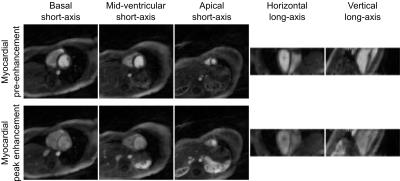
Non-ECG First-Pass Myocardial Perfusion T1 Mapping with Low-Rank Tensor Cardiovascular MR Multitasking
Anthony Christodoulou, Jaime Shaw, Xiaoming Bi, Behzad Sharif, Debiao Li
Quantitative first-pass myocardial perfusion imaging is a potentially powerful tool for diagnosing coronary artery disease. However, quantification is complicated by ECG misfires and the nonlinear response of signal intensity to contrast agent concentration. Here we propose a method overcoming the curse of dimensionality to simultaneously image cardiac motion, contrast dynamics, and T1 relaxation in 2D and 3D, using a low-rank tensor imaging framework for cardiovascular MR multitasking. This non-ECG, first-pass myocardial perfusion T1 mapping method accounts for the signal intensity nonlinearity, allowing direct quantification of contrast agent concentration at any cardiac phase in any cardiac cycle.
|
|
0451.
 |
26 |
T1-T2 Shuffling: Multi-Contrast 3D Fast Spin-Echo with T1 and T2 Sensitivity
Jonathan Tamir, Valentina Taviani, Shreyas Vasanawala, Michael Lustig
Volumetric fast spin-echo (3DFSE) imaging is clinically desirable because of its robustness to off-resonance and its utility for obtaining many types of image contrasts at isotropic resolution. However, its routine clinical use is inhibited by blurring due to long echo trains needed to maintain scan efficiency. Her we present T1-T2 Shuffling, a 3DFSE-based acquisition and reconstruction scheme that mitigates image blur and retrospectively synthesize T1-weighted and T2-weighted image contrasts. The acquisition, an extension of T2 Shuffling, employs a randomizing echo train view ordering with variable repetition times (TRs). The use of short TRs increases scan efficiency while providing T1 sensitivity.
|
|
0452.
 |
27 |
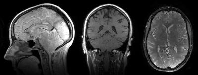
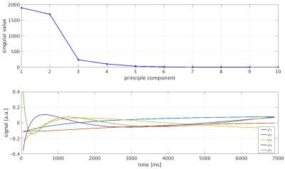
Simultaneous T1/T2 measurements in combination with PCA-SENSE reconstruction (T1* shuffling) and multicomponent analysis
Julian Pfister, Martin Blaimer, Peter Jakob, Felix Breuer
An inversion recovery TrueFISP sequence with a golden angle based radial readout in combination with a view sharing KWIC filter reconstruction is able to produce quantitative M0, T1 and T2 maps within a single shot. However the KWIC filter leads to temporal blurring especially at high spatial frequencies. Here, we propose an alternative reconstruction method based on the principle component analysis (PCA) for providing an improved temporal fidelity. Furthermore this approach is extended for a multicomponent analysis by an inverse Laplace transform. Results from brain measurements demonstrate that this technique can identify different tissue types within a single voxel.
|
|
0453.
 |
28 |
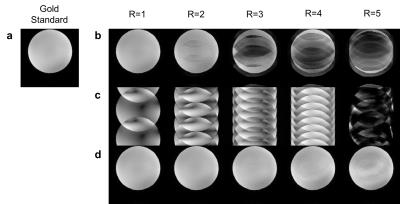
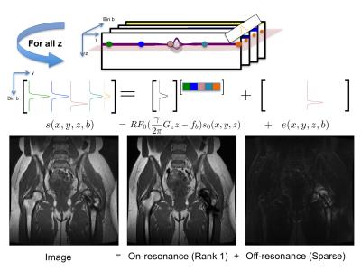
Accelerated 3D Multispectral MRI with Robust Principal Component Analysis for Separation of On and Off-resonance Signals
Evan Levine, Kathryn Stevens, Brian Hargreaves
3D multispectral imaging (MSI) corrects most distortion in MRI near metallic implants at the cost of prolonged scan time by phase encoding to resolve slice distortions. However, existing methods to accelerate 3D MSI do not exploit the redundancy of slice-phase encoding for the dominant on-resonance signal. A novel compact representation of 3D-MSI images based on a decomposition of on- and off-resonance via robust principal component analysis (RPCA) is introduced to exploit this redundancy in a calibration and model-free reconstruction and push the current limits of accelerated 3D MSI. A complementary randomized sampling strategy is used to vary undersampling in different spectral bins to enable the separation. Experiments with retrospective and prospective undersampling show comparable image quality between standard MSI images and 2.6-3.4-fold accelerated RPCA and improvement over bin-by-bin compressed sensing reconstruction.
|
|
0454.
 |
29 |
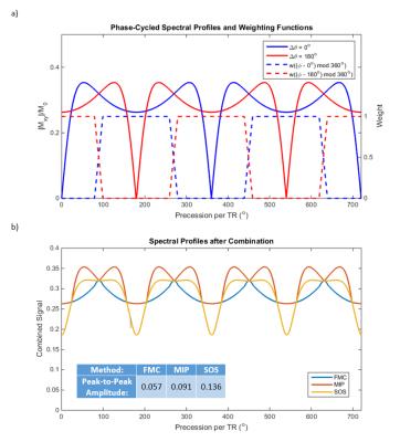
Field Map Combination Method for Multiple-Acquisition bSSFP
Anjali Datta, Dwight Nishimura
FIESTA-C/CISS enables the reconstruction of banding-free bSSFP images, but residual ripple remains in the combined images. Also, in the heart, flow near a stop-band may cause a component image to include significant contributions from out-of-slice spins; this hyper-intense signal persists in images combined using conventional methods. Here, we use a B0 map to combine FIESTA-C/CISS cardiac cine images. Knowledge of B0 enables us to only include pass-band signal in the final image and exclude stop- and bright-bands. In phantom and in vivo studies, the proposed method had less ripple and greatly reduced flow artifacts compared to maximum-intensity projection and root-sum-of-squares.
|
|
0455.
 |
30 |
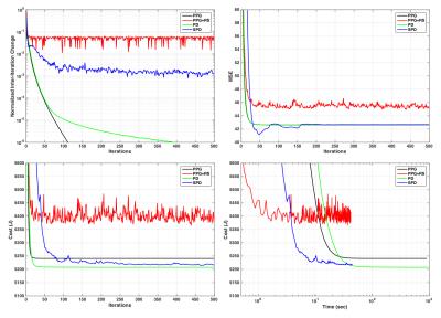
Stochastic Primal-Dual Optimization for Locally Low-Rank MRI Reconstruction: A Stable Alternative to Cycle Spinning
Joshua Trzasko
Locally low rank (LLR) reconstruction is an effective strategy that has found application across a range of MRI applications. In lieu of processing all overlapping image blocks each iteration, most LLR implementations employ “cycle spinning”, where only a random subset of blocks is processed. Cycle spinning improves efficiency, but may compromise reconstruction convergence and introduce artifacts. We propose a primal-dual algorithm for LLR reconstruction and show that stochastically updating the dual variable provides similar computational advantage as cycle spinning but avoids its primary disadvantages. Reconstruction performance benefits are demonstrated on both a numerical phantom and in vivo.
|
|