Poster: Cutting Edge Diffusion
Electronic Power Pitch Poster
Diffusion
Thursday, 27 April 2017
| Exhibition Hall |
14:00 - 15:00 |
| |
|
Plasma # |
|
1082.
 |
1 |
Approaching free intracellular diffusion by diffusion-weighted MRS at ultra-short time scales: initial results in the rodent brain using a 1.5 T/m gradient 
Clémence Ligneul, Marco Palombo, Julien Flament, Julien Valette
At ultra-short time scales, intracellular metabolites are expected to experience less restriction, so that their apparent diffusion coefficient (ADC) as measured by diffusion-weighted MRS should approach the free intracellular diffusivity in a manner which depends on small microstructural features. In this work we use a unique gradient insert capable of reaching 1.5 T/m to measure metabolite ADC in the rat brain up to 665 Hz using oscillating gradients (corresponding to 0.21-ms diffusion time in the Mitra limit), in order to approach and estimate free intracellular diffusion.
|
|
1083.
 |
2 |
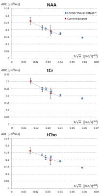
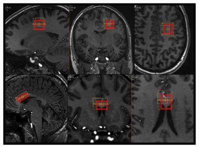
Accurate estimation of intra-axonal diffusivity and anisotropy of NAA in humans at 7T
Henrik Lundell, Carson Ingo, Tim Dyrby, Itamar Ronen
Diffusion weighted spectroscopy offers a unique probe for tissue microstructure and recent studies demonstrate NAA diffusivity as an independent marker of axonal health. In this study we address the problem of macroscopic dispersion of fiber directions and suggest the use of high angular gradient resolution and powder averaging as an experimentally inexpensive and accurate way to solve this problem. We explore the limits of this approach in simulations and in experiments on humans at 7T.
|
|
1084.
 |
3 |
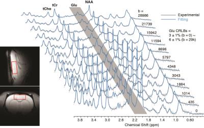
Glutamate diffusion at high b-values in the rat brain in vivo under light and deep anesthesia conditions
Xi Chen, Siddartha Tamang, Fei Du, Dost Ongur
Magnetic resonance techniques are developed to measure brain glutamate (Glu) concentrations but still not able to detect synaptic Glu release. In the currently study, in vivo diffusion-weighted MRS using low to very high b-values was performed on rat brain prefrontal cortex under both light and deep anesthesia conditions. Significant Glu diffusion and concentration changes were observed under different anesthesia levels in the absence of similar changes in NAA or creatine. The slower diffusion and lower concentration under deep anesthesia may reflect more Glu packed into synaptic vesicles with reduced mobility and NMR visibility.
|
 |
1085.
 |
4 |
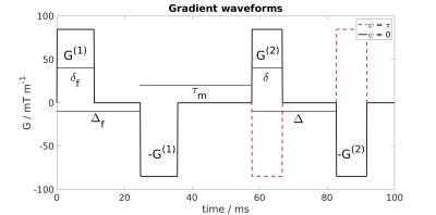
Bias in the apparent exchange rate measurements: insight from numerical simulations
Patricia Ulloa, Vincent Methot, Martin A. Koch
Using double diffusion encoding it is possible to acquire microstructural and water exchange information. Here, simulations are used to study how restriction effects influence apparent exchange measurements. The simulations indicate that at the chosen experimental parameters the restriction effect can be considerable for large pores and small mixing times, τm. In typical exchange rate experiments using clinical MR systems with τm > 40 ms, the restriction effect can probably be neglected if pores are small.
|
|
1086.
 |
5 |
Microscopic anisotropy with spectrally modulated q-space trajectory encoding
Henrik Lundell, Markus Nilsson, Tim Dyrby, Geoff Parker, Penny Hubbard Cristinacce, Fenglei Zhou, Daniel Topgaard, Samo Lasic
Multi-dimensional diffusion encoding can, in contrast to conventional diffusion encoding, disambiguate between isotropic and anisotropic diffusional variance in multicompartment systems. This is done by varying the shape of the encoding tensor, i.e. going from measuring one projection of the diffusion tensors to measuring the trace of the diffusion tensors. Additional morphological features, such as the sizes of cells, are reflected in the diffusion spectrum. In this study we combine encoding tensors with varying spectral content and shape. This augmented protocol demonstrates distinctively different levels of microscopic fractional anisotropy (µFA) and time-dependent diffusion in phantoms and in white matter, cerebral cortex, and cerebellar cortex in a fixed monkey brain.
|
|
1087.
 |
6 |
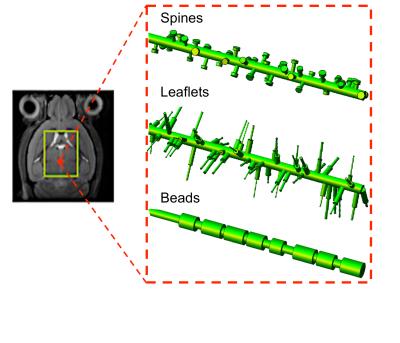
Can we detect the effect of spines, leaflets and beads on the diffusion of brain intracellular metabolites? A confrontation between high b-values and high-frequencies diffusion-weighted MRS in the mouse brain in vivo.
Marco Palombo, Clemence Ligneul, Edwin Hernandez-Garzon, Julien Valette
Prior models used to clarify which aspects of tissue microstructure mostly affect intracellular diffusion and corresponding diffusion-weighted magnetic resonance signal have focused on relatively simple geometrical descriptions of the cellular microenvironment (spheres, randomly oriented cylinders, etc...), neglecting finer morphological details which may have an important role. Neuritis may exhibit beading; some types of neurons present high density of spines; and astrocytes and macroglial cells processes present leaflets, which may all slow impact the diffusion process. Here we use numerical simulations to interpret metabolites diffusion-weighted MRS data in the mouse brain in terms of such fine secondary structures.
|
|
1088.
 |
7 |
Diffusion MRI of axonal degeneration in areas of fiber crossing: Histological correspondence.
Luis Concha, Jorge Larriva-Sahd, Gilberto Rojas-Vite, Ramsés Noguez-Imm, Ricardo Coronado-Leija, Alonso Ramírez-Manzanares, José Luis Marroquín
The tensor model has been widely used to infer characteristics of white matter through diffusion MRI. Unfortunately, this model does not provide reliable information about crossing fiber regions. Several models have been proposed that seem to overcome the limitations of the tensor. However, biological interpretations of such models are limited by the lack of histological conformation. Using an animal model of axonal degeneration, we compare histology to data derived from two approaches (CSD and multi-tensor), in an effort to provide validation of metrics that can bring substantial and clinically useful information about crossing fiber regions.
|
 |
1089.
 |
8 |
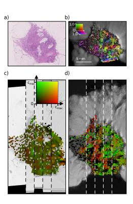
Diffusion anisotropy in breast cancer tissue corresponds to spatial patterns of collagen alignment from structure tensor analysis of histology
Colleen Bailey, Francesco Grussu, Bernard Siow, Thomy Mertzanidou, John Hipwell, Julie Owen, Patrycja Gazinska, Sarah Pinder, Daniel Alexander, David Hawkes, Eleftheria Panagiotaki
Directional and anisotropy measures from a diffusion model composed of VERDICT compartments were compared with directional and anisotropy measures from structure tensor analysis of registered histology images. A significant positive correlation was found between the direction of the Zeppelin component of the diffusion model (assumed to represent the extracellular space) and the predominant direction of the structure tensor from the stroma, where the primary feature is aligned collagen. The correlation of anisotropy measures was weak, which may be due to difficulties in detecting alignment in regions with densely-packed collagen, which have nearly uniform intensity on H&E staining.
|
|
1090.
 |
9 |
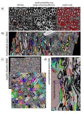
A 3D electron microscopy segmentation pipeline for hyper-realistic diffusion simulations
Michiel Kleinnijenhuis, Errin Johnson, Jeroen Mollink, Saad Jbabdi, Karla Miller
Simulations of the diffusion signal can shed light on how the MR signal is generated from particular tissue microstructure. In our approach we use microscopy data to generate a realistic ground truth for investigating diffusion properties. We have developed a method to automatically segment large volumes of 3D electron microscopy data into individual axons for diffusion simulations. From these segmentations, we can also derive benchmark tissue microstructure characteristics such as axonal diameter, g-ratio and other compartment properties.
|
|
1091.
 |
10 |
Rotationally invariant mapping of microstructural and orientational neuronal tissue parameters in human brain
Dmitry Novikov, Jelle Veraart, Ileana Jelescu, Els Fieremans
We develop a general framework for estimating orientational and microstructural parameters of neurites. By employing a set of rotational invariants, we analytically reveal the nontrivial topology of the parameter estimation landscape, showing that multiple branches of parameters describe the measurement almost equally well, with only one of them corresponding to the biophysical reality. A comprehensive acquisition shows that the branch choice differs for white and for gray matter. We reveal hidden degeneracies in MRI parameter estimation for neuronal tissue, provide microstructural and orientational maps in the whole brain without constraints or priors, and assess commonly used parameter constraints.
|
|
1092.
 |
11 |
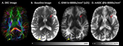
Isotropic Diffusion Weighted MRI (IDWI) – a novel, efficient clinical method for quantifying orientationally-averaged features of water diffusion in tissues
Alexandru Avram, Joelle Sarlls, Elizabeth Hutchinson, Peter Basser
We propose a novel, efficient diffusion method, called isotropic diffusion weighted MRI (IDWI), for measuring orientationally-averaged properties of tissue water diffusion, free from modulations due to anisotropy. Using efficient diffusion gradient sampling schemes, IDWI rapidly and accurately quantifies the mean apparent diffusion coefficient (mADC) over a wide range of b-values, along with other important rotation-invariant intrinsic microstructural parameters, such as the mean t-kurtosis. The ability to efficiently and effectively remove modulations due to anisotropy in images with high-b values may improve existing diffusion MRI techniques and spur the development and clinical translation of new methods with improved biological specificity.
|
|
1093.
 |
12 |
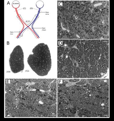
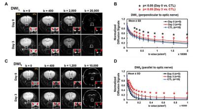
Diffusion MRI differentiated acute inflammation from axonal injury but missed axonal loss
Tsen-Hsuan (Abby) Lin, Michael Wallendorf, Peng Sun, Sheng-Kwei Song
Diffusion MRI with higher b-values and custom-designed diffusion schemes are critical to identify subtle and coexisting pathology in CNS. In the current study, we employed single-axial high-b diffusion-weighted imaging (DWI) and low-b diffusion basis spectrum imaging (DBSI) to assess mouse optic nerve crush acutely. The results suggested coexisting CNS pathology affected apparent diffusion coefficient (ADC), and low-b DBSI was able to reflect axon and myelin integrity as well as inflammatory edema and cellularity even before histological detection. DBSI-detected axon volume correlated with axonal loss negatively, suggesting cytotoxic-edema-associated axonal swelling might mask axonal loss acutely.
|
|
1094.
 |
13 |
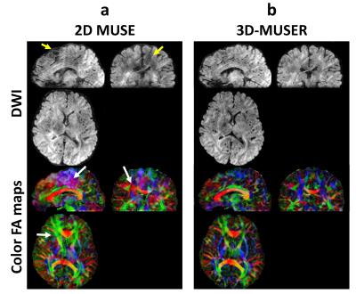
Three-Dimensional Multiplexed Sensitivity Encoding and Reconstruction (3D-MUSER): 3D Phase Correction for 3D Multi-shot DWI
Hing-Chiu Chang, Edward S. Hui, Xiaoxi Liu, Pui-Wai Chiu, Nan-kuei Chen
3D interleaved DW-EPI with 2D multiplexed sensitivity encoding has been shown useful in achieving submillimeter DTI. Similar to other 3D DTI techniques, 2D phase variation is only considered in eliminating aliasing artifacts, thereby limiting feasible slab thickness. 3D phase correction is a potential strategy to significantly improve the image quality and feasible slab thickness of 3D DTI. To enable 3D phase correction, we develop a new reconstruction algorithm, and implement an EVI-based navigator echo for direct 3D phase measurement. Quantitative results show that the proposed algorithm can effectively eliminate aliasing artifacts and signal corruptions due to 3D inter-shot phase variations.
|
|
1095.
 |
14 |
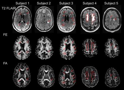
Visualizing Axonal Damage in Multiple Sclerosis Using Double Diffusion Encoding MRI in a Clinical Setting
Grant Yang, Qiyuan Tian, Christoph Leuze, Max Wintermark, Jennifer McNab
Double diffusion encoding (DDE) measurements of microscopic anisotropy show promise as a method of assessing neurodegeneration. Unfortunately, DDE has yet to be demonstrated in a clinical setting due to constraints in SNR and scan time. Here, we used an optimized gradient orientation scheme to show the first DDE measurements of microscopic anisotropy in multiple sclerosis (MS) patients. Five MS patients were scanned using a DDE sequence optimized to run in five minutes. The microscopic anisotropy maps show improved visualization of axonal damage compared to fractional anisotropy (FA) and may provide additional insight into changes in tissue microstructure.
|
|
1096.
 |
15 |
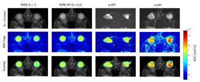
Accelerated Diffusion-Sensitized MR Imaging of the Eye and Orbit at 3.0 T and 7.0 T free of Geometric Distortions Using a Combined RARE-EPI Acquisition Technique
Katharina Paul, Helmar Waiczies, André Kuehne, Till Huelnhagen, Eva Oberacker, Oliver Stachs, Thoralf Niendorf
Diffusion-weighted imaging of the eye and orbit is an emerging MRI application to provide guidance during diagnostic assessment and treatment of ophthalmological diseases. RARE based diffusion-sensitized imaging (ms-RARE) provides images free of geometric distortions. Yet imaging speed, RF power deposition and artifacts by involuntary eye motion remain a concern. Combined acquisition techniques (CAT) merging RARE and EPI within one echo train offer the possibility to shorten acquisition times and relax specific absorption rate constraints. This study examines the applicability of RARE-EPI CAT for diffusion-sensitized imaging of the eye and orbit free of geometric distortions at 3.0 T and 7.0 T.
|
|