ISMRM 25th Annual Meeting & Exhibition • 22-27 April 2017 • Honolulu, HI, USA

Traditional Poster Session: Contrast Mechanisms
1877 -1898 Arterial Spin Labeling Applications
1899 -1923 DSC & DCE
1924 -1943 Relaxation: Mechanisms & Applications
1944 -1968 Electric Property Imaging & Susceptibility Imaging
1969 -1989 All Things CEST/MT
 
Arterial Spin Labeling Applications
Traditional Poster
Contrast Mechanisms

 
Tuesday, 25 April 2017
Exhibition Hall  16:15 - 18:15


1889.  
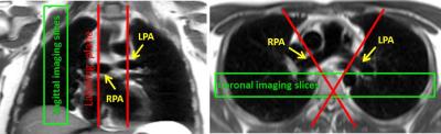
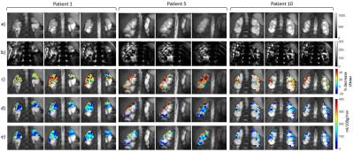
Measurement of lung perfusion using optimized pseudo-continuous arterial spin labeling of pulmonary arteries and fast True-FISP imaging at 3 Tesla
Petros Martirosian, Rolf Pohmann, Martin Schwartz, Thomas Küstner, Wolfhard Binder, Christina Schraml, Ferdinand Seith, Nina Schwenzer, Klaus Scheffler, Konstantin Nikolaou, Fritz Schick
Pseudo-continuous-arterial-spin-labeling (pCASL) has been successfully applied in the brain and kidney providing high signal-to-noise-ratio. The aim of this study was to optimize pCASL for measurement of lung perfusion by optimized labeling of pulmonary arteries and fast signal acquisition. Effective labeling of pulmonary arteries was possible by ECG triggering and an appropriate orientation of the labeling plane. Sufficient signal from lung parenchyma was acquired by True-FISP imaging with TE=1ms. The presented method provides high quality perfusion images of the lung without applying intravenous contrast agents and offers diagnostic imaging of lung diseases such as pulmonary embolism and bronchial carcinoma.


1886.   
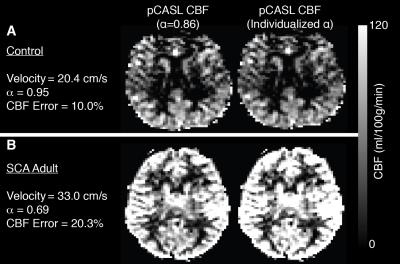
Pseudo-continuous arterial spin labeling considerations in patients with sickle cell anemia
Meher Juttukonda, Lori Jordan, Melissa Gindville, Larry Davis, Jennifer Watchmaker, Sumit Pruthi, Manus Donahue
Pseudo-continuous arterial spin labeling (pCASL) MRI involves labeling of flowing arterial blood water; therefore, the flow velocity of the blood water affects the efficiency of pCASL labeling. This effect has been quantified in healthy subjects but has not been examined in adults with sickle cell anemia (SCA). In this study, we illustrate that cervical flow velocities are elevated in adults with SCA, resulting in a reduced pCASL labeling efficiency of 0.72, and errors associated with this phenomenon are more than twice as large as those associated with bolus arrival time variability when long post-labeling delays times are used.


1883.   
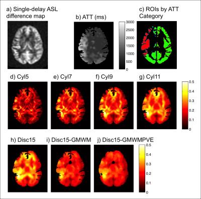
Regional Heterogeneity in Moyamoya Disease: Discovering Local Arterial Transit Time Information from Single-Delay Arterial Spin Labeling
Wendy Ni, Greg Zaharchuk
Arterial transit information provides valuable diagnostic information in steno-occlusive pathologies.  We propose deriving transit information from the regional spatial heterogeneity (ReHet) of standard single-delay pseudo-continuous arterial spin labeling difference images.  With image processing and machine learning, we demonstrate the potential of this technique in identifying regions with slow arterial flow in pre-operative Moyamoya disease patients.  We investigate a selection of 7 different ReHet metrics, and identify trends that will inform better design of ReHet metrics and machine learning models.


1890.  
Quantification of mouse renal perfusion using arterial spin labeled MRI at 1 Tesla
Quyen Do, Ananth Madhuranthakam, Peter Bendel, Robert Lenkinski
The current work demonstrates the use of a 1 Tesla desktop MR system to study mouse kidney perfusion through arterial spin labeling (ASL) technique.  The validity of the implementation was tested by (1) comparing obtained perfusion results with literature values for normal mice and (2) challenging the technique with mice treated with a blood vessel vasoconstrictor drug.  Potential applications include easy assessments of disease state, metabolism, and tissue perfusion using a compact MR system. 


1887.  
Improved reproducibility of longitudinal renal ASL perfusion measurements in children with chronic kidney disease using retrospective motion correction
Fabio Nery, Enrico De Vita, Chris A. Clark, Isky Gordon, David L. Thomas
Arterial spin labelling (ASL) is a unique MR approach for quantifying tissue perfusion non-invasively. However, it is prone to motion-related artefacts which limit its application in the clinical domain, especially outside the brain. In this work, we combine a motion-insensitive ASL acquisition scheme with a specifically tailored retrospective motion correction pipeline. This enabled repeatable renal perfusion measurements to be obtained in the first ASL study in paediatric patients with moderate/severe chronic kidney disease. 


1891.  
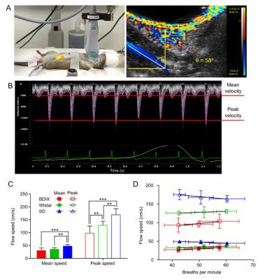
Validation of quantitative pre-clinical pseudo-continuous ASL in rat brain
Manon Simard, James Larkin, Alexandre Khrapitchev, James Meakin, Thomas Okell, Peter Jezzard, Michael Chappell, Nicola Sibson
Guidelines for pre-clinical ASL are lacking. We propose MRI parameters for the use of pseudo-continuous ASL in rats. Carotid artery velocity was determined in three rat strains. Bloch simulations for ASL with this information and with parameters for pre-clinical scanners were used to determine the optimal width of the tagging plane required for blood inversion in this smaller species. The use of multiple post-label delays (PLD) in ASL-MRI generated blood arrival maps indicating that a PLD of 550ms was sufficient. Validation of CBF maps generated from ASL was performed using autoradiography, the current gold-standard technique for pre-clinical perfusion measurement.


1884.  
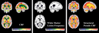
White matter cerebral blood flow in a large healthy cohort from the CARDIA study
Sudipto Dolui, Guray Erus, David Jacobs, Jr., R. Nick Bryan, John Detre
By analyzing cerebral blood flow (CBF) maps generated from arterial spin labeling (ASL) data averaged across 436 cognitively healthy middle-aged subjects from the CARDIA study, we characterized the CBF distribution in white matter. CBF is specifically decreased in periventricular regions in a pattern not reflective of partial volume effects as estimated from the structural MRI segmentation. White matter lesion frequency mapping based on Fluid Attenuated Inversion Recovery (FLAIR) images from the same cohort demonstrates that lesions tend to occur in regions where group averaged CBF is lowest.


1893.  
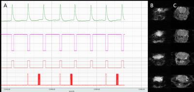
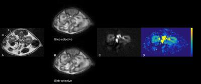
Multiphase pseudo-continuous ASL to image cerebral blood flow in mice at 9.4T
Jessica Buck, James Larkin, Alexandr Khrapichev, Manon Simard, Kevin Ray, Michael Chappell, Nicola Sibson
Pseudo-continuous arterial spin labelling is regarded as the gold standard for clinical ASL, and can be improved in humans using multiphase sequences, but has not previously been implemented in mice. A multiphase pseudo-continuous ASL sequence to measure cerebral blood flow in mice was successfully implemented using respiratory triggering and optimisation of imaging readout, tag placement, labelling bolus duration, and post-label delay. Multiphase pseudo-continuous ASL is sensitive to changes in perfusion in an intracerebral glioma model. 


1885.   
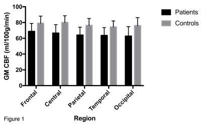
Arterial spin labelling measurements of cortical perfusion in multiple sclerosis show widespread reduced cortical metabolism
Ruth Oliver, Heidi Beadnall, Chenyu Wang, Matthew Kiernan, Todd Hardy, Michael Barnett
Multiple sclerosis (MS)  is primarily an inflammatory demyelinating disease of the central nervous system. However, there is also growing evidence that cortical dysfunction may also be associated with disability in MS. Few studies have investigated cortical cerebral perfusion in MS, and even fewer have utilised arterial spin labelling (ASL)  MRI, which offers noninvasive quantitative assessment of cerebral function using endogenous contrast.   ASL is an inherently low resolution imaging modality known to be affected by the partial volume (PV) effect, leading to an underestimation of grey matter (GM) perfusion. Decreases in GM perfusion could reflect neuronal loss or metabolic dysfunction; PV correction techniques allow decoupling of structure and function. It is hypothesized that reduced regional GM perfusion after PV correction reflects a genuine decreased tissue metabolism, rather than atrophy.


1892.  
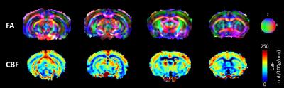
Combined 3D perfusion and diffusion MRI to phenotype the mouse brain: evaluation and application to a model of schizophrenia
Ivy Uszynski, Lydiane Hirschler, Jan Warnking, Cyril Poupon, Jean-Christophe Deloulme, Emmanuel Barbier
Perfusion and diffusion imaging both represent powerful tools in order to phenotype mouse models of brain diseases. Indeed, imaging cerebral blood flow (CBF) may be seen as a surrogate marker of brain metabolism while diffusion imaging provides the structural aspects of brain wiring. To evaluate the potential of combined CBF/Diffusion phenotyping, we evaluated the effect of knocking-out (KO) the microtubule-associated protein 6 (MAP6), which plays a critical role during the development of cerebral axonal tracts. Experiments were performed on homogeneous C57Bl6/129Sv mice using 3D pseudo-continuous Arterial Spin Labeling (pCASL) and 3D diffusion tensor imaging (DTI) at 9.4T.


1894.   
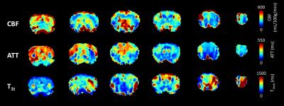
3-Dimensional cerebral blood flow and transit time mouse brain mapping using Dynamic Arterial Spin Labeling (DASL)
Lydiane Hirschler, Ivy Uszynski, Jan Warnking, Emmanuel Barbier
Measuring the arterial transit time (ATT) helps for the optimization and quantification of arterial spin labeling experiments. Moreover, ATT may provide information on potential underlying vascular pathologies. In preclinical perfusion studies, multiple 2D-slices are commonly acquired to measure perfusion. However, this readout limits the number of slices for which ATT can be measured accurately in rodents. In this study, we implemented and optimized a dynamic ASL labeling scheme with a 3D echo planar imaging (EPI) readout to simultaneously map cerebral blood flow, arterial transit time and tissue T1 in the mouse brain at 9.4T.


1895.  
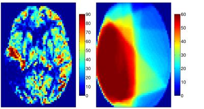
Perfusion decrease during radiochemotherapy is not fully explained by volumetric gray matter changes
Jan Petr, Henri Mutsaerts, Frank Hofheinz, Iris Asllani, Matthias van Osch, Ivan Platzek, Annekatrin Seidlitz, Mechthild Krause, Jörg van den Hoff
Radiochemotherapy in brain-tumor patients was shown to cause gray matter (GM) volume and cerebral blood flow (CBF) changes. The interaction of these two effects, however, remains unclear. Here, we investigated GM volume and ASL CBF changes and their interaction in the healthy hemisphere of 38 glioblastoma patients undergoing radiochemotherapy with Temozolomide. We found a statistically significant CBF decrease with dependence on the RT-dose. PV-corrected results indicated that, while to a certain extent the apparent CBF decrease measured by ASL is caused by GM atrophy, there still remain significant CBF changes that cannot be explained by structural changes alone.


1896.   
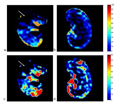
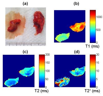
Feasibility and value of VTE-ASL in quantitative evaluation of unilateral renal embolism in rabbits
Hanjing Kong, Fei Gao, Chengyan Wang, Yan Jia, Hui Xu, Xiaodong Zhang, Li Yang, Jue Zhang, Xiaoying Wang, Jing Fang
 Arterial spin labeling with variable echo time (VTE-ASL) is a perfusion imaging technique capable of noninvasive estimating of GFR. But the application of VTE-ASL in renal disease is still lagging behind. The goal of this study was to investigate the feasibility of GFR and RBF using VTE-ASL in evaluation of unilateral renal embolism in rabbits. Compared with normal kidney, embolism area has large decrease in GFR and RBF, and was confirmed by histological findings. 


1877.  
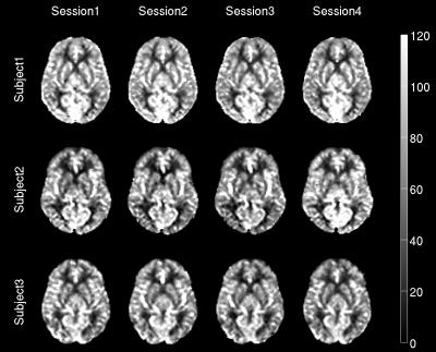
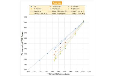
Reliability of Single- and Multi-TI ASL measurements with a clinical product sequence
Antonio Ricciardi, Marco Castellaro, Alberto Miglioranza, Giancarlo Germani, Paolo Vitali, Giuseppe Micieli, Egidio D'Angelo, Fulvia Palesi, Gloria Castellazzi, Claudia Gandini Wheeler-Kingshott, Enrico De Vita, Alessandra Bertoldo
Single-TI Arterial Spin Labeling (ASL) is sensitive to delayed arterial arrival time (AAT) of blood in the tissues. Multi-TI was introduced to overcome this limitation. Moreover, it allows estimating AAT that, therefore, can shed some light in the characterization of the brain haemodynamic processes. In this study, we compare the reliability of the multi-TI approach to the single-TI in a test-retest protocol. The sequence tested was the Siemens product sequence FAIR PASL with 3D-GRASE readout. Results show an overall good level of reliability both in single- and multi-TI, but also a possible sensitivity to the macro-vascular component in multi-TI data.


1880.  
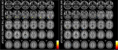
Cerebral Blood Flow and Bolus Arriving Time Changes in Patients with Diabetes Detected by Multi-TI ASL
Yelong Shen, Bin Zhao, Lirong Yan, Kay Jann, Guangbin Wang, Junli Wang, Bao Wang, Josef Pfeuffer, Tianyi Qian, Danny JJ Wang
This study aimed to simultaneously measure cerebral blood flow (CBF) and bolus arriving time (BAT) in a cohort of subjects with type II diabetes, and compared the results with those of matched control subjects using a multi-TI 3D GRASE pulsed-ASL (PASL) sequence. The voxel-based analysis showed that the CBF and BAT values in patients with diabetes presented significant differences compared to healthy subjects, especially in some particular areas of the brain. These differences may be related to functional changes in patients with diabetes, which may have occurred before the onset of the symptoms. 


1898.   
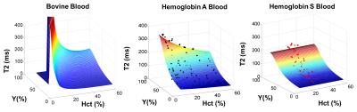
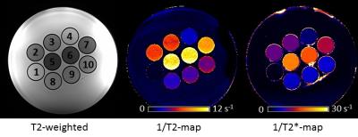
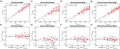
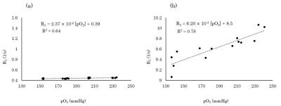
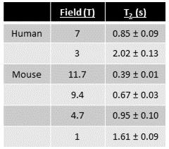
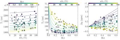
Can We Trust TRUST Venous Oximetry in Sickle Cell Disease?
Adam Bush, Thomas Coates, Herbert Meiselman, John Wood
In this work we derive a de novo T2b oximetry calibration curve for hemaglobin S containing red blood cells.  We then compare predictions made by this calibration and existing T2b calibrations in 84 subjects in vivo using TRUST MRI.  We found that predictions for venous oxygenation saturation and cerebral metabolic rate are widely different depending on the T2b calibration used for oximetry conversion. 


1881.   
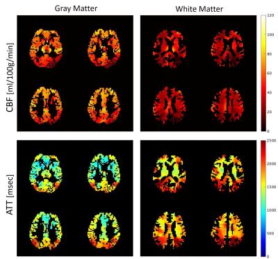
Estimation of Cerebral Blood Flow and Arterial Transit Time Using Partial Volume Corrected Multi-TI Arterial Spin Labeling Imaging
Youngkyoo Jung, Megan Johnston, Christopher Whitlow
A novel PVC algorithm using Multi-TI ASL, acquiring ASL images at multiple PLDs, has been proposed to estimate CBF and ATT in GM and WM separately. A 3D kernel was used to reduce noise sensitivity and improve the estimation power. The proposed method successfully estimated four perfusion parameters (GM CBF, GM ATT, WM CBF, and WM ATT) simultaneously, and may allow region- or voxel-based perfusion analyses in WM, as well as GM.


1882.  
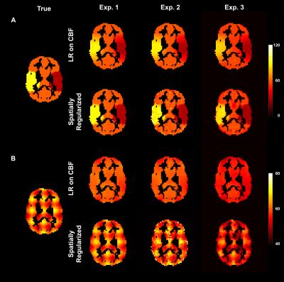
Investigating the Sensitivity to Partial Volume Estimates of Partial Volume Correction for Single Postlabeling Delay Pseudo-continuous ASL
Moss Zhao, Egill Rostrup, Otto Henriksen, Yingyi Xiao, Michael Chappell
This work investigates the sensitivity of partial volume correction methods to partial volume estimates using single-PLD PCASL. Random and biased errors were applied to partial volume estimates to simulate the variabilities in tissue segmentation. The results have indicated that current partial volume correction methods trade off accuracy in spatial variations in CBF against sensitivity to noise and errors in the partial volume estimates.


1888.   
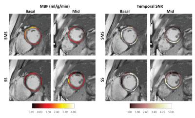
Simultaneous Multi-Slice Cardiac ASL
Terrence Jao, Krishna Nayak
Cardiac arterial spin labeling (ASL) is a promising technique for the quantification of myocardial blood flow (MBF) and has been shown to detect clinically relevant changes in myocardial perfusion under vasodilator stress. However, current cardiac ASL techniques have limited spatial coverage because they cannot be repeated for multiple slices due to limited duration of pharmacologically induced peak stress (~3 min). In this work, we demonstrate the feasibility of using blipped CAIPI bSSFP for cardiac FAIR ASL. 


1878.   
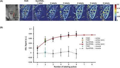
Physiologically synchronized multi-module pulsed arterial spin labeled (SymPASL) MRI
Hung Do, Krishna Nayak
Physiologically synchronized multi-module pulsed arterial spin labeling (SymPASL) involves pulsed labeling that is applied several times prior to pulsations in the arterial blood supply. Simulations and in vivo measurements in human kidneys demonstrate that SymPASL provides superior SNR and SNR efficiency compared to conventional flow-sensitive alternating inversion recovery (FAIR) ASL with a single labeling pulse. Simulations suggest that SymPASL provides comparable SNR and SNR efficiency to pseudo-continuous ASL (PCASL), with lower specific absorption rate (SAR).


1879.   
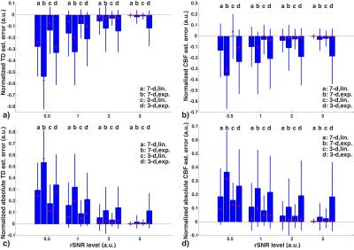
Hadamard-encoded Multi-delay PCASL: Should the Bolus Durations be T1-adjusted?
Jia Guo, Marc Lebel, Samantha Holdsworth, Greg Zaharchuk
It has been hypothesized that in multi-delay arterial spin labeling (ASL), employing T1-adjusted labeling durations (LDs) should provide a more balanced signal-to-noise (SNR) across ASL signals, therefore improving the accuracy of the transit delay (TD) and perfusion estimation. However, this claim has not been thoroughly tested. In this study, we evaluated the effects on the TD and perfusion estimation using LDs both with and without T1-adjusted weighting, using the Hadamard-encoded multi-delay ASL sequence. Using Monte Carlo simulations and in vivo experiments, T1-adjusted weighting was more prone to noise and was likely to underestimate the TD and perfusion measurements compared to that without. 


1897.  
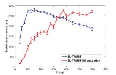
Spatially localised measurements of oxygen extraction fraction using modified T2-relaxation-under-spin-tagging (SL-TRUST)
Caitlin O'Brien, Thomas Okell, Peter Jezzard
An existing MR sequence, TRUST (T2-relaxation-under-spin-tagging), is adapted in order to obtain spatially localised T2 measurements for venous blood. T2-encoding, and hence determination of venous oxygenation, is achieved via localised encoding of the longitudinal magnetisation, that is then decoded in the sagittal sinus. Saturation pulses enable spatial localisation by removing signal from unwanted brain regions. Thus, hemispheric and global T2 measurements are acquired and compared. The delay between labelling and arrival of tagged venous blood in the sagittal sinus is evaluated and shown to increase in the presence of saturation pulses.
 
DSC & DCE
Traditional Poster
Contrast Mechanisms

 
Tuesday, 25 April 2017
Exhibition Hall  16:15 - 18:15


1900.  
Improving Simultaneous T1 and T2* Measurements for Dynamic Susceptibility Contrast MRI using a 3D Distributed Spirals Sequence
Laura Bell, Dallas Turley, Natenael Semmineh, James Pipe, C. Quarles
Multi-echo (ME) DSC-MRI enables the simultaneous assessment of contrast-agent induced T1 and T2* changes, but its sensitivity to and the quantification of these T1 changes could be confounded by long TR and TE. Using simulations, we demonstrate that conventional 2D ME scans underestimate contrast agent concentration and DCE-MRI kinetic parameters. To solve this problem, we propose a 3D ME spiral acquisition that enables lower TRs and minimal TEs for improved T1 quantification and a range of echo times to main T2* sensitivity.  


1901.   
Joint DCE- and DSC-MRI processing using the Gradient correction model
Ondrej Macícek, Radovan Jirík, Zenon Starcuk jr.
The contribution presents a method for simultaneous processing of the DCE- and DSC-MRI perfusion data acquired using a multi-echo sequence. It is an extension of the sequential application of the so-called gradient correction model. In the sequential approach, relaxivity parameters are estimated from the DSC signal based on perfusion parameters calculated from the DCE signal. Here the perfusion and relaxivity parameters are estimated using an iterative alternating optimization strategy. The impact on accuracy and precision is tested on synthetic data. The results show that the suggested approach can yield a remarkable improvement, especially for noisy data. 


1902.  
Correction of R2* Effects in Arterial Input Function of Fast Dynamic Contrast-Enhanced MRI for Accurate Cerebral Blood Flow Measurement
Benoit Bourassa-Moreau, Réjean Lebel, Guillaume Gilbert, Martin Lepage
Signal loss resulting from $$$R_2^*$$$ effects is usually neglected in fast dynamic contrast-enhanced perfusion imaging. A new flow-compensated 3D RF-spoiled dual gradient echo sequence was designed to quantify the $$$R_2^*$$$ variation during a bolus injection of contrast agent and its effect on cerebral blood flow measurement. Moderate $$$R_2^*$$$  effects are found to be amplified by the non-linear relationship between signal ratio and concentration. This is shown to lead to substantial changes in peak arterial input function and measured cerebral blood flow.


1903.  
Improved Arterial Input Function Measurements Using Phase-versus-Time and Modified Look-Locker Inversion Recovery: Phantom Validation Study
Nicholas Majtenyi, Gregory Cron, Hanif Gabrani-Juma, Andreas Greiser, Robert deKemp, Ran Klein, Thanh Nguyen, Ian Cameron
Dynamic contrast-enhanced (DCE)-MRI is used to quantify organ perfusion abnormalities in many diseases, but is prone to errors. This study investigated the accuracy of a new method for measuring the arterial input function (AIF) in a flowing-water phantom using phase-versus-time measurements with pre- and post-DCE Modified Look-Locker Inversion Recovery (MOLLI) T1 measurements (Phase+MOLLI). The Phase+MOLLI technique provides an important improvement over previous methods since it avoids signal saturation and gives correct [Gd] values for the washout of the AIF. The Phase+MOLLI method was validated to be accurate, reproducible, and flow-insensitive so that it may be used for clinical DCE-MRI. 


1899.  
Optimization of Echo Times for CBV Measurements in the Arterial Input Function, Brain, and High-Grade Tumor Tissue using Error Analysis for DSC-MRI
Laura Bell, Mark Does, Ashley Stokes, Leslie Baxter, Kathleen Schmainda, C. Quarles
This abstract demonstrates that the optimal TE for a single-echo DSC-MRI acquisition is 30-35 ms (without a preload) and 23-30 ms (with a standard full-dose preload) for a high-grade glioma population. Optimal TEs were calculated using error analysis on T2* values quantified during the first-pass of contrast agent using dual-echo DSC, and were shown to be the weighted-average of these values. Furthermore, we demonstrate that the optimal TE depends on tissue type (tumor/healthy tissue/AIF) and preload. 


1905.  
Impact of Reference Time Curve Determination on the Correction of Contrast Agent Extravasation in Dynamic Susceptibility Contrast MRI
Mu-Lan Jen, Ping Hou, Jason Johnson, Donald Schomer, Ho-Ling Liu
This study investigated the influence of reference time curve determination on DSC leakage correction. Our findings suggested that the reference time curve obtained with inclusion of leaky tissue could lead a significantly different in K2, which supported that the probable error in lesion blood volume quantification with inaccurate automated segmentation process.


1904.   
Dynamic Susceptibility Contrast MRI at 7T: Tail Scaling Analysis and Inferences About Field Strength Dependence
Linda Knutsson, Xiang Xu, Freddy Ståhlberg, Peter Barker, Pia Sundgren, Peter van Zijl, Ronnie Wirestam
In this study, a dynamic susceptibility contrast MRI (DSC-MRI) protocol for cerebral perfusion imaging at 7T was designed. With reduced contrast agent dose, the obtained perfusion maps showed the same visual appearance as seen at lower field strengths. In addition, a correction method was applied to obtain quantitative estimates of CBF and CBV in order to assess whether previous predictions of a field-strength dependence of the in vivo transverse relaxivity, leading to overestimated perfusion estimates, were supported. We concluded that assumptions of a field-strength dependence were plausible, based on observations of further elevated CBF and CBV estimates at 7T.


1906.  
Robust reference-region DCE-MRI analysis with a vascular component and two-fit analysis
Zaki Ahmed, Ives Levesque
The Extended Reference Region Model (ERRM) can quantify tumour perfusion without needing an arterial input function and includes a vascular term to account for the plasma volume. The addition of the vascular term also leads to larger variability in the estimated parameters. This study notes that one of the ERRM fitting parameters should be the same for all voxels. A two-fit approach is proposed that takes advantage of this constraint to reduce the number of fitting parameters from four to three. Evaluation in simulation and in-vivo found that the proposed two-fit approach resulted in a substantial decrease in variability for KTrans and kep estimates. 


1908.   
Feasibility and value of View-shared Compressed Sensing combined fast DCE-MRI in quantitative evaluation of unilateral renal embolism in rabbits
Hanjing Kong, Bin Chen, Hao Li, Bihui Zhang, Haochen Wang, Xiaodong Zhang, Min Yang, Jue Zhang, Xiaoying Wang, Jing Fang
Dynamic-contrast enhanced MR imaging is widely employed as a clinical tool in kidney imaging and renal function measurements. Some novel works have been made in improve temporal resolution. In this work, we adopt a 3D Cartesian MRI with compresses sensing and variable view sharing sequence to explore its evaluation in renal embolism assessment. GFR was calculated and renal embolism was confirmed by histological results. Fast DCE-MRI is a promising method for renal embolism diagnose. 


1910.   
Influence of parameter initial values on DCE parameter estimates in pharmacokinetic modeling: a simulation study
Charlotte Debus, Ralf Floca, Amir Abdollahi, Michael Ingrisch
In pharmacokinetic analysis of DCE-MRI data, the choice of initial parameter values for fitting has been reported to have a significant impact on the outcome of the optimization and hence, on parameter estimates. In this study, we investigated the influence of initial values by fitting simulated concentration time curves with varying combinations of initial parameters, using the two compartment exchange model. The resulting parameter estimates were visualized and compared to the true values, used for simulation, by means of relative errors. Results showed that the choice of initial values has little influence on the precision of the pharmacokinetic analysis.


1911.   
Nested tracer-kinetic model-based DCE-MRI reconstruction from under-sampled data
Sajan Goud Lingala, Yi Guo, Naren Nallapareddy, Yannick Bliesener, R Marc Lebel, Krishna Nayak
We propose a novel nested tracer-kinetic (TK) model based constrained reconstruction method for DCE-MRI reconstruction from under-sampled data. This approach models the concentration time profiles as a sparse linear combination of temporal bases constructed from TK models of varying complexity. Subspaces from the models of plasma volume, Patlak, and the extended-Tofts are constructed. A spatial mask determining the TK model complexity at every pixel location is derived. Reconstruction involves iteration between data consistency and pixel wise projection of the concentration profiles on one of the three subspaces. We demonstrate its utility in retrospective under-sampled reconstruction of brain tumor DCE-MRI datasets.


1909.   
Comparison of (k,t) sampling schemes for DCE MRI pharmacokinetic parameter estimation
Yannick Bliesener, Sajan Lingala, Justin Haldar, Krishna Nayak
We demonstrate an approach to evaluate and compare (k,t) sampling patterns for DCE-MRI.  We compute Cramér-Rao lower bounds on the variance of pharmacokinetic (PK) parameter estimates, using pathologically- and anatomically-realistic digital reference objects. The framework allows for the optimization of sampling patterns independent of any specific estimator. We apply this framework to a 2D reference object for four sampling patterns: keyhole, TRICKS, lattice, and golden angle sampling. It is shown that TRICKS, lattice, and golden angle sampling enable low variance estimation for low undersampling factors. Out of these, lattice sampling keeps variances lowest with increasing undersampling factors.


1912.  
Measurement of Murine Single-Kidney Glomerular Filtration Rate using Dynamic Contrast Enhanced MRI
Kai Jiang, Hui Tang, Prassana Mishra, Slobodan Macura, Lilach Lerman
A method for noninvasive assessment of mouse glomerular filtration rate (GFR) using dynamic contrast enhanced MRI (DCE-MRI) was developed and validated. The kinetics of gadolinium in the abdominal aorta and two kidneys were measured using a snapshot fast low angle shot based T1 mapping method with a temporal resolution of 1 s. A modified bi-compartmental model was used for quantification of GFR by a least-squares fitting. As a reference standard, GFR was also measured using FITC-inulin clearance. Single-kidney GFR measured from both methods showed a good agreement, suggesting the proposed DCE-MRI method provides an accurate measurement of murine single-kidney GFR. 


1913.  
Patlak analysis of dynamic gadoxetic acid-enhanced MR imaging is an effective and  simpler alternative to compartmental pharmacokinetic modelling for assessing liver function
Matthew Orton, Mihaela Rata, Dow-Mu Koh, Maria Bali, Robert Grimm, David Collins, James d'Arcy, Martin Leach
Liver perfusion and function can be assessed using gadoxetic acid combined with DCE-MRI imaging and pharmacokinetic (PK) modelling.  Whilst compartmental PK models give a good account of the contrast changes over the first five minutes of enhancement, the Patlak graphical approach is a simpler alternative that is more easily implemented.  Patlak evaluation requires the specification of a delay time after which the initial transients in the uptake curves have decayed, so the purpose of this abstract is to present a preliminary evaluation of the sensitivity of liver uptake rate estimates to the Patlak delay time.


1907.  
Quantitative DCE-MRI Analysis using a Reference Tissue and AIF Tail
Zaki Ahmed, Ives Levesque
The reference region model can quantify tumour perfusion without needing an arterial input function (AIF) and provides relative estimates, i.e. KTrans/KTrans,RR and ve/ve,RR which are usually converted to absolute KTrans and ve by using literature-based values for KTrans,RR and ve,RR. However, this approach fails to account for inter-patient variability. This study proposes a method that uses the Reference Region and AIF Tail (RRIFT) to estimate patient-specific KTrans,RR and ve,RR. The AIF tail is the post-peak part of the AIF and is easier to measure than the complete AIF. Evaluation in simulation and in-vivo showed that RRIFT provides comparable results to Tofts model, and even outperforms the Tofts model at slower temporal resolutions.


1915.   
Absolute Quantification of Brain Perfusion using Golden Angle Compressed Sensing DCE-MRI
Radovan Jirík, Marie Danková, Pavel Rajmic, Lucie Krátká, Lenka Dvoráková, Eva Dražanová, Zenon Starcuk, jr.
A DCE-MRI method for absolute quantification of cerebral blood flow (CBF) and volume (CBV) and vessel permeability surface area product is presented. It is based on L+S compressed sensing, the two-compartment exchange model (2CXM) and blind deconvolution estimation of the arterial input function. The method is evaluated on data from a healthy rat.


1916.   
Parameters From Dynamic Contrast-Enhanced Magnetic Resonance Imaging Are Biomarkers Predicting Response after Radiation to Brain Metastases
Zhuo Shi, Lizhi Xie, Peng Wang, Xinming Zhao, Han Ouyang
Dynamic contrast-enhanced (DCE) MRI provides additional information regarding blood-brain barrier integrity, and Ktrans is directly proportional to the level of permeability of the blood-brain barrier. In our study, we found demonstrates that SRS of cerebral metastasis is associated with a reduction of Ktrans values in the early post-treatment period. DCE-MRI derived parameters of Ktrans may be a promising imaging biomarker of tumor aggressiveness.


1917.   
Quantification of tracer kinetic and hemodynamic parameters of human breast tumor and fibro-glandular tissue using DCE-MRI data
Snekha Sehrawat, Pradeep Kumar Gupta , Meenakshi Singhal, Rakesh Kumar Gupta, Anup Singh
Objective of current study was to develop a framework for computing tracer kinetic parameters using GTKM model and hemodynamic parameters using first pass analysis of human breast tissue for characterizing of breast lesion; and also differentiation of the histological grade II and III of breast cancer. A significant difference between benign, malignant and fibroglandular tissues; and also between grade II and III of breast cancer were observed.


1918.  
The Vanishing Shutter-Speed Limit
Ruiliang Bai, Charles Springer, Jr., Peter Basser
Dynamic-contrast-enhanced MRI (DCE-MRI) has been widely used to characterize microvasculature permeability.  Recently, it was shown to reveal metabolic activity using the shutter-speed pharmacokinetic paradigm (SSP), in which steady-state intra/extracellular water exchange kinetics was incorporated into DCE-MRI data analysis. Interesting insights into DCE-MRI signals come from modeling the extravascular tissue MR signal. The questions addressed here are, “When can extravascular 1H2O longitudinal magnetization recovery from inversion/saturation still be described by a single-exponential process, and when can the intra/extracellular water exchange kinetics be accurately determined?”


1919.  
Whole Brain Dynamic Contrast-Enhanced MRI Study of Blood-Brain Barrier Disruption in Systemic Lupus Erythematosus Patients: Implications on the Choice of Tracer Kinetic Model
Dennis Cheong, Mary Stephenson, Sen Hee Tay
Current evidence suggests that blood-brain barrier (BBB) integrity is one of the potential biomarkers to diagnose neuropsychiatric systemic lupus erythematosus patients. We use DCE MRI and performed tracer kinetic analysis using both a distributed parameter (DP) model and the modified Tofts (MT) model. More Leaky BBB in SLE patients with anti-NR2 in their sera than the controls was detected by the permeability related parameters from both models. However, Ktrans of MT model, which is commonly used in DCE MRI of cancer studies, might be less reliable than DP model permeability parameters in this study.


1920.   
Feasibility and Value of Quantitative Dynamic Contrast Enhancement MR imaging in the Evaluation of Lymphoma and Inflammatory Pseudotumor in the Orbit
Liyuan Song, Lizhi Xie, Junfang Xian
This work assessed the feasibility of quantitative parameters derived from dynamic contrast enhanced MR imaging (DCE-MRI) and evaluated the value of quantitative dynamic contrast enhanced MR imaging in the differential diagnosis between lymphoma and inflammatory pseudotumor in the orbit.

From the results we can see that it is feasible that quantitative parameters of DCE-MRI can be applied in the differential diagnosis between lymphoma and inflammatory pseudotumor in the orbit. Thus, it can probably be used as imaging biomarkers to predict prognosis and aggressiveness of orbital lymphoma.


1921.  
Assessment of hydrodynamics and T2 alterations in spontaneously hypertensive rat under short-term hyperhydration
Kun-I Chao, Cheng-He Li, Sheng-Min Huang, Pei-Lun Yu, Kung-Chu Ho, Shang-Yueh Tsai, Ping-Huei Tsai, Fu-Nien Wang
With a fluid infusion of 2% of body weight in the D2O perfusion imaging experiment, the tissues are expected in a hyperhydration state. In this study, we conduct both D2O and H2O infusion experiments on spontaneously hypertension rat (SHR). Fast and slow flow of brain were analyzed by a two-compartmental parallel model. Pre- and post-infusion T2 maps were acquired. The slow flow matched the T2 prolonged regions, which could be due to the CSF production and flow.


1914.   
Vastly accelerated linear least squares fitting with numerical optimization for dual delay compensated quantitative liver perfusion mapping
Ramin Jafari, Yi Wang, Martin Prince, Pascal Spincemaille
Accurate liver perfusion quantification requires correction for dual arterial and portal venous input delays, but such dual delay correction in current nonlinear perfusion methods is computationally too expensive to apply in perfusion mapping. We realize that the kinetic equation is a linear differential equation that would allow fast linear processing. Accordingly, we propose to use linear least squares (LLS) fitting to this kinetic equation with fast conjugate gradient search for processing dynamic contrast enhanced MRI data. Our proposed LLS vastly (~300 times) accelerate computation in perfusion quantification, enabling for the first time accurate liver perfusion mapping with dual delay corrections. 


1922.  
Feasibility study of a Dialyzer as a multi-compartment Perfusion Phantom for microvascular tracer kinetic Modelling
Tanja Gaa, Lothar Schad, Frank Zöllner
Dynamic contrast enhanced MRI combined with tracer kinetic modelling allows for the determination of quantitative perfusion parameters. To achieve standardized examinations and data analysis, phantoms are employed to investigate the reproducibility of perfusion parameters by imitating tissue on capillary level. In this study we used a dialysis filter with a semipermeable membrane of the fibers which enables the simulation of two compartment kinetics and can thus serve as imitation of capillaries and interstitium which might be closer to the anatomical conditions.


1923.  
Feasibility of using Active Contrast Encoding (ACE)-MRI for Assessment of Tumor Treatment Response
Jin Zhang, Willis Chen, Kerryanne Winters, Sungheon Kim
Active Contrast Encoding MRI (ACE-MRI) is a recently proposed method to conduct DCE-MRI experiment without the need to perform separate T1 and B1 measurement. The purpose of this study is to further investigate the feasibility of using the ACE-MRI method for evaluation of tumor treatment response in a mouse model of breast cancer. The results of the ACE-MRI method were compared with conventional DCE-MRI data analysis with separately measured T1 maps. Our preliminary results demonstrate that the ACE-MRI method can be used to evaluate tumor treatment response reliably.
 
Relaxation: Mechanisms & Applications
Traditional Poster
Contrast Mechanisms

 
Tuesday, 25 April 2017
Exhibition Hall  16:15 - 18:15


1937.   
Quantitative relaxation time mapping of axillary lymph nodes and recommended parameters for 3T lymphatic node substructure imaging
Rachelle Crescenzi, Paula Donahue, Vaughn Braxton, Allison Scott, Manus Donahue
A lack of MRI methods exist that are designed with sensitivity to the lymphatics, even though components of the lymphatic system have been discovered in every major organ system of the body and likely play an understudied role in disease. In this work we performed quantitative relaxation time mapping in axillary lymph node substructures, the cortex and hilum, for the first time at 3 Tesla and used these values to optimize structural imaging parameters for the lymphatics. Knowledge of fundamental MR parameters in the lymphatics is the first step to developing novel imaging sequences that exploit lymphatic tissue in vivo.   


1926.  
MR Contrast Effects of Intrinsically Gd-chelated Melanin Nanoparticles at 1.5 and 11.7 Tesla
Soojeong Cho, Weiguo Li, Andrew Larson, Dong-Hyun Kim
The development of polymeric contrast agents exhibiting a high MR relaxivity has been achieved using bio-inspired metal chelating melanin nanoparticle (Mel NP) synthesized with dopamine or L-3,4-dihydroxyphenylalanine (L-DOPA).  In here, we described our simple one-pot synthesis to prepare new Gd chelated Mel NP and their specific features of efficient MR T1 imaging along with their high intrinsic Gd chelation efficiency. 


1933.   
Development and Systematic Analysis of 2D and 3D GRE Myelin Water Imaging
Hyeonggeol Shin, Se-Hong Oh, Jongho Lee
 In this study, we developed high quality GRE-MWI methods for 2D and 3D acquisitions and performed systematic analysis on TR and flip angle. The myelin water images showed that image quality was higher in 2D than in 3D. Over a range of TR (56 ms to 1630 ms), the myelin water fraction was uniform when Ernst angles (assume T1 of 800 ms) were used. The fractions were overestimated when larger flip angles were used.


1924.  
Accurate Tissue Oxygen Level-dependent MRI with true T1-weighted signal
SoHyun Han, HyungJoon Cho, Seong-Gi Kim
Tissue oxygen level dependent (TOLD) MRI utilizes that T1 is directly related with tissue pO2. TOLD MRI is acquired by gradient echo based with a minimized TE. However, T2* contribution may not fully be suppressed. Here, we investigated the modulations of longitudinal and transverse relaxation times with oxygen challenge (OC) and compared TOLD signals from FLASH, UTE, and TSE at 7 T and 15.2 T. At both magnetic fields, T2 did not change with tissue pO2 while T1 did. Spin echo and UTE appear to reflect true pO2 levels due to invariability in T2 during OC and minimized T2* with ultrashort TE, respectively.


1927.  
Effect of vendor specific formalin composition and concentration on post-mortem MRI of human brain tissue
Christoph Birkl, Martin Soellradl, Anna Toeglhofer, Johannes Haybaeck, Lukas Pirpamer, Franz Fazekas, Stefan Ropele, Christian Langkammer
Formalin fixation is common procedure to prevent tissue autolysis by crosslinking proteins. Not unexpectedly this affects the relaxation properties of the tissue. In addition, quantitative relaxation time constants of formalin fixed brain tissue show a broad variation across different studies. To investigate the contribution of the formulation of formalin to this variability we investigated MR relaxation times of pure formalin solutions from different vendors and the effect of formalin concentration on MR relaxation times of fixed brain tissue. Our results showed a strong variation of relaxation times depending on the concentration used and more importantly on the vendor specific composition of the formalin solutions.


1940.   
Determination of Oxygenation Extraction Fraction for People with Sickle Cell Anemia using Calibration Model Specific to SCA Blood
Wenbo Li, Xiang Xu, Peiying Liu, John Strouse, Hanzhang Lu, Peter van Zijl, Qin Qin
For the blood T2-based MRI oximetry methods, the calibration model to convert blood T2 to blood oxygenation (Y) is critical to calculate the brain oxygen extraction fraction (OEF). Here, we established a calibration model specific to people with sickle cell anemia (SCA) using in vitro blood T2 measurements on SCA blood samples under various conditions. The results show that the use of calibration models based on normal blood underestimate Y, and thus overestimate OEF, for individuals with SCA. Using a fast T2 protocol to measure oxygenation in the internal jugular vein (IJV), the whole-brain OEF values of individuals with SCA were determined and compared with healthy volunteers. 


1928.   
Relaxation time shortening by oxygen molecules: Strong enhancement in a viscous solution with cellular viscosity
Masayuki Taguchi, Toru Yamamoto
Since oxygen molecules are paramagnetic, they shorten the relaxation time as well as gadolinium contrast medium. The effect of relaxation time shortening by the paramagnetic substance is enhanced with an increase in viscosity of the solution as in the cell. We investigated the longitudinal and transverse relaxivities of oxygen in viscous solution with cellular viscosity and clarified that the relaxation time shortening by oxygen molecules strongly increases with an increase in viscosity. This effect of oxygen in the cell may be visible by using pulse sequences that enhance the signal from the cellular proton.


1929.  
Characterization of the Four Pool Model in formalin-fixed sheep's brain using NMR spectroscopy
Alan Manning, Alex MacKay, Carl Michal
Despite its importance, T1 relaxation in brain and spinal cord is not well understood. The Four Pool Model gives a fundamental framework for its understanding in white and grey matter tissue. In this work, we characterize the Four Pool Model for the first time in formalin-fixed sheep's brain using NMR spectroscopy. We find this is a suitable system, and our results are consistent with previous studies: T1 is multi-exponential and the values measured result from a convolution of pure relaxation and exchange processes.


1930.   
Transverse relaxation of cerebrospinal fluid depends on glucose concentration
Alexia Daoust, Steven Dodd, Govind Nair, Nadia Bouraoud, Stephen Jacobson, Stuart Walbridge, Daniel Reich, Alan Koretsky

Brain relaxometric properties are widely used by the NMR community. While brain tissue relaxivities are well established, much less work has been done on CSF relaxivities. To clarify this point, we characterized the CSF relaxometric properties at various field strengths in vivo and in vitro. Our results suggest that low field is more optimal to quantify CSF T2 due to smaller residual gradients. There is a significant difference between in vitro CSF T2 vs saline T2 that is mostly explained by the glucose relaxivity. This finding was confirmed in vivo, opening the possibility of studying glucose regulation of CSF at the resolution of MRI.


1931.  
Origin of Dipolar Effects - Achilles Tendon at 3T and 11.7T.
Nikolaus Szeverenyi, Jiang Du, Graeme Bydder
We examined the MR image appearance of human Achilles tendon as a function of orientation to the B0 field at 3 T and 11.7 T.  Images were registered and displayed a remarkable similarity in the fine discernable structural features at both field strengths.   Residual dipolar effects are responsible for the contrast on these images, rather than frequency changes that scale with magnetic field.


1935.   
Spatial distribution of myelin concentration in healthy volunteers measured in GRE myelin water imaging, ViSTa myelin water imaging, quantitative MT and DTI
Dongmyung Shin, Sehong Oh, Jongho Lee
This study investigated the spatial distribution of several myelin imaging methods. Myelin water fraction in GRE-MWI, apparent MWF in ViSTa-MWI, fractional pool size (F) in qMT, MT saturation, MT ratio, and FA in DTI were compared for their spatial distribution in white matter. Strong correlations were measured particularly between GRE-MWI and ViSTa-MWI and also among MT contrasts. FA showed least correlations with the other parameters.


1932.   
Effect of T1 on Multi-echo Gradient Echo based Myelin Water Fraction
Hongpyo Lee, Yoonho Nam, Dong-Hyun Kim, Hongpyo Lee
Myelin Water Fraction uses the property that myelin water has shorter T2* relaxation time compared to axonal/extra-cellular water. Previous studies found that not only the T2/T2* relaxation time but also the T1 relaxation time is difference between these compartments. The T1 relaxation time of myelin water is known to be affected by the cross-relaxation therefore the term “apparent T1” is considered more accurate. In this study, we analyze the effect of this ‘apparent’ T1 in mGRE based MWF. Our results show that the MWF estimation is dependent on the differential T1 of different compartments. T1 effect on the MWF can makes overestimation error. Using the low flip angle could reduce the this error, but it gives rise to insufficient SNR on MWF. Also, increasing TR could be another choice for reducing estimated error, but it leads to inefficient scan time. Thus, GRE-MWF is needed to compensated T1 effect for accurate quantification.


1939.   
Hemodynamic response to respiratory challenge evaluated by dynamic R2’ imaging: application for acute renal ischemia caused by microsphere-induced renal artery embolism
Chengyan Wang, Bihui Zhang, Haochen Wang, Hanjing Kong, Fei Gao, Li Jiang, He Wang, Xiaodong Zhang, Min Yang, Jue Zhang, Xiaoying Wang, Jing Fang
The clinical use of iodinated or gadolinium contrast agents for renal hemodynamic imaging is limited in the presence of renal dysfunction due to its increased risks of exacerbating renal damage. Therefore, we performed a modified HRI technique with a specific-designed magnetic-susceptible sequence that could separate R2’ from the BOLD signals in a unilateral microemboli-induced AKI model. The results show that R2’ in normal or less affected regions reduced after carbogen challenge, while the R2’ in the most affected lesions increased significantly. The dR2’ map could indicate the most affected areas accurately confirmed with the final anatomic T2w image.


1938.   
Relaxation Times and Magnetic Susceptibility of Human Umbilical Cord Blood at 3 Tesla
Sharon Portnoy, Natasha Milligan, Mike Seed, John Sled, Christopher Macgowan
With an increasing proportion of fetal cardiovascular MRI scans being performed at 3 Tesla, there is growing need for an accurate calibration, which characterizes relationships between MRI properties (T1, T2, susceptibility) and blood properties (oxygen-saturation, sO2 and hematocrit, Hct) at 3T. Accordingly, relaxometry measurements were performed at 3T on cord blood specimens (N=89) with a broad range of hematocrits (0.09<Hct<0.82) and oxygen-saturations (7%<sO2<100%). We also measured fetal blood susceptibility, which, to our knowledge, has never been reported. The data were effectively described by a simple, two-compartment model for blood.


1925.   
Direct Assessment of Magnetization Transfer Effects with T1 qMRI
Mitchell Horn, Ning Hua, Stephan Anderson, Hernan Jara
Purpose: To measure inaccuracies on T1 qMRI caused by magnetization transfer effects that are inherent to MRI pulse sequences. Methods: The mixed-TSE pulse sequence was used at several RF power levels to image a series of agarose gels, sucrose solutions, and Gd solutions. Results: Measured T1s are increasingly underestimated as a function of increasing semisolid pool size and RF power level. Conclusion: Uncorrected magnetization transfer effects can cause large underestimation of T1 measurements in tissue imaged with RF intensive pulse sequences. This work could have implications for the design of more accurate qT1 mapping algorithms.


1941.  
Cause of death or caused by death: Differentiation of thromboemboli and post-mortem clots using quantitative MRI
Bridgette Webb, Martin Urschler, Marlene Leoni, Bernhard Neumayer, Thomas Widek, Sylvia Scheicher, Rudolf Stollberger, Thorsten Schwark
MRI is increasingly being used in post-mortem examinations to assist in determining cause of death. Post-mortem changes, such as the formation of post-mortem clots (PMC), present a specific challenge in forensic imaging where differentiation between these alterations and pathological findings (e.g. thromboemboli) is essential. This work imaged thromboemboli and PMC samples collected during autopsy at 3T. K-means clustering was applied to analyse voxel-grouping in the resulting quantitative data. Clusters specific to a single clot type were identified in 3 of the 4 samples. Preliminary findings indicated the existence of at least one common differentiating cluster specific to PMC.


1934.  
Making Myelin Water Imaging Mainstream: Multi-site and Multi-vendor Reproducibility
Emil Ljungberg, Julien Cohen-Adad, Lisa Lee, Alexander Rauscher, David Li, Anthony Traboulsee, Alex MacKay, Chase Figley, Jongho Lee, Shannon Kolind
Myelin water imaging (MWI) is a quantitative T2 relaxation-based MRI technique, measuring the amount of myelin in the central nervous system. We investigated the reproducibility of MWI using GRASE with 3T MR scanners from two different vendors at two sites. Using stimulated echo correction, differences in the refocusing flip angle profile between the two sites were effectively corrected for and myelin estimates between the two sites were found to be highly correlated (slope=1.12, R2=0.96). This is the first multi-vendor and multi-site reproducibility study of MWI using GRASE, encouraging future multicenter MWI studies.


1936.   
Measurement of T1, T2, and flip angle with double-angle inversion-recovery balanced steady state free precession with application to imaging the eye
Eric Muir, Shengwen Deng
Fast measurement of T1 and T2 can be made with high signal to noise using inversion-recovery Look-Locker (LL) bSSFP. However, the LL-bSSFP signal is dependent on the flip angle, which must be known for accurate T1 and T2 calculation. In this study we investigated methods to additionally map and correct for the flip angle with the acquisition of two LL-bSSFP scans with two different flip angles, avoiding the need for a separate flip angle mapping protocol. Simulations and scans of the eye showed that T1, T2, and the flip angle could be measured with the double-angle LL-bSSFP method.


1942.   
An optimised 2D MPRAGE sequence for T1 contrast in the fetal brain: application to slice to volume reconstruction and multiband acceleration
Giulio Ferrazzi, Anthony Price, Rui Pedro AG Teixeira, Francesco Padormo, Lucilio Cordero-Grande, Emer Hughes, Laura McCabe, Mary Rutherford, Maria Kuklisova Murgasova, Joseph V. Hajnal
Ultrafast single-shot T2 weighted images are common practise in fetal MR exams.  However, there is limited experience with fetal T1 acquisitions.

In this study, a 2D gradient echo sequence with an adiabatic inversion was optimized to be robust to fetal motion and to preserve contrast. We also explore slice to volume registration and super resolution reconstruction methods to enhance the resolution, and we show pilot data from a multiband accelerated version of the above-mentioned sequence.


1943.   
Combination of MT and R2* measurements to distinguish between contributions of semisolids and iron to R1
Xu Jiang, Erika Raven, Peter Gelderen, Jeff Duyn
In human brain, the apparent longitudinal relaxation rate (R1) primarily originates from magnetization transfer (MT) effects associated with the macromolecular 1H-proton fraction (f), although in some regions the iron concentration may contribute as well (Rooney, 2007). To quantify their relative contributions at 7 T, we measured f and R1 of water protons (R1,WP) corrected for MT effects using a pulsed, transient MT approach (van Gelderen, 2016). The iron concentration was taken from literature and correlated with R2*. The results indicate that the combination of R2* and MT measurements may provide a sensitive means to quantify R1,WP, f and iron concentration.
 
Electric Property Imaging & Susceptibility Imaging
Traditional Poster
Contrast Mechanisms

 
Tuesday, 25 April 2017
Exhibition Hall 1944-1968  16:15 - 18:15


1944.   
On the feasibility of synthetic sodium MRI based on tissue conductivity
Nazim Lechea, Yu Peng Liao, N. Jon Shah
The recently proposed magnetic resonance electrical property tomography opens new opportunities for sodium ion characterisation. In this study, a model was built by measuring electrical conductivity and sodium MRI in different saline solutions. We exploit this interdependence with additional temperature correction to build a synthetic sodium brain map based on in vivo electrical conductivity. The results were compared to sodium MRI measurements. A statistically significant Pearson correlation (p<0.001; r=0.43) was observed between the two modalities while Bland-Altman analysis revealed discrepancies between them with a mean difference ~4mMol/L in whole brain. The proposed approach facilitates tissue sodium extraction.


1945.   
Advanced 3D simultaneous conductivity and susceptibility imaging with mitigation of nonlinear phase evolution effect.
Kang-Hyun Ryu, Jaewook Shin, Hongpyo Lee, Dong-Hyun Kim
Simultaneous Quantitative conductivity and susceptibility mapping (QCSM) can acquire both 3D conductivity and 3D susceptibility map with a single scan 3D multi-echo GRE sequence. However, error occurs due to nonlinear phase evolution in some regions producing false values especially for conductivity. In this study, we analyzed this effect and propose a way to mitigate this effect.


1946.   
Evaluation of dual-echo EPI for in-vivo current mapping in individual subjects during transcranial Direct Current Stimulation
Mayank Jog, Lirong Yan, Kay Jann, Lucas Parra, Marom Bikson, Danny Wang
Transcranial Direct Current Stimulation (tDCS) is a neuromodulation technique that uses milliampere currents to modulate cortical excitability. Although tDCS has been shown to treat clinical symptoms and improve cognition, the distribution of tDCS currents in the brain remains unknown. Here we show a MRI technique that measures a component of the magnetic field induced by tDCS currents in-vivo. Experimental data acquired using this technique is compared to model-based simulations. Our results demonstrate that mapping the tDCS current-induced magnetic fields in individual subjects is feasible; opening an avenue to map electric currents directly and track target engagement in individual subjects.


1947.   
A stabilized convection-reaction magnetic resonance electrical property tomography (crMREPT) using viscosity-type regularization
Changyou Li, Wenwei Yu, Shao Ying Huang
Convection-reaction MREPT (crMREPT) method is a more general approach to reconstruct an electrical property map based on B1-maps from a magnetic resonance imaging (MRI) scanner compared to other existing methods in the literature, such as electrical property tomography (EPT) and local Maxwell tomography (LMT). However, crMREPT shows global spurious oscillations in the reconstructed maps and persistent artifacts in the region when $$$\small\triangledown|B_1|$$$  is small. We propose a solution to effectively mitigate the artifacts by applying a viscosity-type regularization. This abstract shows that the proposed method significantly increases the accuracy of the reconstructed electrical property maps and reduces the sensitivity to noise comparing to crMREPT.  


1948.  
Three-Dimensional Model-Based Conductivity Mapping with Regularization and a Non-Negativity Constraint
Kathleen Ropella, Douglas Noll
We present a three-dimensional model-based approach to calculating conductivity using MRI. Our proposed method is formulated as an inverse problem based on the phase-based conductivity equation. The algorithm includes an edge-preserving regularization term and we explore the utility of non-negativity constraints. Structural information is also used to inform regions over which to regularize. We present results for simulation, phantom, and human brain data. 


1949.   
Use of Padding to Eliminate Low Convective Field Artifact in Conductivity Maps Obtained by cr-MREPT
Gulsah Yildiz, Gokhan Ariturk, Yusuf Ider
Convection-reaction equation based Magnetic Resonance Electrical Properties Tomography (cr-MREPT) has been developed by Hafalir et.al; however Low Convective Field causes artifacts. This study investigates how padding can be used in practice to eliminate the LCF artifact in conductivity reconstructions. Simulation and experimental results are given to demonstrate that conductivity images are improved by padding.


1950.   
In Vivo Current Density Distribution of Brain during Electrical Stimulation
Nitish Katoch, Bup Kyung Choi, Saurav Sajib, Jin Woong Kim, Hyung Joong Kim, Oh In Kwon, Eung Je Woo
In deep brain stimulation, in vivo mapping of brain response is indispensable to secure its clinical applications. Estimation of current density distribution by stimulating currents can provide alternative way for understanding the therapeutic effects in electrical stimulation. MREIT method enables high-resolved mapping of electromagnetic tissue properties such as current density and conductivity of living tissues. In this study, we experimentally imaged current density distribution of in vivo canine brains by applying MREIT to electrical stimulation. The resulting current pathway and volume activation may provide useful information for adjusting the surgical planning and proving the therapeutic effects of DBS.


1951.   
Conductivity Measurements at 21.1 T using MR Electrical Property Tomography
Ghoncheh Amouzandeh, Jens Rosenberg, Samuel Grant
This study explores the opportunity of using ultra-high field (21.1 T) for producing electrical conductivity maps. Phantoms with known NaCl concentrations were used to compare conductivity maps generated by phase-based MREPT with actual values measured at 900 MHz with a dielectric probe. Phase-based MREPT also was evaluated for in vivo ischemic brain using an MCAO rat model. Increased conductivity values were noted in the stroked region of the rat brain.


1952.  
Current density imaging using novel carbon electrodes proposed for Deep Brain Stimulation (DBS)
Munish Chauhan, Neeta Ashok Kumar, Fanrui Fu, Rosalind Sadleir
Deep Brain Stimulation (DBS) is popular in the treatment of movement disorders. Conventional metal DBS electrodes present MR safety and susceptibility problems. We implemented novel carbon fiber electrodes that produced low susceptibility artifacts for imaging DBS current densities at 7 T. We used Magnetic Resonance Electrical Impedance Tomography (MREIT) to measure the z-component of the magnetic flux density (Bz) resulting from DBS-like pulses and reconstructed projected current density maps (JP) in two objects (agarose-gelatin phantom and ex-vivo piglet brain). We did not observe susceptibility artifact, and reconstructed projected current density maps agreed with simulation in the electrode neighborhood. 


1953.   
Quantitative Susceptibility Mapping of Paramagnetic and Diamagnetic Substances at 3T-MR, 1.5T-MR and CT.
Yasutaka Fushimi, Kyoko Takakura, Tomohisa Okada, Takuya Hinoda, Aki Kido, Kaori Togashi
We compared QSM value of paramagnetic and diamagnetic substances at 3T-MR, 1.5T-MR, and CT in this study. Phantoms with different concentration gadoterate meglumine (Gd) and calciumcarbonate (CaCO3) were created. QSM at 3T and 1.5T and CT imaging were performed for these phantoms. QSM of Gd phantom showed positive susceptibility and QSM of CaCO3 showed negative susceptibility. QSM demonstrated consistent results in paramagnetic and diamagnetic substances at 3T and 1.5T. QSM and CT values were correlated well.


1954.  
Susceptibility Mapping Reveals Inter-Hemispheric Differences in Venous Density in Patients with Brain Arteriovenous Malformations
Emma Biondetti, Alvaro Rojas Villabona, Anita Karsa, Rolf Jäger, David Thomas, Karin Shmueli
Brain arteriovenous malformations (AVMs) are vascular abnormalities characterised by arteriovenous shunting with the lack of a capillary bed. Recent studies have shown that it is possible to create diagrams of the cerebral vein network (venograms) from images of magnetic susceptibility ($$$\chi$$$). Here, we used $$$\chi$$$-based venograms to calculate the hemispheric percentage of venous voxels (venous density) in each hemisphere in AVM patients and healthy subjects. We found larger venous densities in the AVM-containing hemispheres than in the contralateral hemispheres, and more variable venous density in AVM patients than in healthy subjects.


1955.  
Evaluating The Accuracy of Susceptibility Maps Calculated from Single-Echo versus Multi-Echo Gradient-Echo Acquisitions
Emma Biondetti, Anita Karsa, David Thomas, Karin Shmueli
For Susceptibility Mapping (SM), Laplacian-based methods (LBMs) can be used on single- or multi-echo gradient echo phase data. Previous studies have shown the advantage of using multi-echo versus single-echo data for noise reduction in susceptibility-weighted images and simulated data. Here, using simulated and acquired images, we compared the performance of two SM pipelines that used multi- or single-echo phase data and LBMs. We showed that the pipeline that fits the multi-echo data over time first and then applies LBMs gives more accurate local fields and $$$\chi$$$ maps than the pipelines that apply LBMs to single-echo phase data.


1956.   
A Robust 3D Phase Unwrapping Method with Application to Quantitative Susceptibility Mapping
Junying Cheng, Biaoshui Liu, Yingjie Mei, Yihao Guo, Maodong Chen, Xiaoyun Liu, Wufan Chen, Yanqiu Feng
In susceptibility-weighted imaging and quantitative susceptibility mapping, phase unwrapping methods are generally needed to restore the underlying true phase from the principal period (-π, π]. However, current phase unwrapping algorithms are challenged by noise, rapid phase changes and open-end cutlines. In this paper, a 2D phase unwrapping method based on pixel clustering and local surface fitting (CLOSE) was extended to 3D. The simulation and in vivo data is used to test the performance of the proposed method, with a comparison to a region growing method and PRELUDE, which are widely used for human brain phase-related imaging. The proposed method is demonstrated that can accurately unwrap 3D phase data even in the presence of severe noise, rapid phase changes, and open-end cutlines, and will benefit phase-related 3D MRI applications.


1957.   
Iterative Background Phase Correction: Recovering Data for QSM
Johannes Lindemeyer, N. Shah
We present a technique to improve the preparation process of phase data for QSM. In order to compensate for data loss caused by strong local phase gradients near the surface of the brain support, harmonic and dipole-based fitting are used to determine the responsible background fields within an extended brain mask. In an iterative approach, phase data are corrected regarding such contributions prior to further QSM processing steps. This allows for the acquisition of more reliable field maps and larger evaluation masks, which finally leads to more robust susceptibility maps.


1958.  
Incorporating macroscale susceptibility in QSM reconstruction with 3D spiral acquisition.
Giang-Chau Ngo, Berkin Bilgic, Borjan Gagoski, Bradley Sutton
Macroscale magnetic susceptibility creates large variations in the phase images preventing the direct analysis of local tissue-dependent phase in quantitative susceptibility mapping. Unwrapping and background field removal are important steps for calculating tissue phases and susceptibility maps. In this work, a 3D spiral acquisition is combined with an image reconstruction pipeline modelling the macroscale magnetic susceptibility to provide more accurate tissue phases and susceptibility maps, by reducing image distortions and facilitating unwrapping and background phase removal. 


1959.   
Effects of Spatial Resolution on Quantitative Susceptibility Mapping
Timothy Colgan, Samir Sharma, Diego Hernando, Scott Reeder
The purpose of this study was to investigate the effects of spatial resolution on the performance of quantitative susceptibility mapping (QSM).  The combination of magnitude contrast in spoiled gradient echo images and the voxel sensitivity function can create significant errors in the estimated B0 field map.  This work evaluated the use of proton density weighted imaging and joint R2* and field map estimation to reduce the impact of imaging resolution on QSM.  Our results indicate that reducing magnitude contrast in the complex-valued echo images will reduce errors in the field map estimates and, thus, the susceptibility estimates in QSM.


1960.  
Evaluation of Quantitative Susceptibility Mapping for the visualization of the Globus Pallidus Internus and Subthalamic Nucleus at 3T and 7T
Fei Cong, Zhangyan Yang, Xiaohong Zhou, Bo Wang, Yan Zhuo, Lirong Yan
The globus pallidus internus (GPi) and subthalamic nucleus (STN) (Figure 1) were the commonly used nuclei in deep brain stimulation (DBS) for the treatment of the Parkinson disease. In this study, we evaluated the QSM, T2* weighted and SWI methods for the visualization the GPi and STN on both 3T and 7T scanners.  Our results showed the QSM at 7T displayed an excellent delineation of the GPi and STN, compared with T2* and SWI images. 


1961.   
Primal-Dual Implementation for Quantitative Susceptibility Mapping (QSM)
Youngwook Kee, Kofi Deh, Alexey Dimov, Pascal Spincemaille, Yi Wang
We investigate the computational aspects of the prior term in the field-to-susceptibility inversion problem for QSM. Providing a spatially continuous formulation of the problem, we analyze 1) its Euler-Lagrange equation that appears degeneracy and 2) the Gauss-Newton conjugate gradient (GNCG) algorithm that employs numerical conditioning. We propose a primal-dual (PD) formulation that avoids such degeneracy and use the Chambolle-Pock algorithm to solve this alternative formulation; thus numerical conditioning is not required. The two methods were tested and validated on numerical/gadolinium phantoms and ex-vivo/in-vivo MRI data. The PD formulation with the Chambolle-Pock algorithm was faster and more accurate than GNCG.


1962.  
The challenge of phase offset correction for quantitative susceptibility mapping at ultra-high field
Steffen Bollmann, Simon Robinson, Kieran O'Brien, Viktor Vegh , Andrew Janke, Lars Marstaller, David Reutens, Markus Barth
One challenge in quantitative susceptibility mapping (QSM) at ultra-high field (> 3 T) is the combination of phase data from phased array receive coils. We assessed the performance of COMPOSER (COMbining Phase data using a Short Echo-time Reference scan) with separate reference scans and with an intrinsic reference scan, as well as a reference-free single-channel method. Our results show that reference scans can bias QSM results at ultra-high field. We conclude that ultra-short echo-time reference scans reduce quantitation bias and remove the transmit field phase when using COMPOSER to combine phase data at ultra-high field.


1963.  
Accuracy of magnetic resonance based susceptibility measurements
Hannah Erdevig, Stephen Russek, Slavka Carnicka, Karl Stupic, Kathryn Keenan
We examined the accuracy of MR-based susceptibility quantification relative to conventional measurements in preparation of making a standard susceptibility phantom. SQUID magnetometry of tissue mimics provides absolute accuracy of approximately 100 ppb while MR-based techniques give relative accuracy of 10 ppb.


1964.   
Quantification of Dendronized Superparamagnetic Iron Oxide Nanoparticles in Rat Liver Using Quantitative Susceptibility Mapping
Jeam Barbosa, Imen Miladi, Patrick Poulet, Kofi Deh, Yi Wang, Carlos Salmon
Quantitative Susceptibility Mapping was environment independent to quantify iron of SPIONs compared with R2*. 


1965.  
Assessment of melanin content and its influence on susceptibility contrast in melanoma metastases
Sina Straub, Frederik Laun, Martin Freitag, Christian Kölsche, Heinz-Peter Schlemmer, Mark Ladd, Till Schneider
Melanoma metastases can be classified as melanotic or amelanotic based on their T1-weighted magnetic resonance signal. However, the underlying contrast mechanisms have remained unclear and have been attributed to melanin and/or blood products. In this study, non-hemorrhagic cerebral melanoma metastases were investigated using quantitative susceptibility mapping. Susceptibility values for metastases with no, small or high melanin content were very similar (-0.023±0.046 ppm / -0.006±0.02 ppm / -0.018±0.017 ppm). Non-hemorrhagic melanoma metastases show weakly diamagnetic susceptibility values and melanin is not a source of strong susceptibility.


1966.  
A Simple Phase Imaging REconstruction method (ASPIRE)
Korbinian Eckstein, Siegfried Trattnig, Simon Robinson
Combining phase data from multi-channel coils at high field is challenging. Many approaches involve optimization or iterative steps which require offline reconstruction. We present a simple method that avoids the need for unwrapping or other fragile or time consuming steps by using echo times that satisfy $$$T_{E,2}=2\,\cdot\,T_{E,1}$$$. ASPIRE is compared with the Roemer method, MCPC-3D-I and the Hermitian inner product. ASPIRE achieves similar phase matching quality to Roemer but has the advantage that the combined phase values correspond to the B0 field. It is less computationally demanding than MCPC-3D-I and has higher CNR than the Hermitian inner product.


1967.  
Enhancing Quantitative Susceptibility Mapping by Using Gradient L2 Regularization with Morphological Priors
Yihao Guo, Li Guo, Yingjie Mei, Yanqiu Feng
TKD, LSQR and CSC methods have been proposed to reconstruct QSM from field map. However, these three methods result in inconsistent in the magic angle and can be further improved. This work introduces efficient gradient L2 regularization with morphological information from magnitude images for enhancing the QSM reconstructed by TKD, LSQR and CSC methods.


1968.   
Functional and structural ex vivo “MRI staining” using manganese-enhanced MRI (MEMRI), Gd-DTPA and Mn-Gd mixture
Chika Sato, Kazuhiko Sawada, David Wright, Tatsuya Higashi, Ichio Aoki
To reveal brain 3D microstructures noninvasively and microscopically using MRI, we developed “ex vivo MEMRI” and Mn-Gd double-contrast methods, and then compared them with conventional ex vivo Gd-DTPA-doped contrast. Because MEMRI sample loses contrast after perfusion fixation, we examined the stability of Mn2+ accumulation in ex vivo tissue samples. In addition, we tried to improve the contrast of MEMRI in combination with Gd-DTPA. The Mn-Gd double-contrast showed novel contrast and improved visibility. The functional “MRI staining” methods we developed in this study will be useful for visualizing whole brain 3D microstructures with a higher throughput compared to histological staining.
 
All Things CEST/MT
Traditional Poster
Contrast Mechanisms

 
Tuesday, 25 April 2017
Exhibition Hall  16:15 - 18:15


1969.  
A singular value decomposition approach to quantitative magnetization transfer
Riccardo Metere, Samuel Hurley, André Pampel, Karla Miller, Harald Möller
Quantitative magnetization transfer experiments require extensive sampling of the off-resonance spectrum to obtain information of the relaxation properties of non-water protons. To reduce the acquisition times, the off-resonance sampling has been optimized in previous works based on the stability of approximate biophysical model fits. Here, we use a singular value decomposition approach for the analysis of the principal components. In particular we propose a data-driven optimization method for the acquisition scheme and we discuss the potential impact of applying this analysis for parameter estimations, including potential extensions of the classical biophysical models.


1970.  
Highly Accelerated Chemical Exchange Saturation Transfer (CEST) Imaging by Combining Parallel Imaging and Compressed Sensing at 3T
Kyungmin Nam, Namgyun Lee, Ha-Kyu Jeong, Seth Smith, Chulhyun Lee
Chemical Exchange Saturation Transfer (CEST) imaging is an emerging molecular MRI method.  It has been difficult for CEST imaging to adopt into clinical routine since CEST imaging is required relatively long scan time due to multiple saturation offsets. Here, we propose a novel, highly accelerated 3D CEST reconstruction technique by combining parallel imaging and compressed sensing.


1971.   
Fast, Reliable 3D Amide Proton Transfer Imaging of Brain Tumors at 3T with Variably-accelerated Sensitivity Encoding (vSENSE)
Yi Zhang, Hye-Young Heo, Shanshan Jiang, Paul Bottomley, Jinyuan Zhou
The clinical use of amide proton transfer (APT) imaging is hindered by long scan times. Accuracy generally limits the use of conventional sensitivity encoding (SENSE) methods in APT, to an acceleration factor of 2. A novel variably-accelerated sensitivity encoding (vSENSE) method can provide more accurate results and therefore substantially higher overall acceleration factors than conventional SENSE. Here, vSENSE is further developed to eliminate the requirement that one fully-sampled APT frame be acquired, and extended to three dimensions (3D). Furthermore, we combine vSENSE with parallel transmit saturation, and apply it proactively to three normal volunteers and eleven patients with brain tumors.


1972.  
isoAPTR* - a novel method to measure tumour pHi using CEST MRI
Kevin Ray, James Larkin, Michael Chappell, Nicola Sibson
Quantitative CEST MRI studies have so far been hindered by the fact that variations in multiple factors produce identical CEST effect changes. This remains the case when CEST MRI data are analysed using metrics that control for the contaminating effects of T1 and T2, such as APTR*. In this work we introduce isoAPTR*, a novel methodology which, in combination with independent measurement of labile proton concentration, can estimate the change in intracellular pH between two APTR* measurements. We demonstrate the utility of this method by applying it to measure the intracellular pH of U87 glioma in rats. 


1973.   
The comparison of different strategies for transmit field inhomogeneity correction of Amide-CEST and NOE effects at 7T
Vitaliy Khlebnikov, Johannes Windschuh, Jeroen Siero, Moritz Zaiss, Peter Luijten, Dennis Klomp, Hans Hoogduin
We compared three methods for B1 correction of relaxation-compensated Amide-CEST and Nuclear Overhauser Enhancement (NOE) effects at 7T: (1) a linear model; (2) the eight-point interpolation method; and (3) Bloch-McConnell equations (BE) correction algorithm. In the low B1 regime of 0.10 - 0.50 µT, a simple linear model is sufficient to mitigate B1 inhomogeneity of Amide-CEST and NOE effects at 7T.


1974.   
Quantification of Multi-pool Contribution to Endogeneous CEST Effects in Global Ischemia
Iris Zhou, Jerry Cheung, Enfeng Wang, Xiaoan Zhang, Phillip Sun
CEST MRI has been used for quantitative assessment of dilute metabolites and/or pH in ischemic tissue and in tumors. However, conventional asymmetry analysis (MTRasym) may be confounded by concomitant effects such as RF spillover, semisolid macromolecular MT and NOE effects. Therefore, decoupling multiple contributions is essential for elucidating the origins of in vivo CEST contrast for improved quantification. Here we used an Image Downsampling Expedited Adaptive Least-squares (IDEAL) fitting algorithm which is not strongly constrained by image SNR and initial values for fitting. It provides a closer estimation of MTRasym that calculated from acquired data than voxel-wise multi-pool Lorentzian fitting and unravels the major contributors to CEST contrasts between white and gray matters as well as to the CEST changes after global ischemia. 


1975.  
Lorentzian Probabilistic Sum based Z-Spectrum fitting approach for computing CEST and NOE contrast and its Comparison with Lorentzian Sum and Asymmetry Analysis
Ayan Debnath, Anup Singh
In this study, we proposed a Z-spectrum fitting method based upon Lorentzian Probabilistic Sum (LPS) for computing CEST and NOE contrast. Proposed fitting method was tested on multi-pool Z-spectra data acquired using simulations and from in-vivo human brain data at 7T. Proposed fitting results were compared with asymmetry analysis and another fitting method based on Linear Sum (LS) of Lorentzian functions. Results of this study show that both LPS and LS nicely fit z-spectra; however, LPS provide more accurate estimation of NOE and CEST contrast. Therefore, proposed LPS model can be used for improved estimation of separate CEST and NOE components.


1976.  
3D gagCEST of articular cartilage in the knee at 7 T correlates with clinical findings
Sander Brinkhof, Razmara Nizak, Vitaliy Khlebnikov, Dennis Klomp, Bennie ten Haken, Jeanine Prompers, Daniel Saris
The purpose of this study was to assess the sensitivity of 3D gagCEST at 7T in cartilage repair patients with respect to healthy volunteers. Six healthy volunteers were scanned for stability assessments and five patients with cartilage defects were included to assess clinical applicability of the gagCEST sequence. The mean GAG effect size in healthy controls is 10.2 %, which is three times higher than the coefficient of variation in the stability assessments. The results of this study demonstrate the stability of 3D gagCEST at 7T and the results from patients with cartilage defects indicate a correlation with clinical findings.


1977.   
Quantitatively Evaluate the Chemical Exchange Effect in Off-resonance Spin Lock Using Perturbation of Longitudinal Relaxation Rate in Rotating Frame (PLRF) Analysis
Yi Wang, Yang Fan, Jia-Hong Gao
Chemical exchange effect can be evaluated by off-resonance spin-lock sequence (CESL), but it remains unclear whether the effect is accurately reflected. In this study, with the help of perturbation of longitudinal relaxation rate in rotating frame (PLRF) analysis, we quantitatively compared signal from CESL and chemical exchange saturation transfer (CEST) sequences and tested the condition for acquiring high-quality signal from CESL sequence which can accurately reflect chemical exchange effect.


1978.  
Chemical exchange rotation transfer (CERT) using adiabatic hyperbolic secant pulses
Eugene Lin, Zhongliang Zu, Elizabeth Louie, Xiaoyu Jiang, Daniel Gochberg
Chemical exchange rotation transfer (CERT) is an emerging approach for imaging solutes and solute exchange that avoids some of the contributions from the asymmetric background in biological tissues that confound chemical exchange saturation transfer (CEST). To further improve the robustness of CERT methods when there is field inhomogeneity, we examined adiabatic hyperbolic secant pulses for solute saturation. In addition to addressing field homogeneity issues, this new method reveals a new mechanism to generate contrast based on the delay time between pulse.


1979.  
Elegant method to quantify chemical exchange processes for pH CEST imaging
Steffen Goerke, Johannes Windschuh, Moritz Zaiss, Jan-Eric Meissner, Mark Ladd, Peter Bachert
A novel concentration-independent approach is presented to determine the pH-dependence of exchange rates employing a single CEST image of a set of model solutions at different pH. Not only the comparatively short acquisition time, but also the robustness against variations in relaxation parameters makes this modality an elegant way to determine exchange rates in vitro. The calibrated functions are required for accurate pH mapping in vivo using CEST, as well as for design of exogenous CEST contrast agents.


1980.   
Muscular glycogen detection with CEST imaging in living rat at 14.1T
Elise Vinckenbosch, Hongxia Lei, Nicolas Kunz, Masoumeh Dehghani, Rolf Gruetter
Glycogen is the principle intracellular storage for energetic needs in muscle and information about its spatial distribution would be a great additional tool for traumatology and sport sciences. In this study, we aimed to map in vivo muscular glycogen using CEST imaging in rodent. We have shown that optimal B1 saturation power allowed CEST imaging glycogen distinguishable from the neighboring creatine at 14.1 Tesla. We applied our optimized protocol on muscle after exercise session, resulting in 50% reduction of MTRasym comparing to muscle at resting state. This is supporting the specificity of our method and is consistent with literature.


1981.   
Rapid 3D CEST using volumetric reduced field of view imaging
Jianbo Shao, Bing Wu, Hui Lin
A reduced field of view CUBE acquisition was proposed for CEST imaging. It is advantageous in situations where the imaging volume may be constrained to a region of the whole volume. Clinical feasible acquisition (10s per volume) was achieved at 2mm isotropic resolution.


1982.  
The origins of CEST contrast in ischemic tissue: effects of hypotonic stress on the nervous system of Aplysia californica
Tangi Roussel, Pavel Svehla, Denis Le Bihan, Luisa Ciobanu
Recent CEST MRI methods such as Amide Proton Transfer (APT) imaging allow the detection of brain tumors and stroke by generating novel image contrasts which depend on the chemical exchange. In this paper, we are studying the effects of ischemia on the CEST signal at a tissue level using the nervous system of Aplysia californica, a widespread model in neuroscience. A significant change in the Z spectrum was observed at 2.5 ppm after hypotonic shock in the abdominal ganglion and was quantified as a +2.88% MTR increase. Cell swelling, which is a known phenomenon in ischemic tissue, could potentially cause such effect.


1983.   
Simultaneous Acquisition of Multiple Z-spectra using Sinc-Modulated RF Pulse Trains in Gradient Encoded CEST MRI
Hirohiko Imai, Kiyotaka Miyake, Tetsuya Matsuda
We propose a use of sinc-modulated RF pulse train instead of the conventional continuous wave RF irradiation under the presence of constant gradient as a saturation scheme in chemical exchange saturation transfer (CEST) MRI aiming at a simultaneous acquisition of multiple Z-spectra. The proposed method was applied for a glutamic acid solution in water. The multiple Z-spectra could be observed along the gradient encoding direction by repetitive application of the saturation scheme. Thus, the present study shows the potential of the proposed methodology for accelerating the CEST MRI.


1984.   
Investigation of magnitude and phase CEST effects in fixed whole brains and tissue samples: a combined 3T and 9.4T study
Ana-Maria Oros-Peusquens, Nuno Andre da Silva, N. Jon Shah
Saturation transfer effects were investigated in fixed tissue at 3T and 9.4T and a range of saturation powers. Z-spectra on magnitude as well as phase data were studied with high spatial and spectral resolution, also due to the excellent performance of a PCA-based denoising algorithm. The preliminary findings do not support the presence of an APT effect in fixed tissue; however, saturation transfer effects could be enhanced in pathological tissue, similar to in vivo findings in e.g. tumour or stroke. This study opens the way to a systematic investigation of saturation transfer effects in healthy and pathological fixed tissue.


1985.   
An assessment of interdependent chemical exchange saturation transfer (CEST) signals from metabolites with overlapping chemical shift frequencies and proton exchange rates
Masaya Takahashi, Keisuke Ishimatsu, Shanrong Zhang, Kazufumi Kikuchi, A. Dean Sherry
The objectives are to investigate how the chemical exchange saturation transfer (CEST) signal measured at a given frequency is independent of the neighboring CEST signals. We measured the CEST signals from combinations of four metabolites at 3.5, 3, 2 and 1 ppm with 5 different powers and 3 different durations of presaturation pulse to investigate the parameter-dependence and interdependency of each CEST signal in phantoms. The CEST signal of glutamate was less impacted by concentration changes in other exchanging species by subtracting CEST signals at two different power levels. 


1986.   
Modeling the increased inhomogeneous magnetization transfer (ihMT) signal from high amplitude, low duty cycle irradiation
Gopal Varma, Aaron Grant, Olivier Girard, Valentin Prevost, Guillaume Duhamel, David Alsop
Pulsed implementations of RF saturation for MT and ihMT are dependent on the duty cycle of the MT pulse relative to its repetition period. The ihMT signal increases following preparation with high B1, low duty cycle MT pulses. A model was developed to fit ihMT data acquired at different duty cycles, along with the more standard variations in power and offset frequency. Output parameters from the fit were reasonable. The model allowed simulation of the ihMT signal as a function of experimental parameters related to the saturation preparation, and can be used to guide future experiments and/or optimize ihMT.


1987.   
Improved Measurement Precision for AACID CEST MRI of Brain pH using the 2 ppm Amine Resonance
Mohammed Albatany, Robert Bartha
The chemical exchange saturation transfer (CEST) method called Amine and Amide Concentration Independent Detection (AACID) can produce image contrast that is dependent on tissue pH.   The AACID value is calculated by taking the ratio of the 3.5 ppm amide CEST effect to the 2.75 ppm amine CEST effect and varies linearly with pH in the physiological range.  In the current study, we compare the range of the AACID values obtained in 24 mice with brain tumors and normal tissue using the 2 ppm and 2.75 ppm amine resonances.  Using the 2 ppm amine resonance increased the AACID range by 39% compared to the 2.75 ppm resonance and led to reduced measurement variability across the brain suggesting that using the 2 ppm amine resonance could improve AACID based pH measurement in-vivo.


1988.  
Towards Eliminating Magnetization Transfer (MT) Effect on CEST Quantification Using MR Fingerprinting: Simulations
Pei Han, Zhengwei Zhou, Kui Ying, Debiao Li
In this work, a new quantitative chemical exchange saturation transfer (q-CEST) method based on MR fingerprinting is developed to eliminate the influence of MT effect. Signal evolutions are generated by using a series of hard saturation pulses with different amplitudes and durations. The dictionary is constructed using the 2-pool model. Simulation experiments based on the 3-pool model were performed, and simulation results show that good mapping accuracy was achieved even in the presence of the MT effect.


1989.  
Towards an Early Detection of Prostate Cancer using Zinc-Sensitive iCEST MRI
Yue Yuan, Xiaolei Song, Jeff Bulte
As compared to normal prostate tissue, prostate cancer cells exhibit a dramatic reduction in zinc content. We have used 19F-based ion-CEST (iCEST) MRI to sense differential Zn2+ levels between normal and malignant prostate cell lines. We were able to observe clear differences in zinc-induced iCEST signal between normal cells and cancer cells, which was validated by microscopy using a zinc-sensitive fluorescent dye. Hence, iCEST MRI may have potential as a new means to non-invasively detect early prostate malignant transformation.
 

The International Society for Magnetic Resonance in Medicine is accredited by the Accreditation Council for
Continuing Medical Education to provide continuing medical education for physicians.